73
Rheumatoid Arthritis: Metacarpophalangeal Joint Reconstruction Arthroplasty
R. John Naranja, Jr. and Kevin D. Plancher
History and Clinical Presentation
A 53-year-old right hand dominant woman was originally diagnosed with rheumatoid arthritis 14 years ago. Medical management of her disease process has included chronic oral prednisone. Her primary complaint includes pain and deformity of her hands and decreased function secondary to lack of mobility and strength.
Physical Examination
The patient has ∼25 degrees of ulnar drift at the index through small finger metacarpophalangeal (MP) joints. An extension deficit at the MP measures ∼50 degrees (Fig. 73–1). The arc of motion of the digits at the MP joint is 20 degrees. Key pinch strength is 30 N and grip strength is 70 N at position II using the Jamar dynamometer. There is relative preservation of joint motion at the proximal interphalangeal (PIP) joint and wrist articulations without significant collapse or deformity.
PEARLS
- Meticulous detail is necessary when reaming the proximal phalanx of the little finger when there is poor bone stock and especially in patients with juvenile rheumatoid arthritis.
- When silicone synovitis and loss of bone stock occur, a resection arthroplasty should be considered.
PITFALLS
- Inadequate soft tissue balancing and poor hand therapy can lead to recurrence of the deformities and subluxation of these joints.
- Use of silicone implants can lead to an inflammatory response with subsequent erosive changes.
Diagnostic Studies
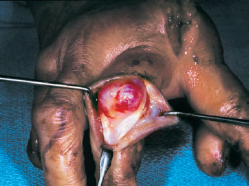
Radiographs of the hand to include posteroanterior (PA) (Fig. 73–2), lateral, and oblique views demonstrate subluxation and joint destruction of the MP joints in the index through small fingers of the right hand. Though there is relative diffuse osteopenia present, adequate bone stock appears present for potential implant arthroplasty.
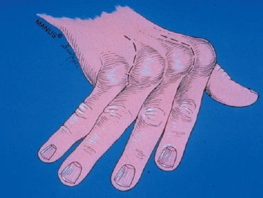
Figure 73–1 A classic ulnar drift and extension deficit in a rheumatoid hand seen at the metacarpophalangeal joints. (Illustration courtesy of The Indiana Hand Center and Gary Schnitz.)
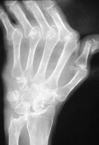
Figure 73–2 Posteroanterior (PA) radiograph of metacarpophalangeal (MP) subluxation and joint destruction.
Differential Diagnosis
Degenerative joint disease
Rheumatoid arthritis
Septic joints
Diagnosis
Rheumatoid Destruction and Deformity of the Index through Small Finger Metacarpalphalangeal Joints
The MP joint in the rheumatoid hand is commonly affected by joint destruction and deformity. Typically, the disease process involves bony articular destruction, and ulnar and volar capsular contracture with concordant radial capsular attenuation. This subsequently results in subluxation of the flexor sheath in an ulnar and volar direction in addition to ulnar displacement of the common extensor tendon. Intrinsic tightness contributes to overall dysfunction. Indications for arthroplasty reflect the disease process and include pain and disability with radiographic evidence of joint destruction, limited MP range of motion secondary to contracted myotendinous and ligamentous supports, ulnar drift that has failed other soft tissue procedures, and stiff distal interphalangeal (DIP) or PIP joints. Arthroplasty of the MP joint predictably addresses these processes and maintains function better than arthrodesis or simple resectional arthroplasty in patients with adequate bone stock, soft tissue coverage, and absence of preexisting infection.
Surgical Management
A transverse or longitudinal skin incision is created dorsally over the region of the metacarpal necks. The dorsal veins are preserved and the extensor hoods to each digit are exposed with care to protect the neurovascular structures radially and ulnarly. The extensor tendon is identified and is usually subluxed ulnarly with attenuation of the extensor tendon at its radial aspect (Fig. 73–3). The extensor hood is longitudinally incised ulnar to the common extensor tendon in the middle and ring fingers, and between the common extensor and the ulnar capsular ligament of each digit. Associated capsular tissue is released distally to adequately allow the base of the proximal phalanx to be dislocated dorsal to the metacarpal. The radial collateral ligament of each digit is preserved if possible. With the MP joint placed into flexion, the metacarpal neck is exposed subperiosteally and transversely osteotomized using a microair saw (Fig. 73–4). Care is taken to leave part of the metaphyseal flare for support of the prosthesis. Proliferative synovium along with the transected metacarpal head is completely removed (Fig. 73–5). Attention is then directed toward the base of the proximal phalanx where all cartilaginous surfaces are removed in addition to any marginal osteophytes. The volar plate is identified and released from its attachment at the base of the proximal phalanx. In all but the index finger it is then completely resected. In the index finger it will be used in conjunction with the radial collateral ligament for later capsular reconstruction. Release and resection of the volar plate allow further identification of the underlying flexor sheath. An incision is made into the flexor sheath with subsequent delivery of the flexor tendon into the wound using a blunt hook. Hypertrophic tenosynovium is then removed. Evaluation of appropriate tendon excursion following the tenosynovectomy will determine whether flexor tendon exploration on the palmar aspect of the hand over the A1 and A2 pulleys is necessary. This is typically done using a palmar/digital zigzag-type of incision with care to preserve the A1 and A2 pulleys in the index and long fingers to avoid further ulnar subluxation of these tendons. The ulnar intrinsic tendon in all but the index finger is also delivered into the wound and incised at its myotendinous junction typically at the level of the MP joint. In the index finger, preservation of the ulnar intrinsic tendon is recommended to help maintain supination in this digit for pinching function. This pinching function is further reinforced by reconstructing the radial collateral ligament of the index and long finger by using the collateral ligaments and capsule. The ulnar collateral ligament, which has previously been divided at its insertion at the proximal phalanx, is now further mobilized by dissection proximally on the metacarpal, with care taken to maintain its attachment. Similarly, the previously preserved radial collateral ligament is dissected as a unit with the overlying capsule from its distal insertion. These capsular tissues may be tagged and retracted for later reconstruction using a 2–0 Dacron suture.