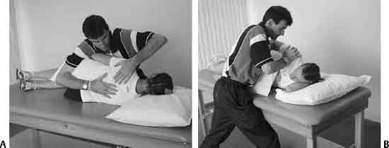
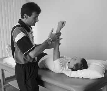
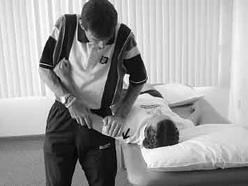
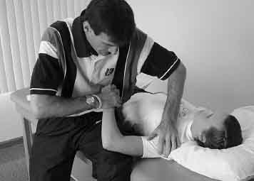
I 1 Rehabilitation of Shoulder Impingement: Primary, Secondary, and Internal 2 Rehabilitation of Micro-Instability 3 Rehabilitation of Macro-Instability 4 Rehabilitation of Adhesive Capsulitis 5 Rehabilitation of Acromioclavicular Joint Injuries 6 Classification and Treatment of Scapular Pathology Todd S. Ellenbecker Primary impingement, also known as compressive disease or outlet impingement, is a direct result of compression of the rotator cuff tendons between the humeral head and the overlying anterior third of the acromion, cora-coacromial ligament, coracoid, or acromial-clavicular joint.1,2 The physiologic space between the inferior acromion and superior surface of the rotator cuff tendons is termed the subacromial space. Measured using anteroposterior radiographs, it was 7 to 13 mm in size in patients with shoulder pain3 and 6 to 14 mm in normal shoulders.4 Flatow et al5 have shown that elevation of the humerus leads to predictable and reproducible patterns of subacromial impingement of the rotator cuff tendons against the overlying acromion and coracoacromial ligament. Biomechanical analysis of the shoulder has produced theoretical estimates of the compressive forces against the acromion with elevation of the shoulder. Poppen and Walker6 calculated this force at 0.42 times body weight. Lucas7 estimated this force at 10.2 times the weight of the arm. Peak forces against the acromion were measured in a range of motion (ROM) between 85 degrees and 136 degrees of elevation.8 This position is a functionally important one for daily activities, sport-specific movements,9,10 and situations commonly encountered on a job as well. The position of the shoulder in forward flexion, horizontal adduction, and internal rotation (IR) during the acceleration and follow-through phases of the throwing motion is likely to produce subacromial impingement due to abrasion of the supraspinatus, infraspinatus, or biceps tendon against the overlying structures.9 These data provide scientific rationale for the concept of primary impingement or compressive disease as an etiology of rotator cuff pathology. Neer1,2 has outlined three stages of primary impingement as it relates to rotator cuff pathology. Stage I—edema and hemorrhage—results from the mechanical irritation of the tendon; this is caused by impingement incurred from overhead activity. Observed in younger, more athletic patients, it is a reversible condition with conservative physical therapy. The primary symptoms and physical signs of this stage of impingement or compressive disease are similar to the other two stages and consist of a positive impingement sign, painful arc of movement, and varying degrees of muscular weakness.2 Stage II compressive disease outlined by Neer is termed fibrosis and tendonitis. This occurs from repeated episodes of mechanical inflammation and can include thickening or fibrosis of the subacromial bursae. The typical age range for this stage of injury is 25 to 40 years. Neer’s stage III impingement lesion, termed bone spurs and tendon rupture, is the result of continued mechanical compression of the rotator cuff tendons. Full-thickness tears of the rotator cuff, partial-thickness tears of the rotator cuff, biceps tendon lesions, and bony alteration of the acromion and acromioclavicular joint may be associated with this stage.12 In addition to bony alterations that are acquired with repetitive stress to the shoulder, the native shape of the acromion is of relevance. The specific shape of the overlying acromion process is termed acromial architecture and has been studied in relation to full-thickness tears of the rotator cuff.11,12 Bigliani et al11 described three types of acromions: type I (flat), type II (curved), and type III (hooked). A type III or hooked acromion was found in 70% of cadaveric shoulders with a full-thickness rotator cuff tear, whereas a type I acromion was only associated with 3% of this group.11 Additionally, in a series of 200 clinically evaluated patients, 80% with a positive arthrogram confirming a full-thickness rotator cuff tear had a type III acromion.12 Impingement or compressive symptoms may be secondary to underlying instability of the glenohumeral joint.13,14 Though relatively common knowledge today, this concept was not well understood or recognized in the medical community even through the mid- to late 1980s. The development of the concept that impingement could occur secondary to instability, rather than as a primary cause, has had significant ramifications altering evaluation methods and treatment/rehabilitation.15,16 Attenuation of the static stabilizers of the glenohumeral joint, such as the capsular ligaments and labrum from the excessive demands incurred in throwing or overhead activities, can lead to anterior instability of the glenohumeral joint. Due to the increased humeral head translation, the biceps tendon and rotator cuff can become impinged secondary to the ensuing instability.13,14 A progressive loss of glenohumeral joint stability is created when the dynamic stabilizing functions of the rotator cuff are diminished from fatigue and tendon injury.14,17 The effects of secondary impingement can lead to rotator cuff tears as the instability and impingement continue.3,14 An additional type of impingement more recently discussed as an etiology for rotator cuff pathology that can often progress to an undersurface tear of the rotator cuff in the shoulder of a young athletic patient is termed posterior, internal or inside, or undersurface impingement.18,19 This phenomenon was originally identified by Walch et al19 upon performing shoulder arthroscopy with the shoulder placed in the 90 degrees of abduction and 90 degrees of external rotation (ER) (90/90) position. Placement of the shoulder in the 90/90 position causes the supraspinatus and infraspinatus tendons to rotate posteriorly. This more-posterior orientation of the tendons aligns them such that the undersurface of the tendons rubs on the posterior-superior glenoid lip and becomes pinched or compressed between the humeral head and the posterosuperior glenoid rim.19 In contrast to patients with traditional outlet impingement (either primary or secondary), the area of the rotator cuff tendon that is involved in posterior or undersurface impingement is the articular side of the rotator cuff tendon. Traditional impingement involves the superior or bursal surface of the rotator cuff tendon or tendons and typically produces anterior and anterolateral pain distributions.20 Conversely, individuals presenting with posterior shoulder pain brought on by positioning of the arm in 90 degrees of abduction and 90 degrees or more of ER, typically from overhead positions in sport or work activities, may be considered as potential candidates for undersurface impingement. The presence of anterior translation of the humeral head with maximal ER and 90 degrees of abduction, which has been confirmed arthroscopically during the subluxation-relocation test, can produce mechanical rubbing and fraying on the undersurface of the rotator cuff tendons. There can be additional harm caused by the posterior deltoid if the rotator cuff is not functioning properly. The posterior deltoid’s angle of pull compresses the humeral head against the glenoid, accentuating the skeletal, tendinous, and labral lesions.18 Walch et al19 arthroscopically evaluated 17 throwing athletes with shoulder pain during throwing and found undersurface impingement that resulted in eight partial-thickness rotator cuff tears and 12 lesions in the posterosuperior labrum. Impingement of the undersurface of the rotator cuff on the posterosuperior glenoid labrum may be a cause of painful structural disease in the athlete practicing sports with overhead movement. Halbrecht et al21 has confirmed via magnetic resonance imaging (MRI) that physical contact of the undersurface of the supraspinatus tendon against the posterior-superior glenoid was found in 10 collegiate baseball pitchers when their pitching arm was placed in the position of 90 degrees of ER and 90 degrees of abduction. Paley et al22 also published a series on arthroscopic evaluation of the dominant shoulder of 41 professional throwing athletes. With the arthroscope inserted in the glenohumeral joint, they found that 41 out of 41 dominant shoulders evaluated had posterior undersurface impingement between the rotator cuff and posterior superior glenoid. In these professional throwing athletes, 93% had undersurface fraying of the rotator cuff tendons and 88% showed fraying of the posterosuperior glenoid. Anterior internal impingement has recently been described as a source of pain in patients with a stable shoulder and positive traditional impingement signs.23 Struhl23 reported this phenomenon during arthroscopic evaluation of patients who had clinical signs of traditional outlet impingement and anterior-based pain presentations. Direct visualization during arthroscopy revealed undersurface tears of the rotator cuff due to the contact that occurs between the anterosuperior labrum and undersurface of the rotator cuff, similar to that described by Walch et al19 in posterior impingement. In a series of 10 patients with traditional impingement signs and anterior-based pain presentations, Struhl23 arthroscopically confirmed contact between the fragmented undersurface of the rotator cuff tendons and the anterosuperior labrum during the Hawkins impingement test, viewed from a posterior arthroscopic portal. The understanding of this new clinical entity is essential for both diagnosis and treatment of patients with the clinical appearance of outlet impingement and an anterior pain presentation. It has been hypothesized that shoulder pain seen in swimmers may be the result of anterior internal impingement; the pain is frequently reported at hand entry into the water–in this position, the humeral position is similar to that of the Neer and Hawkins test.23 It is beyond the scope of this chapter to discuss the complex and comprehensive evaluation methods specifically; however, a detailed and systematic approach to shoulder and upper-extremity evaluation must be undertaken both to identify the specific type of rotator cuff impingement as well as to determine the often-subtle underlying causes. In all types of impingement listed above, scapular dysfunction either can be the underlying cause or can greatly exacerbate the impingement process with altered scapular kinematics in patients with both rotator cuff instability and impingement.24–26 Initial rehabilitation begins with the protection of the rotator cuff from stress but not function. The rotator cuff must be protected against mechanical compression by the overlying coracoacromial arch or posterior glenoid; this can be done by modifying ergonomic, sport-specific postures and movement patterns as well as those related to activities of daily living (ADL). Modalities such as electrical stimulation, ultrasound, and iontophoresis can be applied to promote improved blood supply and decrease pain levels; however, a clearly superior modality or sequence of modalities for the early management of tendon pathology in the human shoulder is lacking. One study highlights the importance of early submaximal exercise to increase local blood flow. Jensen etal27 studied the effects of submaximal [5 to 50% maximum voluntary contraction (MVC)] contractions in the supraspinatus tendon measured with laser Doppler flowmetry. Results showed even submaximal contractions increased perfusion during all 1-minute contractions; but they also produced a postcon-traction latent hyperemia following the muscular contraction. These findings have provided the rationale for the early use of internal and ER isometrics or submaximal manual resistance in the scapular plane with low levels of elevation to prevent any subacromial contact early in the rehabilitation process. A key technique in the early management of rotator cuff impingement is scapular stabilization. Manual techniques allow the clinician to interface directly with the patient’s scapula to bypass the glenohumeral joint and permit repetitive scapular exercise without inducing undue stress to the rotator cuff in the early phase. Figure 1–1A shows the specific technique I use with my patients to resist scapular retraction manually. Solem-Bertoft et al28 has shown the importance of scapular retraction posturing by reporting a reduction in the width of the subacromial space when comparing scapular protraction posturing to scapular retraction. Activation of the serratus anterior and lower trapezius force couple is imperative to enable scapular upward rotation and stabilization during arm elevation.29 Rhythmic stabilization applied to the proximal aspect of the extremity progressing to distal with the glenohumeral joint in 80 to 90 degrees of elevation in the scapular plane (Fig. 1–2) can be initiated to provide muscular co-contraction in a functional position. Additionally, with this technique a protracted scapular position can be utilized to increase the activation of the serratus anterior muscle30,31; several studies have identified decreased muscular activation of this muscle in patients diagnosed with glenohumeral impingement and instability.25,32 Figure 1–2 Rhythmic stabilization performed with scapular protraction. In addition to the early scapular stabilization and submaximal rotator cuff exercise, ROM and mobilization may be indicated based on the underlying mobility status of the patient. Use of examination procedures to assess the accessory mobility of the glenohumeral joint is of critical importance in guiding this portion of the treatment. Patients with secondary rotator cuff impingement due to underlying instability cannot receive accessory mobilization techniques to increase mobility because this would only compound their existing capsular laxity. However, patients with primary impingement often present with underlying capsular hypo-mobility and are definite candidates for specific mobilization techniques to improve glenohumeral joint arthrokinematics. One area that has received a great deal of attention in the scientific literature is the presence of an IR ROM limitation, particularly in the overhead athlete with rotator cuff dysfunction.33,34 To determine the course of treatment for the patient with limited IR ROM, clinical assessment strategies must be employed to determine whether the limitation and subsequent treatment strategy to address the limitation in glenohumeral joint IR should be targeted for the muscle–tendon unit or the posterior capsule. To determine the tightness of the posterior glenohumeral joint capsule, an accessory mobility technique to assess the mobility of the humeral head relative to the glenoid is recommended. This technique is most often referred to as the posterior load and shift or posterior drawer test.35,36 Figure 1-3 shows the recommended technique for this examination maneuver whereby the glenohumeral joint is abducted 90 degrees in the scapular plane (note the position of the humerus 30 degrees anterior the coronal plane). The examiner is careful to utilize a posterolaterally directed force (in the direction of the arrow) along the line of the glenohumeral joint. The examiner then feels for translation of the humeral head along the glenoid face. In the grading technique designed by Altchek,37 grade I is considered normal motion within the glenoid (typically 8 to 10 mm38), and a grade II translation is when the clinician-guided stress produces movement of the humeral head over the glenoid rim posteriorly with relocation of the humeral head into the glenoid when stress is removed. Patients presenting with a limitation in IR ROM who have grade II translation should not have posterior glide accessory techniques applied to increase IR ROM due to the hyper-mobility of the posterior capsule made evident during this important passive clinical test. It should be pointed out that incorrect use of this posterior glide assessment technique may lead to the false identification of posterior capsular tightness. A common error in this exam technique is the use either of the coronal plane for testing or of a straight posteriorly directed force by the examiner’s hand rather than the recommended posterolateral force. The straight posterior force compresses the humeral head into the glenoid because of the anteverted position of the glenoid; this would inaccurately lead to the assumption by the examining clinician that limited posterior capsular mobility is present. The second important test to determine the presence of IR ROM limitation is the assessment of physiological ROM. Several authors recommend measurement of glenohumeral IR with the joint in 90 degrees of abduction in the coronal plane.39–41 During IR ROM measurement (Fig. 1–4), care must be taken to stabilize the scapula, with the patient supine so that the patient’s body weight can minimize scapular motion as the examiner uses a posteriorly directed force on the anterior aspect of the coracoid and shoulder. Bilateral comparison of IR ROM is taken with careful interpretation of isolated glenohumeral motion. One rather consistent finding present during the examination of the overhead athlete is increased dominant arm ER as well as reduced dominant arm glenohumeral joint IR.33,41–43 I have found that this relationship is only identified under conditions where the glenohumeral joint rotation was measured with the scapula stabilized.44 Failure to stabilize the scapula may not produce glenohumeral joint IR ROM limitations even though they are present, possibly due to scapular compensation. It is important to use consistent measurement techniques when documenting ROM of glenohumeral joint rotation. Several proposed mechanisms have been discussed attempting to explain this glenohumeral ROM relationship of increased ER and limited IR.33,45,46The tightness of the posterior capsule as well as the muscle tendon unit of the posterior rotator cuff has been believed to limit internal glenohumeral joint rotation. Additionally, Crockett et al45 have shown unilateral increases in humeral retroversion in throwing athletes, which would explain the increase in ER with accompanying IR loss. Burkhart et al34 have termed this IR loss GIRD-glenohumeral internal rotation deficit—and define it as a loss of internal rotation of 20 degrees or more compared with the contralateral side. Figure 1–3 Posterior glenohumeral joint translation test at 90 degrees of abduction in the scapular plane. Figure 1–4 Technique used to measure more isolated glenohumeral joint internal rotation with the shoulder in 90 degrees of abduction in the coronal plane. To have a numerical representation of the total rotation range of motion available at the glenohumeral joint, the glenohumeral joint IR, and ER ROM measure are added together. Research by Kibler et al47 and Roetert et al48 has identified decreases in the total rotation ROM arc in the dominant extremity of elite tennis players correlated with increasing age and number of competitive years of play. Recently, my colleagues and I measured the bilateral total rotation ROM in both professional baseball pitchers and elite junior tennis players.33 Our findings showed the professional baseball pitchers to have greater dominant arm ER and significantly less dominant arm IR when compared with the contralateral nondominant side. The total rotation ROM, however, was not significantly different between extremities in the professional baseball pitchers (145 degrees dominant arm, 146 degrees nondominant arm). Hence, despite bilateral differences in the actual IR and/or ER ROM in the glenohumeral joints of baseball pitchers, the total arc of rotational motion should remain the same. In contrast, we tested 117 elite male junior tennis players.33 In these tennis players, significantly less IR ROM was found on the dominant arm (45 degrees versus 56 degrees), as well as significantly less total rotation ROM on the dominant arm (149 degrees versus 158 degrees). The total rotation ROM did differ between extremities. Approximately 10 degrees less total rotation ROM can be expected in the dominant arm of the uninjured elite junior tennis player as compared with the nondominant extremity. Table 1–1 contains the descriptive data from the professional baseball pitchers and elite junior tennis players.33 More research including additional subject populations is needed to outline the total rotation ROM concept further. Clinical application of the total rotation ROM concept is best demonstrated by a case presentation of a unilaterally dominant upper-extremity sports athlete. If, during the initial evaluation of a high-level baseball pitcher, the clinician finds a ROM pattern of 120 degrees of ER and only 30 degrees of IR, some uncertainty may exist as to whether that represents a range of motion deficit in IR that requires rehabilitative intervention via stretching and specific mobilization. If measurement of that patient’s nondominant extremity rotation, however, reveals 90 degrees of ER and 60 degrees of internal rotation, the current recommendation based on the total rotation ROM concept would be to avoid extensive mobilization and passive stretching of the dominant extremity because the total rotation ROM in both extremities is 150 degrees (120 ER + 30 IR = 150 dominant arm/90 ER and 60 IR = 150 total rotation non-dominant arm). In elite tennis players, the total active rotation ROM can be expected to be up to 10 degrees less on the dominant arm before an extensive clinical treatment to address IR ROM restriction would be recommended or implemented.
Rehabilitation of
Specific Shoulder Pathologies
1
Rehabilitation of
Shoulder Impingement:
Primary, Secondary,
and Internal
Primary Impingement or Compressive Disease
Secondary Impingement
Posterior, Internal, or Undersurface Impingement
Anterior Internal Impingement
Initial Phase
Total Rotation Range-of-Motion Concept
Total Arm Strength Phase
Discharge Considerations
 Types of Rotator Cuff Impingement
Primary Impingement or Compressive Disease
Neer’s Stages of Impingement
Secondary Impingement
Posterior, Internal, or Undersurface Impingement
Anterior Internal Impingement
 Rehabilitation of Rotator Cuff Impingement
Initial Phase
Total Rotation Range-of-Motion Concept
| Subjects | Dominant Arm (SEM) | Nondominant Arm (SEM) |
| Baseball Pitchers | ||
| ER IR Total Rotation | 103.2 ± 9.1 (1.34) 42.4 ± 15.8 (2.33) 145.6 ± 18.0 (2.66) | 94.5 ±8.1 (1.19) 52.4 ± 16.4 (2.42) 146.9 ± 17.5 (2.59) |
| Elite Jr. Tennis Players | ||
| ER IR Total Rotation | 103.7 ± 10.9 (1.02) 45.4 ± 13.6 (1.28) 149.1 ± 18.4 (1.73) | 101.8 ± 10.8 (1.01) 56.3 ±11.5 (1.08) 158.1 ± 15.9 (1.50) |
Note: All measurements are expressed in degrees.
ER, external rotation; IR, internal rotation; SEM, standard error of the mean.
The loss of IR ROM is significant for several reasons. The relationship between IR ROM loss (tightness in the posterior capsule of the shoulder) and increased anterior humeral head translation has been identified.49,50 The increase in anterior humeral shear force reported by Harryman et al51 was manifested by a horizontal adduction cross-body maneuver, similar to that incurred during the follow-through of the throwing motion or tennis serve. Tightness of the posterior capsule has also been linked to increased superior migration of the humeral head during shoulder elevation.52
Koffler et al53 studied the effects of posterior capsular tightness in a functional position of 90 degrees of abduction and 90 degrees or more of ER in cadaveric specimens. They found, with either imbrication of the inferior aspect of the posterior capsule or imbrication of the entire posterior capsule, that humeral head kinematics were changed or altered. In the presence of posterior capsular tightness, the humeral head will shift in an anterior-superior direction, as compared with a normal shoulder with normal capsular relationships. With more-extensive amounts of posterior capsular tightness, the humeral head was found to shift posterosuperiorly. These effects of altered posterior capsular tension on in vivo posterior glenohumeral joint capsular tightness highlight the clinical importance of utilizing a reliable and effective measurement methodology to assess IR ROM during examination of the shoulder. Additionally, Burkhart et al34 have clinically demonstrated the concept of posterior-superior humeral head shear in the abducted externally rotated position with tightness of the posterior band of the inferior glenohumeral ligament.
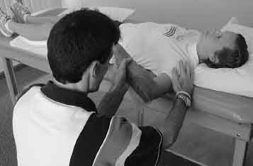
A large spectrum of mobility can be encountered when treating the patient with glenohumeral impingement. Hence, in guiding patients through the rehabilitation process, an accurate ROM measurement and informed decision making are essential to the clinician. To further illustrate the role of ROM and passive stretching during this phase of the rehabilitation, Figures 1–5 and 1–6 show versions of clinical IR stretching positions that utilize the scapular plane and can be performed in multiple and varied positions of glenohumeral abduction. Each utilizes an inherent anterior hand placement; this gives varying degrees of posterior pressure to minimize scapular compensation and to provide a check against anterior humeral head translation during the IR stretch. These stretches can be used in a proprioceptive neuromuscular facilitation (PNF) contract–relax format or following a low–load prolonged stretch–type paradigm to facilitate the increase in ROM.54,55 Figures 1–7 and 1–8 are examples of home stretches given to patients to address IR ROM deficiency. Note the inherent means of scapular stabilization in both methods that are necessary to optimize the value of the stretching procedure. Recent research has compared the effects of the cross-arm stretch to the sleeper stretch in a population of recreational athletes, some with significant glenohumeral IR range of motion deficiency.56 Four weeks of stretching produced significantly greater IR gains in the group performing the cross-body stretch as compared with the sleeper stretch. Further research is needed to better define the optimal application of these stretches; however, this research does show improvement in IR ROM with a home stretching program.56
Related posts:
 Rehabilitation of Adhesive Capsulitis
Classification and Treatment of Scapular Pathology
Rehabilitation of Micro-Instability
Rehabilitation of Macro-Instability
Rehabilitation of Acromioclavicular Joint Injuries
Modification of Traditional Exercises for Shoulder Rehabilitation and a Return-to-Lifting Program
Rehabilitation of Adhesive Capsulitis
Classification and Treatment of Scapular Pathology
Rehabilitation of Micro-Instability
Rehabilitation of Macro-Instability
Rehabilitation of Acromioclavicular Joint Injuries
Modification of Traditional Exercises for Shoulder Rehabilitation and a Return-to-Lifting Program
Stay updated, free articles. Join our Telegram channel

Full access? Get Clinical Tree


