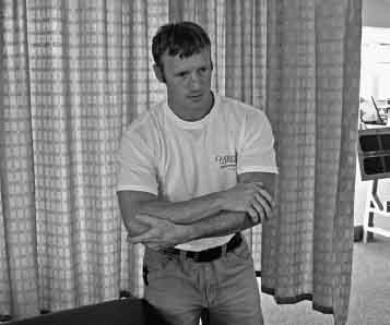
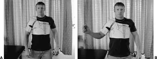
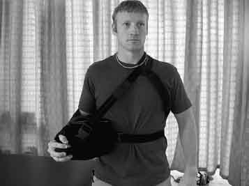
3 George J. Davies, Rob Manske, Robert Schulte, Christine E. DiLorenzo, Jason Jennings, and James W. Matheson Neuromuscular dynamic stability of the shoulder is the key to functional performance. All joints in the body are dependent on stability being provided by static stabilizers (bones, ligamentous/capsular complex, labrum, noncontractile tissue) and dynamic stabilizers (the muscular components contractile unit of muscles and the proprioceptive/kinesthetic system). When these systems work in harmony, the shoulder is one of the more marvelous joints in the body, particularly considering the demands placed upon it. The shoulder is expected to effectively place the hand in a position of function for a variety of tasks, including very delicate procedures such as performing surgery, powerful activities such as lifting hundreds of pounds overhead in Olympic weight-lifting, and sports-specific movements such as throwing a baseball over 90 mph with the shoulder exceeding 7000 to 9000 deg/s in angular velocity.1 However, when there is an injury or dysfunction to any one of the aforementioned components, it disrupts the shoulder complex and creates impairments and functional limitations. Surgical interventions are usually required to correct osseous or ligamentous/capsular problems. Physical therapy interventions can significantly influence the remaining two components of shoulder stability, the muscular and proprioceptive systems, and often are very effective in treating patients with shoulder dysfunction. Coupling the components of the muscular and proprioceptive systems is what is described as neuromuscular dynamic stability.2 Instability of the shoulder is a common clinical entity that is encountered on a daily basis by the rehabilitation specialist. In this chapter, we discuss the unstable shoulder in relation to functional instability that occurs from non-contractile instability (which is the traditional approach) and functional instability that can occur from neuromuscular deficits, including musculotendinous weaknesses or proprioceptive/ kinesthetic deficits. We present the applications of motor-learning principles to rehabilitation, and explore the evidence-based approach to rehabilitation of the unstable shoulder. Additionally, several empirically based concepts that are regularly applied to shoulder instability rehabilitation will be discussed, including the contra-coup concept of shoulder instability, the posterior dominant shoulder, and a phased program with precise recommendations for focusing on neuromuscular dynamic stability. Several concepts and examples of motor-learning principles and how they can be applied to rehabilitation of the unstable shoulder are included. Specifically, we present the concepts related to the examination, evaluation, diagnosis, prognosis, interventions, and treatment outcomes of the unstable shoulder. We also discuss the common mechanisms of injuries because the specifics of the injuries influence the nonsurgical as well as the surgical approaches to treatment, emergency care evaluation, treatment, and disposition. Additionally, we cover the traditional classifications of the unstable shoulder, which includes degree of instability, chronology of the instability, force required to create the instability, patient control over the instability, and direction of the instability. We also will briefly discuss various complications that can potentially influence the rehabilitation program, such as rotator cuff injuries, neurovascular triad injuries (including peripheral nerve injuries), Bankart lesions, bony Bankart fractures, and Hill–Sachs lesions. We present the evolution of immobilization of the unstable shoulder from duration and position in relation to its influence on the rehabilitation program. Recurrence rates and how they impact the rehabilitation process following the immobilization will be considered. In addition, we review the current literature emphasizing the 180-degree paradigm shift in how the anterior inferior instability should be immobilized and the anatomical and biomechanical rationale for the approach. We also present some visionary concepts of where we feel the future of rehabilitation for the unstable shoulder should be heading. In the shoulder complex, particularly in relation to shoulder instabilities or common comorbidities, some terms and conditions include: traumatic, unilateral/unidirectional, Bankart lesion, surgery (TU2BS); atraumatic, multidirectional, bilateral, rehabilitation, inferior capsular shift (AMBRI); acquired ligamentous/capsular laxity (ALL); superior labrum anterior posterior (SLAP) lesions; supraspinatus labral instability pattern (SLIP); glenohumeral internal rotation deficit (GIRD); scapula infera cora-coid dyskinesia (SICK) scapula; anterior labral periosteal sleeve avulsion (ALPSA) lesions; partial articular supraspinatus tendon avulsion (PASTA) lesions; posterior labrocapsular periosteal sleeve avulsion (POLPSA) lesions; humeral avulsion of the glenohumeral ligament (HAGL) lesions; bony humeral avulsion of the glenohumeral ligament (BHAGL) lesions; superior labrum, anterior cuff (SLAC) lesions; avulsion of the anterior inferior glenohumeral ligament (AIGHL); glenoid rim articular divot (GARD) lesions; glenolabral articular disruption (GLAD) lesions; glenoid labrum ovid mass (GLOM) lesions, tensile under-surface fiber failure (TUFF) lesions; and traumatic humeral articular cartilage shear (THACS). The presence of common comorbidities is particularly important to understand from the rehabilitation perspective. Often, the limiting factor in the rehabilitation program may not be the tissues involved from the primary instability problem, but in fact, from the tissue associated with the comorbidity, as in the case of a SLAP lesion.3,4 Several mechanisms of injuries compromise the integrity of the glenohumeral (GH) joint. The positions of the GH joint that usually result in an acute shoulder dislocation are (1) abduction/external rotation, (2) hyper-flexion, (3) hyper-abduction, and (4) hyper-horizontal extension. The only way to identify the actual shoulder dislocation is when a radiograph documents the position of the humeral head relative to the surrounding anatomy. When the shoulder does dislocate, it is important to recognize that the majority of shoulder dislocations are actually intracapsular and create attenuation of the anterior inferior capsule.5 This is one component of the Bankart lesion and has important implications to the postinjury immobilization position. Figure 3–1 Typical position and cluster of signs and symptoms of the patient after a traumatic anterior inferior dislocation. After an acute dislocation, the patient supports the arm away from the body and leans toward the side of the dislocation. There is an observable defect over the deltoid muscle area, often a palpable defect (humeral head) in the axillary area; there is pain from the pressure on the surrounding innervated structures (Fig. 3–1). Glenohumeral dislocations are classified according to the degree of instability, chronology, forces required to create the instability, patient’s control, and the direction of the instability. In the past, immobilization of the GH joint was performed by using a sling or a swathe and sling with the arm in adduction (ADD) and internal rotation (IR). Traditionally, the duration of the immobilization times varied from a few days to 6 weeks. Recently, the trend has been to immobilize the shoulder until it is “ready”; “ready” is undefined. Regardless of the amount of time the shoulder was immobilized, following a rehabilitation program, there was usually a high recurrence rate (30 to 90%). In most studies,8–10 the recurrence rate was related to age: the younger the individual the higher the likelihood of redislocating the shoulder. This was because older individuals have decreased tissue mobility and consequently more limited motion, which makes them less vulnerable to reinjury. Furthermore, the lower redislocation rate in older individuals is also probably related to activity level and specific types of activities. Younger individuals are generally more active and more likely to participate in activities that will stress the joint more; whereas older individuals are less active and participate in activities less likely to place their arms in a compromising position where the arm is likely to redislocate. Numerous shoulder braces are commercially available that try to stabilize the GH joint. This is one of the options available when treating the unstable shoulder. There are limited scientific studies, however, that demonstrate that braces are effective in preventing re-dislocations. There are also few comparative studies demonstrating that one brace is better than another. In addition, taping and strapping techniques have been described in numerous athletic training books, manuals, and courses; but once again, there is limited scientific documentation to support any of the claims of the taping techniques. We have used numerous braces in our 30 years of clinical experience but have found the Dennison Duke-Wyre Shoulder Brace ([Brace International, Atlanta, GA] Fig. 3–2) to be the most effective for those athletes who wear the brace for a season, or who are injured during the season and want to finish the season before undergoing the elective surgery. For a complete discussion of shoulder bracing and taping methods, the reader is referred to chapter 8, “Use of Taping and External Devices in Shoulder Rehabilitation” for more detail. Figure 3–2 Denison-Duke Wyre Brace pictured (A) at rest and (B) with slight abduction and external rotation. The standard of care when someone dislocates the shoulder is to immobilize in adduction and internal rotation (ADD/IR), rehabilitate the patient, and return the person back to activity. Often, the patient will redislocate a second, a third, or more times. Then, the issue of whether surgery should be performed is discussed with the patient. Marx et al11 have demonstrated that a patient who dislocates the shoulder is 10 to 20 times more likely to develop glenohumeral arthrosis in that shoulder than in the normal shoulder. The term dislocation arthropathy describes this condition. Drawing an analogy to a patient who ruptures the anterior cruciate ligament (ACL), we do not immobilize, rehabilitate, and then return the person back to activity and allow reinjury of the knee several more times until a definitive surgical approach is performed. Part of the reason is to prevent additional damage to other surrounding structures within the knee. Yet, based on the Marx et al11 study, a patient who dislocates a shoulder one time has a higher incidence of future degenerative changes. Consequently, not only does the patient have the potential to get future chondrosis in the articular surfaces, but the potential to injure other structures and create additional comorbidities also exists. Therefore, complete rehabilitation and referral for surgical correction of the instability is often needed to prevent reinjury and to prevent injury to other structures in the shoulder complex. Although the focus of this chapter is on nonsurgical rehabilitation, we would be remiss if we did not also acknowledge the landmark article published by Arciero et al.12 In a prospective randomized controlled clinical trials study whereby patients who had initial anterior inferior shoulder dislocations were treated with rehabilitation or an arthroscopic Bankart reconstruction, the initial treatment of early surgical intervention demonstrated significant reductions of redislocations compared with the patients who were treated with nonsurgical interventions (immobilization and rehabilitation). Rowe5 demonstrated the recurrence rate does not depend on how long the shoulder is immobilized or how securely it is immobilized. Hovelius et al8 indicated the shoulder never redislocates in 52% of patients after an initial dislocation, and recurrent dislocations spontaneously cease in 20% of patients with recurrent dislocations. The optimum duration of immobilization, however, still needs to be determined. Moreover, 71% of all recurrent dislocations occur within the first 2 years and 9% from years 2 to 5. Consequently, there have to be other variables that are important in the postinjury treatment and immobilization of patients with traumatic anterior inferior GH dislocations. It is likely that the poor outcomes after shoulder dislocation with rehabilitation have occurred because the position of shoulder immobilization has been inappropriate. Itoi et al13 performed a cadaveric study to identify the apposition of the Bankart lesion. The study demonstrated that the external rotation position at the neutral position provided better coaptation of the Bankart lesion than did the position of the conventional position of ADD/IR. In a follow-up study, Itoi et al14 demonstrated by magnetic resonance imaging (MRI) that the detached soft tissue, known as a Bankart lesion, is better coapted to the bone with the arm in external rotation (ER) than in IR When comparing the ADD/IR position to varying degrees of ER, the apposition of the Bankart lesion structures was very different. In the traditional position (ADD/IR), the Bankart lesion was not approximated at all and consequently compromised the ability of the structures to heal. Whereas, when the arm was placed in 30 degrees ER, there was much better apposition of the Bankart lesion. The less ER, the less contact of the respective structures occurred. There was good apposition at 0 (neutral position), but this significantly decreased the coaptation of the Bankart lesion structures when IR was used. The study’s conclusion was that immobilization of the arm in ER better approximates the Bankart lesion to the glenoid neck than does the conventional position of IR. Miller et al15 followed up with a research study by placing an electronic force transducer between the respective Bankart tissue structures. They confirmed with a force transducer contact study what the original cadaveric, radiographic, and MRI studies demonstrated. The results illustrated the following: With these three studies as a scientific anatomical and biomechanical basis, Itoi et al16 completed a prospective randomized controlled clinical trials study. Forty patients participated and 20 were randomized into the IR position and 20 into the ER immobilization position. Initially, Itoi et al16 immobilized the patients in 30 degrees of ER; however, this much ER was not well tolerated by patients. It is likely that the higher the contact force, the higher the healing rate. However, the less ER the arm was placed in, the more comfortable it was for the patient. Moreover, from a practical standpoint, having the arm externally rotated in 30 or 45 degrees would create an awkward position of the arm for activities of daily living (ADLs). Additionally, Itoi et al16 arbitrarily placed the arm in 10 degrees ER because the Miller et al15 study also showed positive contact of the Bankart lesion via the force transducers. The patients who were positioned in IR were immobilized in the conventional sling and swathe. The patients positioned in ER were immobilized using a wire mesh splint covered with sponge. Because of the healing process with soft tissue, granulation tissue fills the gap and unites the soft tissue within 7 to 10 days and increases in tensile strength within 3 weeks. Therefore, Itoi et al16 empirically immobilized both groups in the respective positions for 3 weeks. The compliance rate for those who were immobilized in IR was 75%. Those immobilized in ER was 80%. The follow-up results at 15.5 months demonstrated the recurrence rate (i.e., redislocation) in all patients in the study for the group immobilized in IR was 30%, whereas those immobilized in ER was 0%. The individuals under 30 years old (who usually have a higher recurrence rate than older individuals) who were immobilized in IR had a recurrence rate of 45%. However, those under the age of 30 who were immobilized in ER had a recurrence rate of 0%. Interestingly, even after a surgical stabilization procedure, there is approximately a 5 to 10% recurrence rate by 1 year.9,10 The ultimate functional outcome is the patient’s ability to return to the premorbid state, particularly with higher levels of activities like sports. The results demonstrated that those who were immobilized in IR had a return to their previous level of sporting activities of 58%, whereas those who were immobilized in ER had an 82% return to sporting activities. The evidence has been universal regarding a high failure rate (defined by recurrent dislocations/subluxations). Consequently, it is time to do something differently: The results of the proper immobilization to create better apposition of the Bankart structures hold a lot of promise based on the scientific anatomical and biomechanical studies as well as a clinical outcome study. Continued research is validating the apposition of the Bankart lesion with the externally rotated humeral position as opposed or in comparison with the ADD/IR position traditionally used. Hart and Kelly17 used arthroscopic visualization to confirm approximation of the Bankart lesion in patients following shoulder dislocation. Based on the plethora of evidence we presently use the UltraSling (Smith & Nephew DonJoy, Carlsbad, CA) to achieve the proper immobilization of the patient. This sling supports the patient’s arm and places the healing tissues into the ER position to provide apposition for the healing structures (Fig. 3–3). This 180-degree paradigm shift in how to immobilize the unstable shoulder is an example of evidence-based practice. Paradoxically, the evidence in the literature indicates that we should not be immobilizing the shoulder in the traditional position of ADD/IR. We expect that over the next several years with proper immobilization resulting in better tissue approximation for healing, the rehabilitation programs for nonsurgical treatments are going to demonstrate significant improvements in outcome. The remainder of this chapter will focus on some of the causes of the shoulder instabilities, rehabilitation techniques, and guidelines to address these conditions. Figure 3–3 UltraSling. Rehabilitation outcomes following shoulder instabilities will improve as we further understand some of the newer concepts of the causes of the occult and more-subtle instabilities. It is important that we identify the cause when we treat shoulder instabilities. Furthermore, it is important that we understand that with shoulder pathologies there often are several comorbidities. Moreover, as we better apply some of the concepts of motor learning principles to shoulder instability rehabilitation, we will see significant improvements in the outcomes. How do we determine the cause of shoulder instability? Functional instability is the patient’s inability to keep the humeral head centered in the glenoid fossa. The cause may be due to one problem or it may be multifactorial, consisting of noncontractile (bones, ligaments, capsule, and labrum) instability, contractile (muscles, tendons) weakness, or neuromuscular dynamic stability (proprioception/kinesthesia) deficits. Each area needs to be tested with the particular deficits identified, and then each deficit needs to be addressed in the rehabilitation program. When considering shoulder stability provided by the static stabilizers, it is important to consider the shoulder as a circle concept. In other words, it is important to check both sides of the joint for any possible injuries that may contribute to the instability. This also introduces the contra-coup concept of shoulder injuries (in other words, the injury may also occur opposite to the side of the obvious injury). Although this chapter is primarily focused on macrotraumatic anterior inferior instabilities because they are the most common, it is also important to check the posterior capsular structures as well because they may also be injured. Ligament stability is dependent on position/specificity of ligament function and the capsular/ligamentous constraint mechanisms. Much of the capsular/ligamentous constraint mechanism is dependent on the position of the arm. When the arm is in the adducted position, the superior GH ligaments and superior capsule are taut. Whereas, when the arm is in the 90-degree abducted and 90-degree ER position, the anterior/inferior capsule and the inferior GH ligament are taut.18 Joint laxity is not the same as instability. Joint laxity is the normal amount of translation of the joint for the individual patient. The patient has control over the joint; it does not subluxate/dislocate and is pain-free. In instability, the humeral head does subluxate/dislocate; the patient senses that and is apprehensive or has symptoms in the joint. We do not treat laxity; we treat instability. The patient’s history, subjective examination, mechanism of injury, epidemiological considerations, high index of suspicion, physical examination,19–22 and imaging studies all contribute to the final diagnosis.23 We primarily focus on a comprehensive physical examination of the shoulder24,25 and use an algorithm-based examination for the special tests of the shoulder because it is clinically efficient.27–29 When performing this portion of the shoulder examination, the following examination criteria are evaluated: The degree of laxity is obviously unique to each patient. The best determination of normal, hypermobility, or hypomobility of the GH joint is to perform a bilateral comparison. However, there will occasionally be the patient who has had injuries to both sides. Then we need to rely on previous clinical experience and descriptive normative data. An example of early descriptive normative data for the GH load and shift test was published by Matsen et al6 where they actually drilled percutaneous pins into the humerus and scapula and performed the testing under fluoroscopy to measure the amount of humeral head translation. They found the anterior load and shift produced 8 ± 4 mm; posterior load and shift = 9 ± 7 mm; and the sulcus sign was 11 ± 3 mm. Moreover, Borsa et al32 studied the in vivo quantification of capsular end points in the nonimpaired GH joint using an instrumented measurement system. They found that it took 9.4 ± .607 pounds of pressure to hit the end point for the anterior load and shift test. The sulcus (inferior) test took 8.4 ± 1.48 pounds to reach the capsular end point. This study found that not much force is required when performing a physical examination of the shoulder. The American Shoulder and Elbow Society’s Classification33 of shoulder laxity/instability is predicated on many of the previous concepts, such as edge loading and end feels (Table 3–1). The physical examination of the shoulder using all the aforementioned concepts helps the clinician determine whether the ligamentous/capsular laxity or a labrum injury is contributing to the “instability.” In the case of macro-traumatic injuries, as described in this chapter, ligamentous/capsular laxity or a labrum injury is often the patient’s primary problem. We primarily use the algorithm-based examination for the special tests of the shoulder as described by Davies et al26 Schulte and Davies28 and Ellenbecker31 (Figs. 3–4, 3–5).
Rehabilitation of Macro-Instability
Common Instabilities and Comorbidities
The Mechanism of Injury
Patient’s Clinical Signs and Symptoms
Classifications of Shoulder Instabilities
Complications of Shoulder Instabilities
Immobilization: Past and Present
Noncontractile Instability
Contractile Stability: Muscle Strength, Power, and Endurance
Neuromuscular Dynamic Stability
Ligamentous/Capsular Instability
Musculotendinous Weakness
Exercises for Rehabilitation of the Shoulder Complex
Principles of Exercise and Their Application to Rehabilitation
What Is the Optimum Dosing for Therapeutic Exercise Programs?
Unique Concepts of Exercise and Their Application to Rehabilitation
Stage 1: Proprioception and Kinesthetic Exercises (Baseline for Dynamic Stability)
Stage 2: Dynamic (Proactive) Stabilization Exercises
Stage 3: Reactive Neuromuscular Control
Stage 4: Functional Skill Movements and Activities
 Instability of the Shoulder
Common Instabilities and Comorbidities
The Mechanism of Injury
Patient’s Clinical Signs and Symptoms
Classifications of Shoulder Instabilities
Complications of Shoulder Instabilities
 Nonsurgical Treatment of Shoulder Instability
Immobilization: Past and Present
 Causes of Shoulder Instability
Noncontractile Instability
Examination of the Glenohumeral Joint for Instability
Special Tests for Joint Instability
| Grade | Glenohumeral Translation |
| Trace | Small amount of humeral head translation |
| I | Humeral head rides up the glenoid slope, but does not subluxate over the rim |
| II | Humeral head rides up and over the glenoid rim and dislocates, but spontaneously reduces when the stress is removed |
| III | Humeral head rides up and over the glenoid rim and dislocates, but remains dislocated on removal of the stress |
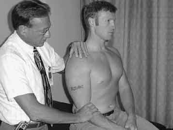
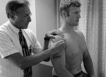
Figure 3–4 Physical examination—sulcus sign at zero degrees.
Contractile Stability: Muscle Strength, Power, and Endurance
Contractile stability is the functioning of the musculotendinous unit (MTU). Muscles provide more-dynamic stability at the middle of the ROM where the ligaments are lax. The MTU also provides stability at the end-ROM along with the ligaments/capsule due to the MTU tightening. As the MTU is lengthened, it provides a prestretch to the muscles based on the length:tension ratio and actually facilitates a muscle’s ability to generate force.
The contractile stability, muscle strength, power, and endurance are commonly assessed with a variety of muscle performance testing, including manual muscle testing, hand-held dynamometry, cable tensiometers, isokinetic testing (Fig. 3–6), etc.34–37
Davies et al34–37 and Ellenbecker and Davies38 have published extensively on muscle performance, data analysis, and applications to clinical practice, particularly relative to the shoulder complex. Examples of data analysis include:
Related posts:
 Rehabilitation of Adhesive Capsulitis
Classification and Treatment of Scapular Pathology
Rehabilitation of Micro-Instability
Rehabilitation of Shoulder Impingement: Primary, Secondary, and Internal
Rehabilitation of Acromioclavicular Joint Injuries
Modification of Traditional Exercises for Shoulder Rehabilitation and a Return-to-Lifting Program
Rehabilitation of Adhesive Capsulitis
Classification and Treatment of Scapular Pathology
Rehabilitation of Micro-Instability
Rehabilitation of Shoulder Impingement: Primary, Secondary, and Internal
Rehabilitation of Acromioclavicular Joint Injuries
Modification of Traditional Exercises for Shoulder Rehabilitation and a Return-to-Lifting Program
Stay updated, free articles. Join our Telegram channel

Full access? Get Clinical Tree


