Rehabilitation of Persons with Parkinson’s Disease and Other Movement Disorders


Rehabilitation of Persons with Parkinson’s Disease and Other Movement Disorders


Ron Hirschberg

Nutan Sharma

Donna Moxley Scarborough


INTRODUCTION

Parkinson’s disease affects roughly 1% of the population older than age 50 in the United States, incidence increases with age, and the peak onset occurs in the sixth to eighth decade, making it one of the most common chronic diseases of adulthood (1). Life expectancy is near normal; however, there is increased risk for morbidity and mortality due to associated complications, such as falls, orthostatic hypotension, or aspiration pneumonia.

Rehabilitation has a vital and unique role in movement disorders. Parkinson’s disease is a chronic, progressive, neurological disease, which at various stages and manifestations affects all aspects of a patient’s functional state. The goal of this chapter is to review the clinical presentation and neuropathology of Parkinson’s and other movement disorders, with specific emphasis on the collaborative process of rehabilitation. The disablement process in Parkinson’s is based, just as in other medical and neurological disease states, on the continuum of impairments and how they affect functional limitations and subsequent disability (2, 3). The impact of multifaceted treatment is covered in this chapter as well, placing high importance on the role of exercise with and without pharmacological management.

The shaking palsy or paralysis agitans was the description James Parkinson used in 1817 to represent what would later be referred to as “Parkinson’s Disease,” the progressive, central nervous system impairment that is characterized by the presence of two or more cardinal signs: bradykinesia, resting tremor, rigidity, and postural instability. The diagnosis of Parkinson’s disease can be challenging and is first and foremost a clinical diagnosis. The typical individual afflicted with Parkinson’s disease presents with symptoms on one side of the body, referred to as asymmetric onset. Typically, individuals with Parkinson’s disease can be subdivided into two groups: those with a predominance of rigidity and akinesia, with minimal or no rest tremor; and those with rest tremor predominant disease. However, other signs and symptoms may be present as well (Table 26-1). Parkinson’s disease can be incorrectly diagnosed as much as 25% of the time if based solely on clinical features and not on diagnostic trials, specifically responsiveness to dopamine treatment (4, 5). The rate of progression of Parkinson’s varies significantly from one person to another.

Although the exact cause of Parkinson’s is not known, environmental and genetic factors have been implicated. One theory regarding the pathogenesis of Parkinson’s disease is that unidentified environmental triggers cause accelerated destruction of the dopamine-producing neurons in the substantia nigra in a genetically predisposed individual. Secondary parkinsonism refers to those disorders that have the same combination of signs and symptoms that are seen in primary Parkinson’s disease but result from a clearly identifiable insult to the central nervous system. Examples of such insults include exposure to 1-methyl-4-phenyl-1,2,3,6tetrahydropyridine (MPTP) (6, 7, 8), which causes the death of dopamine neurons and ischemic infarcts involving the vascular supply to the midbrain that can also result in the death of dopamine neurons. Dopamine-antagonist medications, (specifically antagonists of D2 receptors), such as the typical neuroleptics haloperidol and thioridazine, and other D2 antagonists, such as metoclopramide, have been known to exacerbate or even induce parkinsonism. Infectious conditions, such as St. Louis encephalitis, and von Economo’s and other influenza-related encephalitides, have also been associated with secondary parkinsonism.


ANATOMY AND PATHOPHYSIOLOGY

Motor impairment in Parkinson’s and other movement disorders stems from a common principle: There is a disruption in the modulation of the cortical and subcortical structures of the brain. The anatomic structures involved in the control of movement are referred to as pyramidal and extrapyramidal structures, or motor cortices and the basal ganglia, respectively (9, 10, 11, 12). A basic understanding of these structures and their physiology forms the basis of symptomdriven therapy in the management of patients with movement disorders.

The several interconnected nuclei of the basal ganglia make up a large portion of the extrapyramidal system. The
nuclear groups of the basal ganglia include the caudate and putamen (together called “the striatum”), the subthalamic nucleus (STN), internal and external portions of the globus pallidus (Gpi, Gpe), and the pars compacta and pars reticulata of the substantia nigra (SNc, SNr).


TABLE 26.1 Clinical Presentations of Parkinson’s Disease


Clinical Signs and Symptoms of Parkinsonism


Symptoms


Clinical Features


Positive phenomena


Tremor


Most common symptom. Distal involvement frequency: 3.5 Hz. Suppressed by sleep and activity, increased by fatigue or stress. EMG: rhythmic alternating bursts in agonist and antagonist muscles.


Rigidity


Increase in muscle tone during passive limb movement equal through entire range of motion; increases if contralateral limb is engaged in volition.


Flexed posture


Dominance of progravity flexor muscles (bowed head, chin toward chest, kyphotic thorax, protracted shoulders, internally rotated arms, flexed elbows, knees, and hips).


Negative phenomena


Bradykinesia


Slowness of movement, masked facies, decreased eye blinking, inability to move. Fatigue. EMG: delayed motor unit recruitment, pauses once recruited, inability to increase firing rate.


Loss of postural reflexes


Tendency to fall to the side (lateral pulsion) or backward (retropulsion); sitting en bloc (collapses in the chair when attempting to sit down).


Postural instability


Freezing phenomenon


Transient inability to move.


EMG, electromyography; Hz, hertz.


Adapted from Jain SS, Francisco GE. Parkinson’s disease and other movement disorders. In: DeLisa JA, ed. Rehabilitation Medicine: Principles and Practice. 3rd ed. Philadelphia, PA: Lippincott Williams & Wilkins; 1988:1035-1056.


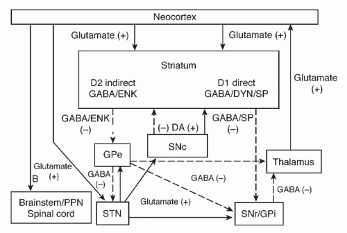
The primary afferent nuclei of the basal ganglia are the striatum and the STN. The striatum receives excitatory input from the cerebral cortex, midline thalamic nuclei, hippocampus, amygdala, and primary olfactory cortex. The striatum also receives dopaminergic input from the substantia nigra pars compacta (SNc) and serotonergic input from the midbrain raphe nuclei. The primary efferent nuclei of the basal ganglia are the globus GPi and the substantia nigra pars reticulate (SNr). These output nuclei project inhibitory gamma-aminobutyric acid (GABA)-mediated control over the thalamus and brain stem targets (pedunculopontine nucleus). Basal ganglia output indirectly influences cortical activity by inhibitory control of the thalamus. A detailed hypothesis set forth by Mink (13) described that the net effect of basal ganglia activity during a voluntary movement is twofold: “Inhibition of competing motor patterns and a focused facilitation of selected voluntary movement pattern generators.” Physiologically, the substantia nigra and the globus pallidus tonically inhibit the motor nuclei of the thalamus, which have excitatory influences at the motor cortex. Pathologically, the thalamic motor nuclei are disinhibited, resulting in increased cortical motor output activity (Fig. 26-1). Electrophysiologic studies have demonstrated that the basal ganglia do not initiate or program movements but appear to modulate a motor pattern initially generated by cortical neurons (13). With the understanding that the essence of basal ganglia influence on motor activity is inhibitory control of the thalamus, the theoretic net effect of basal ganglia activity on voluntary movement is the inhibition of competing motor patterns and the focused facilitation of the cortically selected voluntary movement (13).


FIGURE 26-1. Circuitry of direct and indirect pathways and pharmacology of the basal ganglia. The direct pathway projects gamma-aminobutyric acid (GABA)-mediated inhibitory input directly to the output nuclei of substantia nigra reticulata (SNr) and globus pallidus interna (GPi). The indirect pathway sends an inhibitory GABA projection to the globus pallidus externa (GPe), which subsequently projects GABAmediated inhibitory input on the subthalamic nucleus (STN). The STN output to the SNr/GPi is an excitatory glutamate projection. Solid arrows reflect excitatory pathways and dashed arrows, inhibitory pathways. DA, dopamine; D1, D1 dopamine receptor; D2, D2 dopamine receptor; DYN, dynorphin; ENK, enkephalin; PPN, pedunculopontine nucleus; SNc, substantia nigra zona compacta; SP, substance P. Modified from Alexander GE, Crutcher MD, Functional architecture of the basal ganglia circuits: neural substrates of parallel processing (10).


REHABILITATION AND COLLABORATION

Neurologists and, to some extent, primary care physicians largely oversee the care of persons with Parkinson’s and medially manage the patient. What has become increasingly clear in neurological and neuromuscular disease management is the necessity of the multidisciplinary approach, in order to offer holistic and comprehensive medical and functional care of the patient. A rehabilitation team should ideally incorporate neurology, physiatry, physical and occupational therapy, speech and language pathology, social work, psychology and/or psychiatry, and nutrition. It, of course, promotes a patient-centered approach with collaboration of all disciplines with the expertise to diagnose and treat various impairments (14).

Physiatrists often see patients with movement disorders, mainly those with Parkinson’s, in consultation. This practice may be in the form of a multidisciplinary clinic with neurologists, therapists, social workers, and psychiatrists or psychologists, or in a stand-alone physiatric practice. Physiatrists can be helpful for specific therapy prescription and analysis and treatment of gait abnormalities. Although a relatively low number of studies address the optimal therapeutic management plan, a collaborative effort by physiatrists and neurologists should logically provide the best therapeutic structure for the movement disorders population. Physiatrists who treat patients with various neurologic diagnoses should be able to assume a more active role in managing movement disorders.

The role of physiatry may be to assist with nonoperative orthopedic problems or basic musculoskeletal and pain impairments typical in this population. Physiatrists in the outpatient and inpatient settings collaborate and communicate with physical therapists, occupational therapists, and speech and language pathologists in order to optimize the function of the patient with a movement disorder. Therapists who subspecialize in Parkinson’s treatment and other movement disorders are invaluable links between patient and physician. Therapists offer not only highly skilled assessments and treatments of functional impairments but also longitudinal education of the patient and family that is essential for carryover and functional gains.

Therapists and physiatrists may encounter a person with Parkinson’s admitted to the rehabilitation unit for another condition, such as stroke, or after an orthopedic procedure or after the implantation of deep brain stimulators, which are used to treat some of the complications of Parkinson’s disease (discussed later in this chapter) (15). These patients pose a unique challenge to the rehabilitation team, who are cognizant of baseline parkinsonian impairments and the additional neurological, orthopedic, or other impairment (16). One of the vital roles of the rehabilitation team is to help the patient and his or her primary medical or surgical team understand how the new illness or procedure will likely influence his or her preexisting parkinsonian impairments.

The team designs a rehabilitation program in order to set appropriate therapeutic goals. The deconditioning syndrome in and of itself certainly affects the hospitalized patient with a movement disorder while in the acute-care setting and can impact upon the patient’s short- and long-term functional goals. Nursing staff should be aware of the key importance of timely administration of anti-Parkinson drugs in order to minimize fluctuations in blood levels. Optimal pharmacological management impacts proper daytime participation with therapists and exercise (17). Those with Parkinson’s are at risk for nutrition impairment, constipation, and low-energy states, and therefore a nutritionist or dietitian is required to focus on preparing the appropriate caloric and supplement needs for the patient.


IMPAIRMENTS AND FUNCTIONAL LIMITATION IN PARKINSON’S

The concept of disability or disablement has been demonstrated in various ways, with the two most common being the International Classification of Impairments, Disabilities and Handicaps (ICIDH), and the Nagi (Saad Z. Nagi) framework in the 1960s. Nagi introduced the principle of “disablement,” which is a process initiated with pathology leading to impairment that causes function limitation and in turn disability. What is intriguing about this framework is that disability can be positively affected at all four conceptual levels of the continuum. In Parkinson’s, pathology resides in the substantia nigra at the cellular level and results in the inability to create sufficient dopamine. One’s impairment may be muscle rigidity or bradykinesia, for instance, with functional limitation manifesting in gait dysfunction. Disability is the inability to optimally participate in community activities or carry a job, which occurs due to the functional limitation (2, 3).

Physiatrists and the rehabilitation team assist in the care of persons with Parkinson’s by managing the various impairments that result from the condition (Tables 26-2 and 26-3). This section highlights ways that rehabilitation professionals can help minimize the impact that these impairments, and therefore function limitations, have upon well-being and disability of those with Parkinson’s.


TABLE 26.2 Impairments Resulting From Parkinson’s Disease


Motor


Gait


Movement initiation and execution


Bradykinesia


Tremor


Rigidity


Sensory/pain


Autonomic dysfunction


Orthostatic hypotension


Constipation


Cognitive


Difficulty in switching from one task to another


Behavioral and affective


Depression


Anxiety


Psychotic features, such as hallucination


Gastrointestinal


Dysphagia


Constipation


Bladder dysfunction


Sexual dysfunction


TABLE 26.3 Therapeutic Plan for Patients With Parkinson’s Disease


Medical and Nursing


Firm bed to decrease contractures and improve bed mobility


Gradual changing of positions, elastic stockings, abdominal binder, sodium tablets, and possibly pseudoephedrine, midodrine, and/or fludrocortisone for orthostasis


Regular meals with proper diet (low protein); nutritional consultation


Measure vital capacity and enforce incentive spirometry to prevent atelectasis and pneumonia


Bowel program for gastrointestinal hypomobility (stool softeners, bulk-forming agents, cisapride, and suppositories may be required)


Bladder evaluation and urodynamics; anticholinergics (e.g., oxybutynin chloride [Ditropan]) for hyperreflexic bladder


Artificial tears for lack of blinking


Sexual dysfunction evaluation


Anticholinergic medications before mealtime to help facilitate oral and pharyngeal movements


Physical Therapy


Relaxation techniques to decrease rigidity


Slow rhythmic rotational movements


Gentle range-of-motion and stretching exercises to prevent contractures, quadriceps and hip extensor isometric exercises


Neck and trunk rotation exercises


Back extension exercises and pelvic tilt


Proper sitting and postural control (static and dynamic); emphasize whole body movements


Breathing exercises stressing both the inspiratory and expiratory phase


Functional mobility training, including bed mobility, transfer training, and learning to rise out of a chair by rocking; may require a chair lift


Stationary bicycle to help train reciprocal movements


Training in rhythmic pattern to music or with auditory cues such as clapping may help in alternating movements. Standing or balancing in parallel bars (static and dynamic) with weight shifting, ball throwing


Slowly progressive ambulation training (large steps using blocks to have patients lift legs, teaching proper heel-to-toe gait patterns, feet 12-15 in. apart, arm swing; use inverted walking stick, colored squares, or stripes as visual aids)


Use of assistive devices (may need a weighted walker)


Aerobic conditioning (swimming, walking, cycling)


Frequent rest periods


Family training and home exercise program


Occupational Therapy


Range-of-motion activities of upper extremity with stretching


Fine motor coordination and training, hand dexterity training using colored pegs or beads


Hand cycling to help train reciprocal movements


Rocking chair to help with mobilization


Transfer training


Safety skills


Adaptive equipment evaluation, including Velcro closures, raised toilet, grab bars, eating utensils with built-up handles, and key holders


Family training and home exercise program


Speech


Deep breathing and diaphragmatic breathing exercises


Articulatory speech training for dysarthria


Facial, oral, and lingual muscle exercises


Swallowing evaluation, including a modified barium swallow as needed


Teaching compensatory strategies for safer swallowing


Psychology


Psychological support for patient, family, and caregivers


Cognitive assessment


Adapted from Jain SS, Francisco GE. Parkinson’s disease and other movement disorders. In: DeLisa JA, ed. Rehabilitation Medicine: Principles and Practice. 3rd ed. Philadelphia, PA: Lippincott Williams & Wilkins; 1988:1035-1056.


Gait Dysfunction

Gait dysfunction is the primary presenting symptom in as many as 18% of patients with Parkinson’s disease (18, 19). Persons with Parkinson’s report that one of the leading causes for diminished quality of life is difficulty walking (20). Shortened step length is the focal disturbance of gait in a person with Parkinson’s, and the classic gait pattern is characterized by shuffling (21, 22). Reduced hip, knee, and ankle flexion angles, decreased arm swing, limited trunk rotation, and increased forward flexed trunk are typical kinematic changes in gait among persons with Parkinson’s disease. Parkisonian gait becomes progressively inefficient and unstable as a result of multiple impairments (23, 24). There are two stereotypical variations of parkinsonian gait patterns. Many persons demonstrate changes in the fluidity of gait with episodes of hesitation to initiate gait or instances of “freezing” when the person stops and cannot initiate movement while walking (25). These difficulties in performing efficient gait patterns are often observed when the person is turning around, walking through a doorway threshold, or stepping around or over objects (23, 26, 27). The second parkinsonian gait pattern is festination, characterized by small steps increasing in speed and frequency with a forward trunk posture (26). These gait abnormalities place persons with Parkinson’s at varying risk for loss of balance or falls (24, 28).

Although people with Parkinson’s may be able to perform straight-line walking, their overall gait is hampered by hypokinesia, rigidity, postural imbalance, and the fear of falling, especially in advanced stages of the disease (24, 29, 30, 31). Deficits have also been implicated, thus aggravating the problem and may be the reason why people with Parkinson’s disease rely strongly on visual cues (32, 33). The loss of proprioceptive feedback from the knee extensor load receptors is suspected to cause a reduction in the activation of leg extensor muscles (34). Other problems that contribute to the gait abnormality include impairment in programmed muscle activation in the lower limbs and diminished postural reflexes (33). These impairments result in progressive difficulty adapting postural responses and stability during gait, especially on uneven surfaces (24, 33, 35, 36, 37).


TABLE 26.4 Gait Disturbance in Parkinson’s Disease


Gait Disturbance


Parkinsonian Direct Impairment


Shuffling gait pattern: Decreased step and stride length, decreased cadence and velocity


Bradykinesia and hypokinesia; movement initiation and execution impairment


Festination


Impaired automatic motor task performance; movement execution impairment; impaired postural responses


Stooped (flexed) posture


Rigidity


Freezing, “start hesitation”


Impaired automatic motor task performance; hypokinesia; movement initiation and execution impairment


“Cautious” gait (fear of falling)


Impaired postural responses; rigidity


Impaired balance and unsteadiness due to lightheadedness


Impaired postural responses; orthostatism; ANS impairment


Dystonia, dyskinesia


Medication effect; Parkinson’s disease—direct impairment related movement disorder


During early stages of Parkinson’s, primary impairments of hypokinesia, rigidity, proprioceptive deficits, decreased postural reflexes, and movement execution may have subtle effects on gait. During progression of the disease, the primary impairments are typically magnified and result in new secondary impairments (Table 26-4) (38). Muscle weakness due to the disuse and contractures from rigidity are examples of secondary impairments that further contribute to gait dysfunction. These abnormalities affect balance, alter gait, and increase the patient’s risk for falls (24, 28). Another factor contributing to gait instability is orthostatic hypotension, which will be discussed further in this section.

Gait dysfunction in patients with Parkinson’s may be due to either the disease itself or as a side effect of pharmaceutical therapy (see Table 26-4). Levodopa therapy affects parkinsonian gait and movement, in that although the medication may facilitate gait during the “on” phase, deterioration may be seen as part of motor fluctuations during the “off” state. Its effect is largely due to the control of force and amplitude of limb movement, rather than improved automaticity or rhythmicity (39). Levodopa may also cause dystonia, which can negatively affect ambulation.

Direct and secondary impairments of Parkinson’s can lead to further decline if patients respond to these impairments and limitations by decreasing activity levels. This results in further muscle weakness, joint contractures, and worsened endurance. Discussion with the rehabilitation team and prompt referral to therapy can facilitate early treatment approaches to “break the cycle” and prevent resulting functional limitation in gait and other activities. Advances in technology are exploring ways to help monitor changes in gait and functional mobility in the home environment among persons with Parkinson’s using wearable sensors (40, 41). In the future, such devices should offer clinicians the ability to titrate medication and make referrals to appropriate therapies based on these objective data.


Movement Initiation

The person with Parkinson’s typically possesses slow movement initiation that may result from delayed activation of the motor cortex, impeding one’s ability to initiate and execute normal movement (42). Although the corticospinal system is intact, the abnormal motor commands result in bradykinesia in the implementation and execution of a motor task. These impairments are exacerbated with high-complexity tasks. Highly refined motor activities such as handwriting require switching between motor components, and the limited ability of people with Parkinson’s to adapt to changing task conditions makes this difficult. Indeed, it appears that slow initiation of movement may be a strategy adopted by those with the disease to facilitate motor tasks (43, 44).


Dyskinesia and Dystonia

Dyskinesias, the excessive movement of muscles in the trunk or limbs that cannot be controlled voluntarily, are a common complication that typically develops after 7 or 8 years of levodopa therapy. It is thought to result from a complex interplay between the imperfect supplementation of dopamine, in the form of oral carbidopa/levodopa tablets, and the underlying, progressive loss of dopamine neurons that is the hallmark of Parkinson’s.

Most commonly, dyskinesias are seen when the plasma concentration of levodopa is at its highest. This is referred to as “peak-dose” dyskinesia. It is believed to result from abnormal neuronal firing patterns in response to pulsatile stimulation of the dopamine receptors (45). In the early stages, it can be managed by reducing the dose of levodopa or taking levodopa less frequently. However, this typically results in a worsening of the symptoms of Parkinson’s, such as rest tremor, bradykinesia, and rigidity. It is important to remember that if dyskinesias are relatively mild and not bothersome or dangerous to the patient, then most people prefer to move too much rather than too little. If dyskinesias become large in amplitude, to the extent that they risk injury to the patient, then they must be treated. Treatment options include decreasing the dose of levodopa, decreasing the frequency with which levodopa is taken, and adding amantadine to the medication regimen.

Dystonia, the involuntary contraction of a single muscle or multiple muscles that cause an abnormal posture, is also commonly seen in those with Parkinson’s. The most common site for dystonia in those with Parkinson’s is the foot. Typically, at the end of a dose of levodopa, when plasma concentrations are at their lowest, the foot begins to contract with flexion of the toes and inversion of the entire foot. With the ingestion of another dose of levodopa, the dystonia is relieved. Dystonia may also occur as a peak-dose effect, when plasma concentrations of levodopa are at their highest. Unfortunately, peak-dose dystonia is not easily treated. If dystonia presents as a focal problem, such as involving the neck muscles or the facial muscles, then local therapy with botulinum toxin injection is an option.


Orthostatic Hypotension

Up to 20% of the population with Parkinson’s may experience orthostatic hypotension, which may be the result of central or peripheral mechanisms. This usually results from sympathetic outflow dysfunction that leads to impaired peripheral vasoconstriction. Intravascular volume depletion due to poor fluid intake or other mechanisms magnifies the problem. Because Parkinson’s usually occurs in the elderly, other age-related medical conditions, such as cardiovascular diseases and use of drugs that cause hypotension, should be taken into consideration (46, 47, 48).

Lifestyle modification and education are important aspects of treatment. For example, patients should be counseled that warm or hot baths increase peripheral vasodilatation and may contribute to orthostasis. A heavy meal may also result in splanchnic vasodilatation and “steal” blood volume from elsewhere. Excessive straining while defecating and other tasks inducing the Valsalva maneuver should be avoided, and high-fiber diets and stool softeners should be incorporated as well. Nonpharmacologic methods include the use of compression leg stockings and abdominal binders. Management also includes eliminating unnecessary antihypertensive drugs or other medications that may cause hypotension and (47) potentially providing blood pressure support with fludrocortisone or midodrine.


Gastrointestinal Problems


Swallowing Dysfunction

Dysphagia typically results from loss of lingual control and inability to propel the bolus due to delay in the contraction of pharyngeal muscles. Up to 75% of people with Parkinson’s experience dysphagia, and esophageal dysmotility may also occur (49, 50, 51, 52, 53, 54). Abnormalities in striated muscles under dopaminergic control and smooth muscles under autonomic influence contribute to this complicated impairment. Videofluoroscopic swallowing evaluations help to determine which specific phases of swallowing are impaired. Modified barium swallows under fluoroscopy have shown that the most common abnormalities are motility problems, hypopharyngeal stasis, aspiration, and deficient positioning of the esophagus (55, 56).

A speech pathologist may help by teaching oral-motor exercises and providing education on compensatory strategies to prevent penetration and aspiration. A recent randomized controlled trial evaluating the efficacy of the chin-tuck maneuver versus thickened liquids only for Parkinson’s and dementia patients demonstrated that a chin-tuck cohort and thickened-liquids-only group equally prevented the development of pneumonia at 3 months follow-up; however, aspiration during videofluorographic assessment was prevented more readily with thickened liquids only (not using chin-tuck) (57). Importantly, when an individual cannot meet his or her caloric and fluid requirements, a gastrostomy feeding tube should be considered.


Nutrition

Parkinson’s patients are at risk for weight loss and should be closely evaluated for nutritional losses (52). Protein intake should be monitored and limited, in that amino acids compete with levodopa for absorption. Clinicians may advise patients to take levodopa 1 hour before or after a meal in order to facilitate proper absorption. This is of particular concern to those in the late stages of the disease who have severe dyskinesias. Vitamin supplements should be considered for those who are unable to have well-balanced and adequate nutrition. The precise amount of vitamin supplementation needs to be monitored carefully because vitamin B6 supplementation may result in reduced absorption of levodopa from the intestines (58).


Delayed Gastric Emptying

Parkinson’s patients experience early satiety or may experience nausea and vomiting due to delayed gastric emptying. Reduced peristalsis and gastroesophageal reflux present as complaints of “heartburn” or indigestion. This also puts the patient at risk for poor nutritional intake and therefore can exacerbate absorption of levodopa and other medications. Anti-Parkinson drugs themselves may also contribute to gastric emptying. On the other hand, promotility agents, such as metoclopramide (Reglan), may worsen dyskinesias (55).


Constipation

Constipation is a frequent obstacle in people with Parkinson’s. Its cause is multifactorial, including altered sympathetic innervation of the gastrointestinal tract, various concomitant medications, overall limited mobility, and impaired hydration. Nonpharmacological treatments include adequate hydration, increased physical activity, and high-fiber diets. If these tactics do not resolve the problem, the use of a daily osmotic agent such as miralax or lactulose can be helpful.


Bladder Problems

One of the most common and earliest bladder abnormalities in patients with Parkinson’s is nocturia (45). This, along with urgency and frequency, is thought to result from detrusor hyperreflexia. In some patients, detrusor hyporeflexia and urinary sphincter problems occur. Urodynamic studies may be needed to diagnose the nature of the problem before appropriate treatment can be instituted. Management includes timed voiding, intermittent catheterization, and pharmacologic agents, such as peripherally acting anticholinergics. Redistribution of fluid intake, such that the bulk of fluid ingestion occurs earlier in the day, may be necessary to avoid nocturia.


Cognition

The onset of the cognitive symptoms in those with Parkinson’s is usually slow. The cognitive domains that are most often affected include attention, memory and learning, executive functions, and visual-spatial functions. Verbal function and the ability to reason seem to be spared, although information processing may be slower. In a study of people who were nearly diagnosed with Parkinson’s disease, researchers found that 36% showed some form of cognitive impairment (59). Specifically, these patients had difficulty with visual processing and executive functioning.

There is no specific medical treatment for the cognitive dysfunction seen in those with Parkinson’s. However, minimizing the use of medications that may cause confusion is an important precaution, as those with baseline cognitive dysfunction are more likely to become transiently worse if treated with medications that are known to cause some confusion as a side effect.


Psychiatric Manifestations

Many people with Parkinson’s experience depression. About half of the Parkinson’s patients have dysthymia. Estimates of prevalence of depression in those with Parkinson’s disease have ranged from 40% to 50% (60). There are unique characteristics to the depression found in those with Parkinson’s. Depressed patients with Parkinson’s experience higher rates of anxiety, are more often sad without feeling guilt or self-blame, and have lower rates of suicide despite higher rates of suicidal thoughts. The most commonly prescribed antidepressants for people with Parkinson’s disease are the selective serotonin reuptake inhibitors (SSRIs). Dopamine replacement, the cornerstone of treatment for Parkinson’s disease, can also result in psychiatric complications (61). As levodopa replacement is increased to ease worsening motor symptoms, some people may experience hallucinations, delusions, agitation, mania, or confusion.

As many as 40% of people with Parkinson’s experience anxiety and panic attacks. They may result from central neurotransmitter dysfunction or as a reaction to Parkinson’s and its complications. Thus, management includes optimization of an anti-Parkinsonian drug regimen, discontinuation of offending drugs, and institution of anxiolytic therapy.

Depression occurring in Parkinson’s has been shown to be independent of disease severity and duration (62, 63, 64, 65). The prevalence of depression in the Parkinson population has been shown to be roughly 30% as indicated by a 1996 review of 45 studies. Twenty percent of persons demonstrated moderate to severe depression on the Beck Depression Index in a community-based study (66). Many features of depression, such as psychomotor retardation, stooped posture, lack of initiation, decreased affect, poor sleep and appetite, are in fact similar to symptoms of Parkinson’s (67, 68). Thus, depression in people with Parkinson’s may be underrecognized due to some of the similar somatic complaints that exist in both conditions. Those with depression and Parkinson’s typically have more anxiety and suicidal ideation and less guilt. It is unclear as to the extent to which “reactive depression,” or one’s depressive symptoms as related to the chronic disease, exists as compared to neurochemically based or intrinsic depression; however, it is likely that both play a role. Those with Parkinson’s indeed exhibit more depressive symptoms than do other chronic disease counterparts, showing that intrinsic processes likely predominate in Parkinson’s (69, 70). Regarding
treatment for depression in Parkinson’s, there is limited data pointing to specific medications. One report showed efficacy, albeit with a less desirable side effect profile, in a tricylcic antidepressant versus fluoxetine trial (71). Ideally, patients should be followed in a multidisciplinary clinic where Parkinson’sspecific counseling can be offered, as this can be a crucial adjunctive treatment option.


Pain and Parkinson’s

Roughly two thirds of people with Parkinson’s experience pain during the course of the disease (72). Pain in patients with Parkinson’s is either primary (central processes) or secondary to other conditions. Primary pain typically presents as an aching pain in the affected limb. It is more likely to occur in younger patients with predominant dystonia and akinesia and involves the side more affected by rigidity.

Limb rigidity is the most common cause of pain in patients with Parkinson’s and can be incorrectly diagnosed as cervical or lumbar radiculopathy (73). Many people with Parkinson’s have “pseudo-rheumatic disorders” that result in limb and joint deformities, and mechanically derived pain. Fractures should be ruled out whenever a person with Parkinson’s experiences a fall. Patients may also suffer from restless leg syndrome, a condition characterized by an unpleasant sensation in the legs accompanied by an overwhelming need to stretch or walk (72). Other manifestations of primary pain in people with Parkinson’s are headaches, characterized by deep, throbbing, occipital or neck pain. Oral and genital pain has also been reported (72).

Typical analgesics may not be effective. Treatment of the parkinsonian symptoms pharmacologically and nonpharmacologically, with the goal being to improve mobility and flexibility of the affected limbs, can help alleviate primary pain. Secondary pain may present as abdominal discomfort resulting from constipation, or shoulder and limb pain due to complex regional pain syndrome (formally known as reflex sympathetic dystrophy, RSD), an autonomic nervous system dysfunction. Painful limb dystonia is also an important cause of secondary pain and may be helped by botulinum toxin injections.

Only gold members can continue reading. Log In or Register to continue

Stay updated, free articles. Join our Telegram channel

May 25, 2016 | Posted by in PHYSICAL MEDICINE & REHABILITATION | Comments Off on Rehabilitation of Persons with Parkinson’s Disease and Other Movement Disorders

Full access? Get Clinical Tree

Get Clinical Tree app for offline access