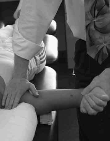
4 Terry Malone and Charles Hazle Authors do not use the same definition for frozen shoulder; this may account for the difference of care and success presented related to this pathology. We recommend dividing these patients into idiopathic (primary) and trauma/immobilized (secondary) groupings. Idiopathic patients often do not present until they notice significant loss of motion, whereas the trauma-based subjects develop significant “stiffness” after immobilization. These two groups require very different approaches to management. Although not well received, education of the idiopathic patient is critical to reasonable expectations as to treatment efficacy and timeframes for interventional success. We will address each group with pathogenesis, treatment concepts, and evidence-based clinical outcomes after providing a history of this condition. The first defined reference to this pathology may be attributed to Duplay who in 1872 described “periarthrite scapulohumerale”—the onset of which was attributed to subacromial bursitis.1 The condition was better defined and treatment described by Codman in 1934.2 He stated that these patients exhibited a slow onset, pain-disrupted sleep, painful and significant restriction of elevation and external rotation yet had a normal radiographic picture. Interestingly, he noted that these patients recover, with even the most difficult cases responding in about 2 years.2 Although many changes have occurred over the past 75 years, his observations are still relevant today. In 1945, Neviaser3 introduced the term adhesive capsulitis when he discovered dense thickening of the capsule, particularly in the axillary fold. He also believed that there were “intraarticular adhesions”; hence, his naming of the condition. DePalma4 reported that this syndrome was caused by bicipital tenosynovitis. In 1975, Reeves defined frozen shoulder as patients presenting with restricted motion in all directions, spontaneous onset of pain, and no apparent causation.5 The natural history has often included the description of symptom resolution over time. It should be noted that Codman’s early work2 is cited for this concept but his definition of resolution was not of an asymptomatic state but rather that the patients did not have permanent damage or deformity and that they were not disabled through the development of arthritis. Shaffer et al6 evaluated 62 patients with long-term follow-up to determine the expected outcome. Interestingly, they found that patients returned to function but approximately half did so with mild pain and/or stiffness. More than half of the patients demonstrated restriction of active motion, with the greatest restriction seen in external rotation. These authors then questioned the long-held belief of this condition being self-limiting because more than half of these patients do have persistent loss of motion, although the loss does not appear to significantly impact shoulder function. This is reinforced by the work of Griggs et al7 who followed 75 patients with idiopathic adhesive capsulitis treated with a four-direction stretching program providing a mean follow-up of 22 months. They found significant positive outcomes with high patient satisfaction, but there existed retained differences in pain and motion when compared with the uninvolved shoulder. The reason for these persistent changes may be related to the observations of Bunker.8 He examined via arthroscopy 35 recalcitrant idiopathic frozen shoulder patients. The consistent finding (31 of 35) was one of abnormal villous fronding of the synovium arising from the sub-scapularis bursa; the other four patients were very longstanding and actually had dense scarring of these same areas of the synovium/capsule. After a second arthroscopy that followed manipulation in 13 of these patients, 12 patients demonstrated avulsion of the capsule in the infraglenoid region. These findings are consistent with Wiley,9 who used arthroscopic evaluation of 37 patients with primary frozen shoulders demonstrating a granulation scarlike appearance in the same region as described by Bunker. Segmuller et al10 likewise found proliferative synovial tissue beneath the biceps and encompassing the subscapular recess in 24 primary frozen shoulder patients. The common feature in all of these studies is the development of proliferative synovial tissue in the subscapular recess extending to the rotator interval, which may ultimately become scarlike; this explains the loss of capsular volume and resultant decrease in range of motion (ROM). It also may help explain the ultimate outcome of a return to function but with residual loss of motion because the scar tissues may lack normal capsular extensibility. Numerous concomitant conditions may predispose one to idiopathic frozen shoulder. Bunker8 presents a compelling case for these patients having “Dupuytren’s like disease.” He shows many relationships with the links of frozen shoulder patients to Dupuytren’s (fibromatoses conditions) because they found more than half in his series presenting with both conditions. Many authors have described a link to diabetes, with the most accepted values being a 10 to 20% incidence of frozen shoulder in diabetics. There is an approximate doubling of this incidence in insulin-dependent patients. Patients are typically 40 to 60 years of age, and there is a higher frequency in women than men, with a 2 to 5% incidence in the general population.11 Also, those who develop bilateral frozen shoulders are even more likely to have diabetes.8,11 These findings indicate that some type of capsular fibroplasia is the causative agent of idiopathic primary frozen shoulder. This condition is often marked by an acute onset associated with trauma that is followed by immobilization (desired and designed via cast or other intervention) or unintentional as in the patient restricting use and movement volitionally, resulting in loss of motion. Harryman et al12 reiterate this through their recommendation of the designation “posttraumatic stiff shoulder.” They further state that this group can be subdivided into the causative trauma: injury, disease, or surgery. Treatment concepts for these patients will vary significantly; prevention is the rule in secondary designated subjects, whereas it is often not possible to prevent development in primary idiopathic patients. It should be noted that clinicians may exacerbate its development in primary idiopathic patients if lengthy immobilization in response to pain is imposed. Importantly, the trauma-related secondary designated patients typically present with a specific direction of limitation rather than the more complete or global loss exhibited by the primary frozen shoulder. We will address treatment concepts and provide the existing evidence of their efficacy specific to patient type (primary versus secondary). Unfortunately, although numerous treatments have been espoused, the evidence of efficacy is limited and further clouded by the previously mentioned level of recovery that is the rule rather than the exception: The primary designated patients regain function regardless of treatment if adequate time is provided—although still exhibiting measurable loss of motion and having some discomfort with use. Idiopathic primary patients have a sequence of progression of symptoms; therefore, the process can be divided into stages marked by the predominant symptoms at particular times. We prefer to use a four-stage sequence: painful, freezing, frozen, and thawing. In the painful stage, the patient complains of pain with motion and use of the shoulder. This pain is exacerbated by active use and can be quite significant, particularly at the end ROM. In response to the pain, patients have a tendency to restrict use, particularly at end or extreme ROM, thus setting in place the initiation of loss of motion. In the freezing stage, there is a progressive loss of motion. Patients lose significant amounts of external rotation (ER), elevation, and internal rotation (IR). They lose the motion actively and passively, with capsular shortening or restraint becoming obvious and having global restriction. The patient will exhibit pain at the end of the available ROM, which then facilitates a continued loss of motion. The frozen stage is a mature form of the freezing stage with a residual loss of motion but without significant pain at the end of the available range of movement. The patient can move and use the shoulder pain-free in the residual ROM. The patient is not experiencing additional loss of motion but rather is in transition to the next stage. The final stage is thawing. Patients begin to see an increase in their ROM; during this stage, physical therapy interventions can be quite helpful in assisting the return of motion. The great challenge is enabling patients to accept that these stages will occur sequentially and that they will regain their motion and function, but it requires months and years rather than weeks. This is not the information desired by a patient experiencing limited motion—particularly when pain is still a significant factor. An interesting observation has been that male patients often seem to recover more quickly than female patients do. We believe this is related to the time of presentation to the clinician. Women present when they lose the ability to reach clothing buttons or snaps behind their back (loss of IR), which is earlier in the progression. Men usually do not present until they are unable to get their wallet from their rear pocket; that is when they are closer to the thawing phase, later in the sequence or progression. Treatment must be stage-specific and include significant patient education. Patients must understand that the expression, “no pain, no gain” is not appropriate for their condition. Each stage will be approached related to the predominant problems and expected tissue reactions to intervention. We will present some common overall themes and then present recommendations for treatment associated with each stage. Multiple authors have recommended the use of analgesics and antiinflammatories, with some level of success in alleviating the overall pain associated with shoulder pain.11,12 Our experience supports the observation of Lee et al13; patients did better when these agents were combined with gentle exercises. The use of intraarticular injection has been shown to have mixed results in published studies8 and in our experience. No definitive recommendations can be made regarding their use; pain relief seems to be more obtainable than increased motion or function. Oral agents may be used throughout the treatment stages, particularly to assist with sleep disturbances. Some clinicians have attempted to increase the capsular volume through injecting fluid under pressure until the constrained capsule is distended and ruptures. In our experience this process has been of limited long-term success and must be done in later stages (frozen or thawing) to be successful. Stretching must be very carefully applied during the painful and freezing stages. It has been our experience that gentle stretching within the pain-free ROM is quite useful in all stages; more aggressive or forceful end-ROM exercise, however, can only be used during the frozen and thawing stages. In most patients, several sets or a short series of motion exercises should be done throughout the day rather than attempting to do a lengthy single session. In our experience, a program of three to five repetitions in five or more short sets dispersed throughout the day is more efficacious and better tolerated by most patients, particularly during the first two stages. For patients who are awakened by shoulder pain, a gentle motion program performed within the pain-free ROM facilitates their return to sleep. We use low loads, or stretch intensities, and longer durations when treating patients clinically, but this is typically during the thawing stage to facilitate the greatest possible return of ROM. The isolated use of heating modalities is probably used less today than in the past. Heating modalities were used to modulate pain and thus promote increased shoulder movement by the patient. The use of these modalities today is as a precursor to stretching to attempt to increase tissue temperature and thus extensibility. If you are attempting to use moist heat, we advise that you be sure to place it in the axilla (use a cervical-sized heating pack) to reach the anterior inferior fold most effectively. Some clinicians recommend using strengthening activities within the available pain-free ROM. It is our recommendation that the patient be encouraged to use the arm as much as possible but that independent strengthening exercises are of limited value. They may have a greater role during the late-thawing stage as patients reengage in higher levels of function. Many authors have recommended the use of manipulation in the treatment of recalcitrant frozen shoulder patients.11,12,14–16 The success of the treatment is dependent on patient selection and timing of intervention. Because of complications (fractures, tendon ruptures, etc.), manipulation under anesthesia is best applied only in the most severe cases and, importantly, only after the patient has reached the frozen stage. Early manipulation during the freezing stage results in a proliferative response and continued loss of motion; however, it is effective when performed in the frozen stage. Diabetic patients have an inconsistent response to manipulation. Most patients manipulated during the frozen stage do experience a significant increase in motion, with release typically of the inferior capsule. Harryman et al,12 Roubal et al,14 Placzek et al,15 and Ekelund and Rydell16 describe a number of appropriate manipulation techniques. Our recommended approach is translational with constant controlled loading; it is encouraging when we perceive a crepitant release with an immediate significant increase in motion. Figure 4–1 illustrates a modified technique for small–amplitude oscillations at the end ER range. In recalcitrant cases, typically after failed manipulation, surgical release of the capsule may be necessary. Open procedures were used until the improvement in arthroscopic techniques; the majority of cases today are performed using the arthroscope. Harryman et al17 and Warner et al18 both demonstrated excellent outcomes via arthroscopic intervention in these difficult patients. For a detailed description of surgical intervention, refer to Harryman et al.12
Rehabilitation of Adhesive Capsulitis
Treatments of Primary Frozen Shoulder
Treatments of Secondary Frozen Shoulder
 Idiopathic or Primary Frozen Shoulder
 Secondary Frozen Shoulder
 Treatment Concepts
Treatments of Primary Frozen Shoulder
Algorithm of Care
Analgesics and Anti-inflammatories
Distension of the Capsule
Stretching
Heating Modalities
Strengthening Exercises
Manipulation
Arthroscopic or Open: Surgical Release
Related posts:
 Use of Taping and External Devices in Shoulder Rehabilitation
Rehabilitation of Micro-Instability
Rehabilitation of Macro-Instability
Rehabilitation of Shoulder Impingement: Primary, Secondary, and Internal
Rehabilitation of Acromioclavicular Joint Injuries
Modification of Traditional Exercises for Shoulder Rehabilitation and a Return-to-Lifting Program
Use of Taping and External Devices in Shoulder Rehabilitation
Rehabilitation of Micro-Instability
Rehabilitation of Macro-Instability
Rehabilitation of Shoulder Impingement: Primary, Secondary, and Internal
Rehabilitation of Acromioclavicular Joint Injuries
Modification of Traditional Exercises for Shoulder Rehabilitation and a Return-to-Lifting Program
![]()
Stay updated, free articles. Join our Telegram channel

Full access? Get Clinical Tree


