Rehabilitation after Joint Preservation Surgery
Marc J. Philippon
Diana Patterson
Sean Garvey
Karen K. Briggs
Introduction
Excellent outcomes for arthroscopic procedures of the hip are allowing active patients to return to high-level athletic competition and active lifestyles at ever-increasing rates (1,2,3,4,5,6). With arthroscopy being performed for a growing number of hip pathologies, such as labral tears, femoroacetabular impingement (FAI), and other etiologies such as gluteus medius repairs, ligamentous laxity, and congenital hip dysplasia, there must be a larger focus on the design of rehabilitation protocols specific to hip arthroscopy and distinctly different from those for open surgical procedures of the hip (7,8,9,10,11,12,13,14). Contrasting surgical techniques and patient populations place emphasis on different aspects of the recovery following arthroscopy. For example, the arthroscopic approach does not require dislocation of the hip joint, so the postoperative range-of-motion (ROM) limitations of open surgeries are not necessary for patients undergoing arthroscopy (15). In addition, those patients undergoing arthroscopic procedures are more likely to be from a younger and more physically active population with a desire to return to high-level athletic participation. Thus, the postoperative rehabilitation process should be designed with those goals in mind and be emphasized in the preoperative planning period as an integral part of the procedure and its potential outcome.
Rehabilitation Goals
The most important factor of the post-arthroscopy rehabilitation process is that it be specific to the procedure performed, the patient’s healing time, discomfort tolerance, desired sport-specific activities, and postoperative athletic participation goals. Knowledge of hip biomechanics, the healing constraints specific to the affected tissues, and the type of surgical repair are necessary to develop an appropriate physical therapy protocol. The weight-bearing progression and ROM limitations in the immediate postoperative period are specific to the surgery performed and differ between types of procedures. Ideally, advancement of exercises in the early phases of rehabilitation is similar among different procedures, but actual progress is dependent on the patient and must be tailored to his or her specific situation. Surgical candidates should be educated preoperatively that rehabilitation is an involved, not particularly rapid, process (16). Its success depends highly on the patient’s expectations, procedure performed, and the patient’s level of commitment. Return to high-level activity following arthroscopy for FAI is rarely less than 12 weeks, but can be longer, and even in the precocious patient, the rehabilitation process must never be shorter than the necessary time for natural tissue and osseous healing (15). An ideal outcome for the procedure and rehabilitation is not determined by the speed with which the patient returns to their prior level of activity or competition, but their overall satisfaction and longevity of function (17). The major components of the individualized rehabilitation program are protection, with procedure-specific restrictions, ROM movements, strengthening exercises, and functional progression of sport-specific activities.
Four-Phase Model of Rehabilitation
Specific exercises for each component of the rehabilitation process are discussed in more detail later in this chapter, but the model used for rehabilitation protocol has four stages and levels of intensity. These four phases—maximum protection and mobility, controlled stability of movement, reclamation of strength, and return to sport—provide an overarching framework for the process. However, ensuring the program is individualized to the patient and monitoring his or her progression through the steps is also imperative. The mobility and protection phase is dependent on soft tissue and osseous healing, and thus necessarily covers approximately the initial 6 to 9 weeks postoperatively, but the other steps are reliant on the patient’s ability, commitment to reaching the goals of each phase, and meeting the criteria required to advance, and therefore exist on a more variable timeline.
Phase one, the immediate postoperative period, aims to protect the integrity of newly repaired tissues, diminish inflammation, prevent muscular atrophy, contraction and inhibition, and restore passive ROM (18). Tools utilized may include flat foot weight bearing (FFWB), passive
circumduction, stationary biking, and continuous passive motion (CPM) machines. The requirements to progress are experiencing minimal pain or pinching with all exercise and achieving ROM ≥75% of the nonoperative side; full weight bearing must also be permitted by the surgeon depending on the procedure performed (18).
circumduction, stationary biking, and continuous passive motion (CPM) machines. The requirements to progress are experiencing minimal pain or pinching with all exercise and achieving ROM ≥75% of the nonoperative side; full weight bearing must also be permitted by the surgeon depending on the procedure performed (18).
Phase two focuses on regaining pelvic and trunk stabilization and weight-bearing gait. Stabilization exercises may begin simultaneously with the exercises of phase one once in the second postoperative week and progress according to protective restrictions (19). Goals of this phase are to normalize the gait, restore full ROM, improve neuromuscular control, balance, and proprioception, and initiate sport-specific functional exercises, with a focus on core and pelvic stability (17). Weight-bearing gait can begin as soon as 2 weeks postoperatively for some nonmicrofracture surgeries, but must wait until 8 weeks postoperatively for surgeries including a concomitant microfracture to ensure appropriate fibrocartilage healing. Exercises possible in phase two include standing or prone-resisted internal and external rotation, 1/3 knee bends, wall sits with an abductor band for resistance, sidesteps with an abductor band for resistance, and two-legged bridging for core strength (20). Late in phase two, when all surgery-specific restrictions are lifted, functional movements, such as light jogging for field and court sports, skating for ice sports, and dance movements for dancers can be initiated. Aquatic therapy may be useful in the progression of weight bearing and ambulation, as the buoyant environment allows for controlled gait training (15). However, treadmill use is not recommended at any time, even if underwater, as it introduces a secondary shearing force to the surgically repaired hip because of the movement of the tread (19). Requirements to progress to the next phase include demonstrating a normal pain-free gait, full ROM, successful initiation of exercises without pain, and exhibiting good control and stability of the pelvis and hip musculature (19).
Phase three aims to restore full preoperative muscular strength, endurance, and cardiovascular fitness, and continue improving neuromuscular control, balance, and proprioception (18). The ultimate goal is that hip flexion strength is of 70% and adductor, abductor, extension, and rotational strength are 80% of the contralateral hip, and the patient is able to demonstrate proper body mechanics and muscle firing patterns on basic agility drills (18). Sample exercises include standing resisted hip external rotation, walking lunges, lunges with trunk rotation, plyometric bounding in water, resisted sport cord walking in the forward, backward, or sideway direction, and progressive exercise ball work for increasing core strength. If the patient does not desire to return to a high level of athletic activity, he or she can progress with their normal daily activities as desired at this point (19). If he or she is an elite athlete, passing the sport test, as detailed below, is necessary for the athlete to progress to the final phase of rehabilitation.
Phase four consists of sport-specific training to restore the patient’s prior level of power, explosiveness, and agility to enable return to play. Another important element of the conclusion to rehabilitation is to assist the patient in becoming independent in and educated about a maintenance program to ensure the future health of the surgically treated hip.
Protection in Immediate Postoperative Period
Hip arthroscopy techniques performed during surgery can include but are not limited to capsular plication, labral repair, labral reconstruction, osteoplasty, chondroplasty, microfracture, iliopsoas lengthening, iliotibial band (ITB) release, gluteus medius repair, for conditions such as hypermobility, hip dysplasia, soft tissue repair, chondral defects, and FAI. Each procedure has a specific set of postoperative restrictions designed to protect the particular surgical repair in the days and weeks immediately after the procedure.
Patients undergoing an osteoplasty or rim trimming, for cam and/or pincer-type FAI, should be limited to 20 lb (9 kg) of FFWB for the first 3 weeks after surgery (Fig. 52.1). In postoperative weeks 3 to 4, weight bearing can begin to be increased to 50% of the patient’s body weight and increase 10% to 15% per day as the patient tolerates progression. The FFWB limitation is designed to reduce the risk of a stress fracture to the newly altered femoral neck, reduce inflammation more quickly within the joint, and reduce the shearing force on the anterior acetabulum created by heel-strike motion. FFWB technique is preferred over toe-touch weight bearing as to not create a flexion contracture; it also assists with venous return and attempts to regain correct gait neuromuscular firing patterns of the hip musculature. There is no passive ROM limitation. Exercises immediately after surgery to regain passive ROM can prevent adhesions in the surgical area, especially between the acetabular rim and the capsular attachment. Passive circumduction exercises performed
at neutral and at 70 degrees of flexion are done to prevent adhesions between the circular fibers of the zona orbicularis and the femoral neck (12,14,19). Implementation of circumduction exercises in the postoperative period has been shown to reduce the occurrence of capsular adhesions from 4% without its use to 1.4% with its use (20).
at neutral and at 70 degrees of flexion are done to prevent adhesions between the circular fibers of the zona orbicularis and the femoral neck (12,14,19). Implementation of circumduction exercises in the postoperative period has been shown to reduce the occurrence of capsular adhesions from 4% without its use to 1.4% with its use (20).
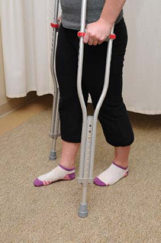 Figure 52.1. Flat foot weight bearing is recommended for the first 3 weeks after surgery. Flat foot weight bearing is preferred over toe-touch weight bearing to limit flexion contracture. In addition it assists with venous return and helps the patient regain correct gait neuromuscular firing patterns of the hip musculature. |
Patients requiring a labral repair should avoid sitting at 90 degrees. These restrictions are to reduce stress on the femoral neck during early healing; however, these motions are toward the end of most patients’ range and generally takes 1 to 2 weeks before they are comfortable attempting these ROMs. There are no other restrictions to ROM, as there are no muscular attachments directly to the labrum. However, because the surgical repair is deep within the soft tissues of the hip, it is recommended that the overall volume of active movements be limited. There are no weight-bearing restrictions. Patients who have had a labral reconstruction should follow the same postoperative restrictions as a labral repair.
If a microfracture procedure was performed, the most significant limitation is FFWB of 20 lb (9 kg) for 8 weeks following the procedure. This is to protect the clot that has formed in the cartilage defect following microfracture. They should also use a CPM machine for at least 6 to 8 hours per day during their weight-bearing restrictions to prevent adhesions and possibly promote fibrocartilage growth (22,23,24). If only a chondroplasty, there are no weight-bearing restrictions, and the CPM should be used for 4 to 6 hours per day during their weight-bearing restrictions.
Restrictions following procedures on selected pelvic muscles generally consist of avoiding contraction of the operative area. Following an iliopsoas lengthening, the patient should limit firing of the surgically altered hip flexor by using the contralateral limb, brace or clothing to assist with movement, and a brace to prevent hyperextension. Following ITB or greater trochanter bursa release, the patient should engage in gentle stretching until weight bearing is permitted and more aggressive stretching can take place to avoid adhesions and a tightening of the ITB. After a gluteus medius repair, the hip should be contained to a ROM from 15 to 45 degrees of abduction to avoid any stress to stretch to the muscle. To do so, the patient should be braced at 15 degrees abduction and use the CPM machine while at the same 15 degrees. Abduction restrictions should not be reduced until 6 weeks postoperatively to allow for adequate muscle healing and reduction of inflammation. At that time, abduction restrictions can be reduced 5 degrees per week until 0 degree and beyond. When strengthening exercises are begun at approximately 6 weeks post surgery, the patient should begin with isometrics to prevent overloading of the repaired muscle and to reduce the risk of tendonitis development. Patients may progress as tolerated.
Following surgery for hip hypermobility, patients are restricted from external rotation and hyperextension for 21 to 28 days postoperatively. To prevent hyperextension, the brace should be locked at 0 degree of extension. External rotation boots or bracing may be used while sleeping or lying supine to prevent natural inclination of the hips to roll into external rotation (Fig. 52.2). Capsular plication recipients should externally rotate or hyperextend the hip for the 18 to 21 days following surgery depending on quality of capsular tissue, presence of hypermobility, and dysplasia. Progression to neutral extension while lying supine and with brace to prevent naturally occurring external rotation is recommended by the end of the first postoperative week to prevent hip flexor contractures (19). It is also important to achieve extension to 0 degree to normalize gait even while on crutches. There are no weight-bearing restrictions following a capsular plication.
 Figure 52.2. Natural external rotation. |
Last, following surgery for the treatment of hip dysplasia, the patient should be braced at 15 degrees of abduction to increase the stability of the joint during early healing. Starting at week 3, the abduction restriction can be decreased 5 degrees per week to prevent contraction of the abductors.
Related posts:

Stay updated, free articles. Join our Telegram channel

Full access? Get Clinical Tree


