Fig. 8.1
Anatomy of the quadriceps muscle. (a) Deep plane, (b) superficial plane. VL vastus lateralis, VI vastus intermedius, VM vastus medialis, RF rectus femoris. Reproduced from Pasta G, Nanni G, Molini L, Bianchi S. Sonography of the quadriceps muscle: Examination technique, normal anatomy, and traumatic lesions. J Ultrasound. 2010;13:76–84. Copyright© 2010 Elsevier. All rights reserved

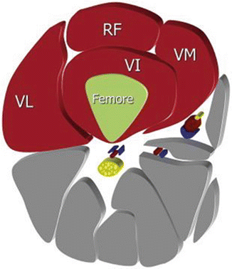
Fig. 8.2
Anatomy of the quadriceps muscle. Axial plane (diagram): VL vastus lateralis, VI vastus intermedius, VM vastus medialis, RF rectus femoris. Reproduced from Pasta G, Nanni G, Molini L, Bianchi S. Sonography of the quadriceps muscle: Examination technique, normal anatomy, and traumatic lesions. J Ultrasound. 2010;13:76–84. Copyright© 2010 Elsevier. All rights reserved

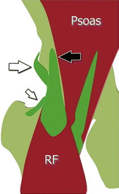
Fig. 8.3
Anatomy of the proximal rectus femoris. RF rectus femoris, black arrow = direct tendon and its insertion on the anteroinferior iliac spine; white arrow = indirect tendon and its insertion on the lateral aspect of the acetabular rim; small white arrow = reflected tendon and its insertion on the anterior articular plane. Reproduced from Pasta G, Nanni G, Molini L, Bianchi S. Sonography of the quadriceps muscle: Examination technique, normal anatomy, and traumatic lesions. J Ultrasound. 2010;13:76–84. Copyright© 2010 Elsevier. All rights reserved
As stated above, the proximal tendon of the rectus femoris has both superficial and deep components [4]. The direct head forms the superficial anterior fascia that covers the ventral aspect of the proximal 1/3 of the muscle. The tendon of the indirect head travels parallel and deep to the direct head ending in the distal 1/3 of the muscle belly. This terminal extension of the indirect head forms an intramuscular muscle-tendon junction and a “muscle within a muscle” [4, 5] that has been described by Hughes et al. [4] and Hasselman et al. [2]. The rectus femoris muscle is unipennate in its proximal third. The fibers arise from the direct head and travel distally to insert onto the distal tendon. Distal to the direct head, it is a bipennate muscle. Muscle fibers arise from the indirect tendon and extend medially and laterally to insert distally on the tendon of insertion [2, 4].
The quadriceps muscle group is innervated by the femoral nerve, and its blood supply comes from the femoral artery. The action of the muscle group is to extend the leg at the knee. The rectus femoris is the only one of these muscles that crosses two joints, and it also flexes the pelvis on the femur and stabilizes the pelvis during weight bearing [5]. Gait analysis has shown that during the heel strike of running, the action of the quadriceps is to decelerate knee flexion [4].
Quadriceps Strains
Quadriceps strains are common in sports that require a lot of eccentric contraction such as soccer, track and field, basketball, rugby, and football [2–6]. Activities that require eccentric contraction of the quadriceps include jumping, sprinting, and kicking [2–6]. Three large prospective studies of quadriceps muscle injury did not find any association between a person’s age and muscle injury [5]. Previous injury to the quadriceps muscle and recent hamstring strain increase the risk of quadriceps strain [5, 7]. Leg dominance has also been shown to be a risk factor for quadriceps muscle strain with the dominant leg being injured more than the nondominant leg [5]. A dry playing surface and ground firmness have also been shown to be risk factors for quadriceps muscle strains [5, 7]. Other proposed risk factors for quadriceps as well as other muscle strains include low muscle strength, muscle fatigue, age, lack of warm-up, muscle temperature, and poor flexibility [6, 7].
Medications that predispose to quadriceps tendon tears include anabolic steroids, statins, locally injected corticosteroids, and prolonged use of systemic corticosteroids; however, many of the studies done on quadriceps tendon tears are focused on the distal, not proximal, tendon injury [8]. Fluoroquinolones and androstenediol supplements have been associated with ruptures in other tendons [8]. Systemic diseases that can predispose to ruptures include renal disease, diabetes, hyperparathyroidism, rheumatoid arthritis, systemic lupus erythematosus, gout, obesity, and infection [8].
The rectus femoris is the muscle most commonly injured in this muscle group [4–7]. Sixty percent of rectus femoris strains occur in the proximal 1/3 of the muscle [3]. Factors that are thought to contribute to rectus femoris strains are the high percentage of type II muscle fibers (approximately 65 %) that the muscle contains [2, 4–6], the fact that it crosses two joints, its eccentric contraction as it decelerates the hip and knee, and its complex musculotendinous architecture (the “muscle within a muscle,” as described above) [2, 6]. Injuries of the vastus muscles are typically due to extrinsic factors such as being struck by another player or by an object, and they are less likely to be a result of the intrinsic factor of eccentric loading as are rectus femoris strains [3].
Athletes who sustain an acute quadriceps strain typically notice a sharp pain in their quadriceps associated with a loss of function of the muscle. They often point to the area of the pain in their distal quadriceps, but several studies have shown that quadriceps strains commonly occur at the mid to proximal portions of the rectus femoris [6]. Many times athletes will be able to continue to play, and the pain will intensify later in the day [6].
Strain of the quadriceps may be associated with a bulge or defect in the muscle belly. Bruising may not occur until 24 h after the injury. Pain is often felt by the patient with resisted active muscle activation, passive stretching, and direct palpation of the muscle [6]. In a recent study by Cross et al. [7], it was found that the site of maximal tenderness was often 3 cm or more away from the site of maximal strain on MRI. They also found that tenderness over the rectus femoris didn’t always mean that the injury was in the rectus femoris [7].
The treatment of muscle strains to the quadriceps muscles is not unlike strain of any other muscle. There is little scientific evidence for the majority of treatment protocols [6]. Within 24–72 h after a muscle strain, the treatment focuses on minimizing bleeding into the muscle, and hence avoiding further complications, with rest, ice, compression, and elevation (RICE) [6]. A US Naval Academy study showed that immobilization with the knee at 120° in the first 24 h was associated with a return to activity in an average of 3.5 days [9]. A review by Bleakley et al. [10] did not show that the use of ice facilitated faster healing and return to sport; however, they did comment that there are not many high-quality studies evaluating this and more quality research needs to be done. Ice can decrease the pain associated with a muscle injury [6, 10]. If the athlete has a lot of pain, crutches may be needed to help with ambulation immediately after the injury [6]. NSAIDs may be used for a short period (3–7 days), but corticosteroids should be avoided due to possible delayed healing and reduced strength of the muscle [6, 11]. After the first 3–5 days, active rehabilitation is started. Prolonged immobilization has been shown to be associated with longer disability [9]. Rehabilitation includes stretching, strengthening, and range of motion, aerobic fitness, proprioception, and functional training [6]. The intensity of the rehabilitation should be done to the level of patient discomfort but not to pain. Rehabilitation should also be done within a pain-free range of motion [6, 9].
Grading of quadriceps muscle strains is not much different than any muscle strain (Table 8.1). The severity (grade) of a muscle strain has been defined in many ways. They are typically divided into three grades. A grade 1 strain involves disruption of a few of the muscle fibers within a muscle fascia [3]. Grade 1 strains are also associated with no or minimal loss of strength and no palpable muscle defect [6]. In a grade 2 strain, the surface of the damaged fascia represents less than 3/4 of the total section of the muscle [3]. There is a moderate loss of strength and there may be a small palpable muscle defect in grade 2 strains [6]. In a grade 3 strain, the surface of the rupture extends to more than 3/4 of the total section and may extend to the entire muscle belly resulting in a complete rupture [3]. There is significant or even complete loss of strength in grade 2 strains and there is usually a palpable mass [3]. In adults, muscle strain injury is almost always located at the musculotendinous junction [6].
Table 8.1
Grading for quadriceps strainsa
Grade | Pain | Strength | Physical exam |
|---|---|---|---|
1 | Mild | No or little loss of strength | No palpable muscle defect |
2 | Moderate | Moderate loss of strength | Possible small palpable muscle defect |
3 | Severe | Near to complete loss of strength | Palpable muscle defect |
The rectus femoris is the muscle of the quadriceps muscle group that is most commonly strained [4–7]. With grade 1 proximal strain, there is hemorrhagic infiltrate around the muscle tissue where it attaches to the central tendon. On ultrasound this will appear hyperechoic. Grade 2 lesions show partial rupture of the muscle fibers and a fluid collection (hematoma) around the central tendon. The hematoma will appear as a hypoechoic area on ultrasound. The muscle tissue around the central tendon will also become infiltrated with blood [4]. This will cause enlargement of the muscle belly. There is a complete disruption of the fibers from the central tendon in grade 3 strains, and the muscle bellies may retract. Acutely there will be a hematoma formation around this area, but chronically, healing may result in fibrosis resulting in local retraction of the muscle fibers. On ultrasound, this will appear as hyperechoic areas within the muscle with surrounding muscle retraction [3].
Cross et al. found that the prognosis of acute quadriceps strains was significantly dependent on both the site and size of the injury. Cross-sectional area percentage and length were independently predictive of prognosis [7]. Injuries to the central tendon of the rectus femoris have the least favorable prognosis [7]. In a 3-year causal comparative study by Cross et al. in 40 Australian Rules football players, 25 clinical quadriceps strains were recorded, and none of these were the injuries that have been classically described in literature as the grade 3 complete disruption of the distal rectus femoris. Cross et al. suggest that distal quadriceps strains may not be as common as previously thought. In Cross’s study, all 25 athletes who had a muscle strain complained of pain in the midline of the anterior thigh despite seven of the injuries being due to a vastus muscle and three of the injuries having no MRI findings of strain. Also, the site of maximal tenderness was often 3 cm or more away from the site of strain on MRI [7].
Hughes et al. coined the term “bull’s-eye” lesion to describe a muscle strain injury associated with enhanced signal around the central tendon on T1-weighted MRI images after IV gadolinium [4, 7]. This high signal is associated with increased vascularity due to chronic inflammation and vascular infiltration of a fibrous scar [4]. This is an incomplete intrasubstance tear occurring at the muscle-tendon junction formed by the deep tendon of the indirect head of the muscle and those muscle fibers taking their origin from this tendon to insert distally into the quadriceps tendon [4]. Each of the injuries in Hughes’ study had presented for medical care at least 4 weeks after injury and as long as 156 weeks after injury and are considered more chronic injuries [4]. Cross et al. did a study that included patients who presented within 24–72 h of injury, and they found increased signal, best seen on axial T2 images, about the central tendon. They hypothesize this signal is from edema, hemorrhage, and muscle debris. Cross et al. termed these lesions as “acute bull’s-eye” lesions.
Stay updated, free articles. Join our Telegram channel

Full access? Get Clinical Tree


