Fig. 1.1
Illustration of the posterior thigh demonstrating the hamstring gross anatomy. The hamstrings lie in the superficial muscle layer of the posterior thigh, with the semitendinosus (A) and semimembranosus (B) on the medial side and the long head (C and E) and short head (D) of the biceps femoris on the lateral side of the posterior thigh
Biceps Femoris
The biceps femoris muscle is located on the posterolateral aspect of the thigh. It originates from two locations, with the long head originating from the medial facet of the ischial tuberosity and the short head arising from the lateral supracondylar ridge of the femur and the middle third of the linea aspera (Fig. 1.2). The short head of the biceps femoris follows a path distally and laterally at a 30° angle to the coronal plane of the femur and a 45° angle to the sagittal plane of the femur when the knee is flexed to 90° (Fig. 1.3) [1]. The origin of the short head of the biceps femoris on the femur is often used as a landmark to classify a hamstring injury as proximal or distal [2]. The short head of the biceps femoris muscle is innervated by the common peroneal division of the sciatic nerve, while the long head is innervated by the tibial division of the sciatic nerve (both L5 and S1 nerve roots) [3].
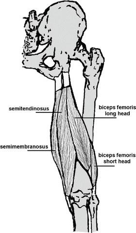
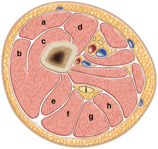
Fig. 1.2
Illustration of the hamstring origins. The semitendinosus, the semimembranosus, and the long head of the biceps femoris originate from the ischial tuberosity. The short head of the biceps femoris originates from the lateral supracondylar ridge of the femur. With kind permission from Springer Science + Business Media: Skeletal Radiology, MR observations of long-term musculotendon remodeling following a hamstring strain injury, 37(12), 2008, Amy Silder
Fig. 1.3
Cross-section of the gross anatomy of the mid-thigh. The quadriceps muscle complex [rectus femoris (A), vastus lateralis (B), vastus intermedius (C), and vastus medialis (D)] runs along the anterior and lateral aspect of the femur. The hamstring muscle complex [short (E) and long (F) head of the biceps femoris, semitendinosus (G), and semimembranosus (H)] runs along the posterior aspect of the femur surrounding the sciatic nerve (I)
An understanding of the tendinous insertion points of the long and short heads of the biceps femoris is crucial for appreciating the biomechanics of the posterolateral corner of the knee. Acute injuries to the biceps femoris muscle and subsequent insertion points can lead to acute knee instability [4–6]. The tendinous insertion components of the long head of the biceps femoris begin to form proximal to the knee and then divide into two tendinous components (the direct and anterior arms) and three fascial components (the reflected arm, the lateral aponeurosis and the anterior aponeurosis) at the knee [1]. Terry et al. have shown that the direct arm inserts at the posterolateral edge of the fibular head at a point lateral to the fibular styloid, while the anterior arm inserts on the lateral edge of the fibular head [1]. The reflected arm of the fascial components originates from the tendon just proximal to the fibular head and inserts to the posterior edge of the iliotibial tract [1]. The lateral aponeurotic expansion attaches to the tendinous anterior arm and covers the fibular collateral ligament, while the anterior aponeurotic expansion covers the anterior compartment of the leg [1]. The short head of the biceps femoris has a muscular attachment to the anterior and medial sides of the distal long head tendon. There are also tendinous attachments to the posterolateral joint capsule at the level of the posterior horn of the lateral meniscus (a capsular arm), the fibular head (direct arm), and the lateral tibial tuberosity 1 cm posterior to Gerdy’s tubercle (anterior arm) [1, 7]. The other significant insertion of the short head is an attachment of the capsuloosseous layer to the iliotibial tract that forms a biceps–capsuloosseous iliotibial tract complex [1].
Understanding the anatomical relationships of the components of the biceps femoris muscle complex is very important in developing an understanding of their role in stability of the knee. Studies have shown that injuries to the biceps femoris tendons have been seen in conjunction with lateral ligamentous injuries of the knee and anterolateral–anteromedial rotary instability of the knee [8, 9]. A study by Terry et al. [1] has shown that the short head of the biceps femoris muscle is more commonly injured than the long head. The injuries most commonly seen include avulsions of the capsular arm followed by injuries to the biceps–capsuloosseous complex. Other avulsion injuries are seen at the insertion of the anterior arm of the short head at the lateral tibial tuberosity [1]. It has also been proposed that the dual innervation of the biceps femoris muscle may lead to desynchronized firing of this muscle, and this could be one of the underlying reasons that it is the most commonly injured muscle in the hamstring muscle complex [10, 11]. The biceps femoris muscle complex is a very important dynamic knee stabilizer and repair or reconstruction of the tendinous and facial insertion components should be taken into account when patients present with knee instability.
Semitendinosus Muscle
The semitendinosus muscle receives its name from the substantial tendonous component to the overall size of the musculotendinous unit. This muscle originates from the inferomedial side of the ischial tuberosity as part of a conjoint tendon that also includes the long head of the biceps (see Fig. 1.2) [3]. The conjoint tendon is oval in shape and measures 2.7 cm superoinferiorly and 1.8 cm transversely on average [12]. The semitendinosus muscle has a complex tendinous intersection that separates the muscle into inferior and superior regions that are innervated by separate branches of the tibial nerve [13]. It has been postulated that the superior region may specifically function in driving motion at the hip while the inferior region may specifically function in driving motion at the knee [14].
At the more distal aspects of the semitendinosus, the muscle forms a long round tendon at the midpoint of the thigh that runs along the medial side of the popliteal fossa. The tendon follows a path around the medial tibial condyle and then passes over the medial collateral ligament, and inserts on the superomedial surface of the tibia. The semitendinosus, gracilis, and sartorius muscles all contribute to the pes anserinus on the anteromedial surface of the proximal tibia, and their corresponding bursae can be a source of pain due to pes anserinus bursitis [3]. The long length of the tendon of this muscle has been thought to predispose this muscle to rupture [15]. Additionally, the semitendinosus tendon is often harvested for anterior cruciate ligament (ACL) reconstruction, and though it has been shown to regenerate to a certain degree, the distal aspect appears to reinsert on the gastrocnemius fascia rather than the tibia.[16]. This can occasionally lead to ineffective scar formation and, in turn, hamstring weakness and recurrent injury in the high-level athlete [16, 17].
Semimembranosus Muscle
The semimembranosus muscle originates from the ischial tuberosity at a point that is superior and lateral to the biceps femoris and semitendinosus muscles. The origin of the semimembranosus tendon is crescent shaped and extends superoinferiorly over 3 cm and transversely over 1 cm (see Fig. 1.2) [12]. The proximal tendon follows a course that travels medial and anterior to the other muscles of the hamstring complex (see Fig. 1.3). The proximal tendon is an elongated structure with fibrous connections to the origin of the biceps femoris and adductor magnus tendons [3]. The tendinous origin of the semimembranosus is the longest of the proximal hamstring tendons, averaging 31.9 cm in cranial–caudal length, and becomes aponeurotic soon after its origin [13]. Distally the muscle is composed of numerous short unipennate and multipennate fibers, which maximize the number of muscle fibrils per unit area [18]. The semimembranosus muscle belly is the largest of the hamstring muscle complex averaging 15.7 cm2 in its midsubstance. This large area allows for the muscle to generate the greatest force but at the slowest velocity of all the hamstring muscles [19]. Distally, the muscle inserts primarily on to the posterior medial aspect of the medial tibial condyle, with multiple tendon slips that expand across the medial aspect of the knee and attach to various soft tissue support structures of the knee.
The multiple insertion points of the semimembranosus are important contributors to the stability of the posteromedial corner of the knee [18, 20, 21]. There are discrepancies as to how many insertion points or “arms” are formed from the distal tendon of the semimembranosus. LaPrade et al. [22] have attempted to create a common terminology and description of the anatomy of these insertion points. Generally there is agreement on three arms: the direct arm, the anterior arm, and the expansion in the oblique popliteal ligament [22]. The direct arm of the insertion follows an anterior course deep to the anterior arm and inserts on the posterior medial aspect of the tibia. The anterior or tibial arm extends anteriorly under the posterior oblique ligament and inserts on the proximal tibia inferior to the tibial collateral ligament [22]. The oblique popliteal ligament is a broad, thin lateral continuation of the semimembranosus tendon that becomes part of the posterior medial capsule [22].
LaPrade and colleagues [22] have also described multiple additional insertions: the distal tibial arm (also referred to as popliteal aponeurosis), the components of the posterior oblique ligament, the meniscal arm, and the proximal posterior capsular arm. The distal tibial arm is an expansion of the semimembranosus that forms a facial layer over the popliteus muscle belly [22]. The fibrous sheath of the semimembranosus tendon extends anteriorly and contributes to the posterior oblique ligament and is thought to act as a secondary stabilizer to posterior tibial translation [23]. The meniscal arm is described as a short band-like connection between the tendon and the meniscotibial band at the posterior horn of the medial meniscus, and is thought to prevent impingement of the posteromedial meniscus during flexion [24]. The proximal posterior capsular arm has been shown to course along the superior aspect of the oblique popliteal ligament and ends with several fine attachments to the posterior capsule [22, 25]. These insertions all work together to stabilize the posterior medial corner of the knee and should be taken into account when evaluating the patient with posteromedial knee instability.
Muscle Composition
The muscles of the hamstring complex all have distal tendons that originate from deep within the muscle belly and run close to the entire length of the muscle and then emerge at the distal end of the muscle–tendon unit as distinct tendonous structures [20]. These long tendons help to develop a “spring” effect that accentuates performance during athletics; however, this ability may also be a detriment as the “spring” effect leads to increased susceptibility to injury [26]. The tendons are attached to the muscle fibers in a pennate arrangement on the central tendon [20]. The distal myotendinous junction has been described as the weakest link in the muscle–tendon–bone complex, and as a result is a common region of injury [2].
The muscle and tendon structure of the hamstring complex creates three distinct areas within each muscle–tendon unit where the different physical properties of tissues interact, resulting in areas susceptible to eccentric injuries [10, 27–29]. The first of these distinct areas is the myotendinous junction, the point where the distal and proximal tendons emerge from the muscle belly. The second is the myofascial junction, the location at which the muscle fibers connect to the aponeurotic fibrous layer surrounding each of the muscle bellies. Lastly, the intramuscular myotendinous junction runs along a large portion of the muscle belly [2]. It is important to know that in the skeletally immature athlete the ischial apophysis is the weakest point of the hamstring muscle–tendon unit until the secondary ossification center of the ischium is closed sometime between the 15th and 25th years of life [26]. Understanding the basic anatomy and the locations predisposed to injury can assist the treating physician with appropriate diagnosis and treatment.
Variant Anatomy
There are a number of commonly described anatomic variants to the hamstring complex. The biceps femoris muscle has been described with variant origins on the ischial tuberosity [21]. The semimembranosus muscle has also been documented as hypoplastic or absent in some cases, while others have documented hypertrophic tendon slips [30, 31]. Injury may result due to weakened muscle–tendon units or decreased flexibility secondary to hypertrophic tendon slips. Hamstring variant anatomy is typically diagnosed on ultrasound or magnetic resonance image (MRI) and knowledge of such variations is crucial for guiding diagnosis and must be appreciated if operative intervention is to be conducted [21, 30].
Biomechanics
The muscles of the hamstring complex work together to both extend the hip and flex the knee during the gait cycle. During the swing phase of the gait cycle the hamstring muscles coordinate extension at the hip and prevent extension at the knee [21]. The hamstring complex is also involved in external and internal rotation of the leg due to the obliquity of the biceps femoris and semitendinosus, respectively, when the knee is in a flexed position [21].
The hamstring complex also plays an important role in stabilizing the hip joint. The inferior origin of the hamstring muscles within the pelvis, but at the level of the hip joint, also assists in stabilizing the hip joint. The length of the hamstring muscle complex limits the range of motion of the hip in a way that prevents full hip flexion unless the knee is in the flexed position. As a result, forward swing of the thigh results in passive flexion of the knee and protects the hamstrings from strain due to overextension injuries during the swing phase of the gait cycle [32]. This mechanism of protection breaks down when an athlete is sprinting at full stride, attempting to over stride, or when his or her foot hits the ground. At this vulnerable position the hamstring muscle complex is at its point of both maximal lengthening and maximal muscle unit contraction, leading to eccentric firing of the muscle and increased risk of injury [33, 34].
The hamstring muscle complex plays a large role in the stabilization of the posterolateral corner of the knee. In knee flexion the insertion of the biceps femoris on the iliotibial tract and fibular collateral ligament helps to keep the iliotibial tract tight during knee flexion. This assists with knee stability under varus loads [35]. The hamstring insertion at the fibular collateral ligament has been shown to help keep the collateral ligament taut during the full range of knee flexion, while the insertion at the posterior capsule contributes to the posterior pull of the capsule during knee flexion contributing to capsular stability [35].
The multiple insertion points of the semimembranosus are also paramount for stabilization of the posteromedial corner of the knee [23]. These insertions strengthen the stability of the knee under valgus load and have a small but mentionable role in prevention of ACL and posterior cruciate ligament reconstruction failure if injuries to the posteromedial corner are recognized preoperatively [23]. The contribution of the semimembranosus insertion on the posterior oblique ligament (POL) has a large effect on the stability of the posteromedial corner of the knee during the full range of knee motion [36, 37]. It is a primary stabilizer against internal rotation of the knee in full extension, but also contributes to the stability through all knee flexion angles [37]. The POL functions by sharing the forces on the medial collateral ligament (MCL) with internal and external rotation, valgus stress, and anterior and posterior tibial translation of the knee [36, 37].
The hamstring muscles also act in conjunction with the ACL to prevent anterior tibial translation during the heel strike portion of the gait cycle. A study by Li et al. has shown that the combination of both hamstring and quadriceps co-contraction can reduce the in-situ forces on the ACL at various points during the gait cycle [38]. Increasing the strength of the hamstring muscle complex has been shown to decrease ACL strain, and as a result some have advocated for training programs focused on hamstring strengthening to prevent ACL ruptures [39, 40].
Injury to the hamstring muscle–tendon unit during the gait cycle is due to the contraction of the antagonistic hamstrings and quadriceps muscle complexes and the imbalance in muscle strength, with quadriceps muscle being stronger. These conditions result in forced hip flexion with the knee held in extension [21, 28, 41]. The sudden functional change of the muscle complex from dynamic stabilizer to rapid contractor has been thought to be the cause of hamstring injuries at this stage of the gait cycle [42]. Another possible cause for hamstring injury is the aforementioned biarticular design of the hamstring muscle complex. Due to this design the hamstring muscles are not able to act on one joint without stabilizing the other joint. This results in the muscle complex having to absorb and counteract these multiple competing forces, leaving the hamstring muscle complex susceptible to injury [15, 42]. Compensatory injury to the hamstring complex has also been described in athletes with deficiencies in the surrounding joints. In cases of athletes with femoroacetabular impingement, the demand for motion at the hip may exceed the functional range of motion. In this case the athlete may recruit adjacent muscles and joints to provide the necessary excess motion, which can injure the hip joint and adjacent muscles [43].
Quadriceps
Normal Anatomy
The quadriceps muscle complex consists of the rectus femoris, the vastus medialis, the vastus lateralis, and the vastus intermedius (Fig. 1.4). The quadriceps are the primary extensors of the knee and all are innervated by the femoral nerve (L2-4 nerve roots) and receive their blood supply from the common femoral and deep femoral arteries [3]. The quadriceps muscle complex is one component of the extensor mechanism. The additional extensor mechanism components include the quadriceps tendon, patella, patellar tendon, and patellar retinaculum. All of these structures work in concert to provide knee extension.
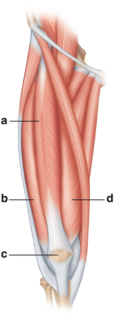
Fig. 1.4
Illustration of the anterior thigh demonstrating the quadriceps gross anatomy. The quadriceps muscle complex lies along the anterior thigh. The rectus femoris (A) is the most superficial with the vastus lateralis (B) and the vastus medialis (D) on the lateral and medial sides of the thigh, respectively. The quadriceps muscle complex inserts on the patella (C) and ultimately onto the tibial tubercle via the patellar tendon
Rectus Femoris
The rectus femoris muscle is the only muscle of the quadriceps muscle complex to cross two joints. One head of the muscle originates from the anterior inferior iliac spine (the direct head) and the other head (the reflected or indirect head) originates from the ilium superior to the acetabulum [3]. The rectus femoris lies on top of the vastus intermedius and is the most superficial muscle of the anterior compartment (see Figs. 1.2 and 1.3). The true insertion of the rectus muscle is the tibial tubercle through confluents with the patellar ligament, but in some literature the insertion is described to be at the patella as a common tendon with the vastus muscles [44]. The rectus femoris inserts on the anterior portion of the base and the superior third of the anterior surface of the patella, with some portions contributing to the patellar tendon [45–47].
Vastus Medialis
The vastus medialis runs along the medial side of the extensor compartment of the anterior thigh (see Fig. 1.4). The muscle originates from the intertrochanteric line and the medial lip of the linea aspera of the femur, and the muscle inserts on to the superior medial border of the patella via the quadriceps tendon. The muscle is composed of an oblique portion and a longitudinal portion referred to as the vastus medialis obliquus (VMO) and vastus medialis longus (VML), respectively. The VMO arises from the tendon of the great adductor muscle and inserts on the medial and superior borders of the patella at an angle between 50° and 55° [48]. Weakness, atrophy, and variations in the attachment location of the VMO have all been shown to be causes of patellofemoral instability and maltracking [47, 49, 50]. This often leads to anterior knee pain and instability, thus strengthening of the VMO must be an integral part of the physical therapy and rehabilitation protocols when such pathology is suspected.
Related posts:

Stay updated, free articles. Join our Telegram channel

Full access? Get Clinical Tree


