Fig. 9.1
Quadriceps relaxed
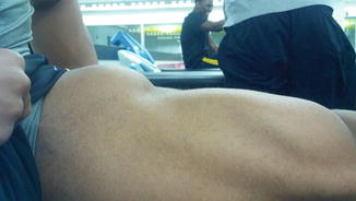
Fig. 9.2
Quadriceps contracted
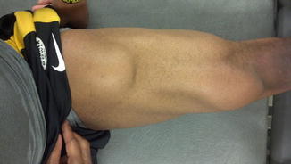
Fig. 9.3
Quadriceps anterior
Imaging is needed to help confirm the presence of MO. Modalities can include plain radiography, ultrasound, or MRI. Plain radiographs can start to show changes of faint periosteal bone reaction as early as 7–10 days after injury but usually take 3 weeks or longer to become visible. The more characteristic findings of a peripheral rim of calcification will begin to be visible as early as 6–8 weeks after injury and will progress over a few months to appear more like mature, lamellar bone [17]. Figures 9.4 and 9.5 show radiographs from a collegiate baseball player who presented with mild aching thigh pain 1 year after a quadriceps contusion. He was diagnosed with MO, treated with physical therapy, and was able to return to full participation without deficit.
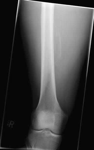
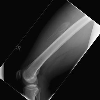
Fig. 9.4
Myositis ossificans: anterior posterior radiograph
Fig. 9.5
Myositis ossificans: lateral radiograph
Musculoskeletal ultrasound is beneficial for imagining of MO as the calcification will appear as an acoustic shadowing earlier than expected changes on plain radiographs [19]. The main caution is that early changes can appear similar to sarcoma. Typically the changes of MO are more rapidly progressive than a slow growing sarcoma, but serial imaging is needed to help confirm the diagnosis. As MO progresses, ultrasound will show central decreased echogenicity and occasionally an outer lamellar hyperechoic rim [20]. As peripheral ossification ensues and the lesion matures, acoustic shadowing will become apparent.
MRI of MO can also be useful. Initially the imaging shows an ill-defined mass with surrounding edema, which like ultrasound can also have the appearance of a sarcoma. As the lesion matures it will more clearly appear ossified without surrounding edema.
Treatment for MO is mostly close observation. Initially rest, ice, compression, and elevation are generally the treatments of choice, followed by physical therapy and rehabilitation. There is some mixed belief regarding nonsteroidal anti-inflammatory drug (NSAID) use in the acute setting. Some have recommended NSAIDs by extrapolating the data supporting the use of indomethacin or naproxen to prevent heterotopic bone formation following hip replacement surgery [21]. NSAIDs may be helpful in the short term to prevent MO from developing initially, but overall evidence for NSAIDs and corticosteroids shows impaired healing muscle response that could potentially interfere with normal muscular healing. Given the anticoagulant properties, it may be that NSAIDs can also increase the degree of hematoma formation. Therefore, some authors suggest avoidance of NSAIDs for 48–72 h, to avoid hematoma formation, followed by regular use of indomethacin or naproxen to help prevent myositis ossificans [22].
A few case reports and one case series have evaluated the potential for extracorporeal shockwave therapy (ESWT) to benefit in the treatment of MO. Buselli et al. demonstrated that three treatments with ESWT every other week for a duration of 3 weeks showed improvement in range of motion and pain within 48 h of the first treatment. The majority of athletes were able to resume full participation within an average of 11 weeks. Despite decreased pain and improved range of motion, Buselli’s study was unable to show a decrease in the size of the ossification with ESWT treatment [23]. Buselli’s group also used an intensive rehabilitation program with physical therapy visits six times per week, 80 min at a time [24].
Others have suggested, with little evidence, the use of platelet-rich plasma injections into the area of myositis early in the process to help prevent the overall severity and degree of MO.
Surgical excision, if needed, should not be performed until the ectopic bone has fully matured which may not occur until 12–14 months after the initial injury. King proposes surgical intervention when a mechanical block persists after all of the clinical signs of pain, inflammation, tenderness, and swelling have resolved [25].
Compartment Syndrome
Compartment syndrome of the thigh is a condition more commonly associated with a greater severity of injury, such as underlying femoral fracture. However, it can also be seen from a direct blow to the anterior thigh (in sports such as soccer, rugby, football, and martial arts) [26]. The inexperienced clinician may be led to the precocious diagnosis of compartment syndrome in the first 24 h following a thigh contusion because of the acute, severe pain and swelling. It is imperative in these cases to examine and reexamine the patient in the first 24 h.
The increasing pressure is secondary to vascular trauma leading to bleeding and hematoma formation but can also be seen due to increased capillary permeability with third spacing of fluid within the compartment. Therefore, as noted above, minimizing the swelling after the injury is of primary importance by reducing activity, compression dressings, and icing before beginning more aggressive rehabilitation.
The natural course of compartment syndrome involves increasing the pressures with any of the three compartments of the thigh, triggering muscular ischemia, acidosis, skeletal muscle death, and occasionally neurovascular compromise. Of the three compartments of the thigh, the anterior compartment by far is the most commonly involved. There are some reports of compartment syndrome appearing as late as 8 days after injury, but usually symptom onset is within the first 24 h following injury [27]. One hallmark of compartment syndrome is pain that is out of proportion relative to the severity of the injury. Passive quadriceps extension will increase the pressures of the compartments and athletes will often present with the knee in nearly full extension, as this helps to mitigate pain. Paresthesia in the saphenous nerve distribution (anterior aspect of the knee and medial aspect of the leg and foot) can be a sign of more severe injury [28].
It is important to consider in compartment syndrome in the differential diagnosis and recognize early through compartment pressure testing and imaging to avoid poor outcomes. Whitesides et al. developed the classic method of compartment pressure testing in 1975 [29]. It is important to note that most of the work on compartment pressure measurement has been done on the leg, and not the thigh; therefore, all the available literature needs to be taken with that consideration. Measurement should be near the site of injury as differences as little at 5 cm can cause tissue pressure differences. It is imperative to determining pressure thresholds for treatment and diagnosis. Szabo et al. showed the relationship between pressure and physiologic nerve dysfunction [30]. In healthy muscle this is approximately 10 mmHg less than the diastolic blood pressure and is 20–30 mmHg less than DBP in damaged muscle. Others have suggested fasciotomy when pressure exceeds 30–35 mmHg. Robinson et al. treated six athletes with acute compartment syndrome of the anterior thigh following contusion and noted intraoperative pressures of 68–88 mmHg [31]. Rothwell managed nonoperatively 60 cases of quadriceps hematoma due to blunt trauma. Pressure readings were not done in this series; however, thigh circumference was increased up to 4 cm compared to contralateral side, suggestive of significantly increased pressure. All patients recovered without quadriceps weakness or decreased range of motion [32].
Fasciotomy and decompression should be done for acute-onset severe disease with associated neurovascular compromise and should be considered in the treatment of symptoms of ongoing pain despite conservative treatment. Anterior compartment fasciotomy should be performed through a mid-lateral longitudinal incision and wounds should be packed open. After 48 h there can be consideration for delayed primary wound closure versus skin grafting if required.
In the absence of progressively worsening symptoms or the development of neurovascular compromise, mild to moderate elevations of compartment pressures can be treated conservatively with observation, passive ROM, ice, heat, elevation, and analgesia [28]. Hyperbaric oxygen (HBO) increases tissue oxygenation and has been shown to have benefit in the treatment of injuries resulting in tissue ischemia, such as crush injuries or compartment syndrome [33]. The majority of the HBO treatment literature to date has been more in the treatment of trauma rather than athletic injuries. While HBO remains a promising treatment, further randomized, controlled trials are needed to extrapolate its use for the athletic population [34].
One final complication that can accompany thigh contusion or crush injury is a Morel-Lavallee lesion. This is triggered when the skin and subcutaneous fatty tissue abruptly separate from the underlying fascia. This is also referred to as a “closed degloving injury.” These effusions are most common in the trochanteric region and proximal thigh. Imaging with MR can present in a variety of ways. They can present as a hematoma with capillary ingrowth. More established lesions can appear like encapsulated cysts or seromas and have quite varied signal intensity. Based on the vascularity of these lesions, they can sometimes be difficult to distinguish from soft tissue neoplasm with increased vascularity. Treatments for Morel-Lavallee lesions have included compression, percutaneous aspiration with debridement, irrigation, and suction drainage. In recalcitrant cases either talc sclerosis or doxycycline sclerodesis has been successful [35].
Related posts:

Stay updated, free articles. Join our Telegram channel

Full access? Get Clinical Tree


