Physical Inactivity: Physiological and Functional Impairments and Their Treatment


Physical Inactivity: Physiological and Functional Impairments and Their Treatment


Eugen M. Halar

Kathleen R. Bell


While physical activity and exercise are generally well-accepted concepts in healthy persons, the consequences of inactivity and bed rest are less well understood by clinicians and the general public. The deleterious effects of immobility and physical inactivity are common and may affect multiple body systems and the stealthy onset of these effects may minimize the awareness of the dangers and the timely response for prevention and treatment.

Bed rest and immobilization were widely used before 1950 in the management of trauma and acute and chronic illness, before the physiologic effects were well understood. It was generally assumed that rest fostered healing of the affected part of the body. What was not appreciated was that immobility and inactivity could be harmful to the unaffected parts of the body. For example, the immobilization of long bones with a rigid cast has a beneficial effect on bone healing after fractures. However, it may also result in undesirable effects, such as joint contracture and atrophy of the healthy muscles and bones.

Clinical studies on enforced bed rest in normal subjects and on astronauts in microgravity conditions (in which their bodies rest from the effects of gravity) have shown significant undesirable effects that may override the therapeutic effects of bed rest in subacute and chronic conditions, impacting complexity and cost of medical treatment as well as functional outcome.

A review of randomized controlled trials on the effects of bed rest and immobility did not demonstrate improvement or better outcome of primary medical conditions for those on extended periods of bed rest. In many cases, worsening occurred if early mobilization was not provided (1). Persons who are chronically sick, aged, or disabled are particularly susceptible to the adverse effects of immobility. A patient with motor neuron disease and its accompanying limb weakness or spasticity would be expected to develop the same musculoskeletal complications but at a much accelerated rate. The healthy subject on prolonged bed rest may only show some degree of atrophy and weakness; the neurologically impaired subject will likely also lose a significant amount of independent functioning. Many of these complications could be easily prevented or, if they occur, easily treated once they are recognized. Therefore, the prevention of such complications should be one of the basic principles of any rehabilitation management plan.

Negative effects of immobility or inactivity are rarely confined to only one body system (Table 48-1). Immobility reduces the functional reserve of the musculoskeletal system, resulting in weakness, atrophy, and poor endurance. Metabolic activity and oxygen utilization in the muscle are reduced, degrading the functional capacity of the cardiovascular system as well (Fig. 48-1). In addition, postural hypotension and deep venous thrombosis (DVT) are commonly encountered in bedridden patients. Immobilization osteoporosis is yet another complication that has been well documented in the studies of astronauts and individuals exposed to prolonged bed rest. Over time, clinical experience has dictated a move toward earlier mobilization and provision of functional training with a resulting decrease in the length of hospitalization and in the incidence of major morbidity associated with prolonged inactivity (2).

The most deleterious effects of inactivity can be grouped together under the general term “deconditioning,” which is defined as reduced functional capacity of musculoskeletal and other body systems. It should be considered a distinct diagnosis from the original condition that has led to a curtailment of normal physical functions (3). This chapter describes the widespread effects of immobility and physical inactivity, reviews the evidence-based therapeutic and prophylactic approaches to counteract these complications, and redirects attention to the benefits of physical activity and exercises in maintaining a good health and independence.


MUSCULOSKELETAL EFFECTS OF IMMOBILITY AND INACTIVITY

Moving the body and limbs freely in the environment is a principal physical function requiring that the muscles, nerves, bones, and joints be in an optimal physiologic state. Disuse weakness and reduction of free joint motion can initially cause minimal functional limitations that can be easily overlooked or
neglected. However, advanced contractures and disuse weakness can cause a loss of mobility and decrease in activities of daily living (ADL) functions (4).


TABLE 48.1 Adverse Effects of Immobility and Inactivity


System(s)


Effect(s)


Musculoskeletal


Muscle weakness, fatigue, and atrophy


Muscle and joint contractures


Muscle stiffness and pain


Osteoporosis


Hypercalcemia


Cardiovascular and pulmonary


Redistribution of body fluids


Dehydration


Orthostatic intolerance


Reduction of cardiopulmonary capacity


Reduction of VO2max


Elimination of bronchial secretions


Hypostatic pneumonia


Genitourinary and gastrointestinal


Urinary stasis, stones, and urinary infections


Loss of appetite


Constipation


Metabolic and endocrine


Glucose intolerance


Electrolyte alterations


Increased parathyroid hormone production


Other hormone alterations


Immune system


Impaired wound healing


Reduction in cellular immunity


Resistance to infection reduced


Anti-inflammatory suppression reduced


Cognitive and behavioral


Sensory deprivation


Confusion and disorientation


Anxiety and depression, memory


Decrease in intellectual capacity


Impaired balance and coordination


Cellular/Genetic


Diminished gene expression


Mitochondrial dysfunction


For the neurologically impaired or multiple trauma victim, considerations such as preserving functional range of motion (ROM) may seem trivial; however, neglect of these simple factors can be responsible for prolonging hospital stays, increasing the use of health care resources, and prolonging dependency in mobility and ADL (5).


FIGURE 48-1. Inactivity, immobility, and prolonged bed rest influence total body functioning.

Three main types of adverse effects from inactivity are encountered in the musculoskeletal system: muscle atrophy and weakness, joint contracture, and immobilization osteoporosis (see Table 48-1).


INACTIVITY OF SKELETAL MUSCLE


Physiological Impairments


Disuse Atrophy

Decrease in the size of muscle fibers and reduction of muscle mass are the hallmark of muscle atrophy. In a lower motor neuron lesion, the atrophy is regional and related to the particular nerve or root. Atrophy associated with muscle disease is more pronounced in the proximal muscles. The atrophy of disuse is generalized or localized to the immobilized limb(s) and more prominent in the antigravity muscles. It is a consequence of the limited physical activity and musculoskeletal loading that occurs during immobilization, immobility, bed rest, or exposure to microgravity during space flight. As a general rule, increased muscular activity leads to hypertrophy, whereas limited physical activity leads to disuse atrophy and weakness. The changes in muscle fiber size either in atrophy or in hypertrophy and phenotype changes of muscle fibers are the results of remarkable muscle plasticity and adaptability to external physical demands to generate adequate or peak contraction and endurance.

Disuse atrophy is defined as an alteration of metabolism and muscle cell homeostasis in response to muscle inactivity. Recent studies indicate that muscle protein synthesis as well as whole body protein production is significantly reduced during immobility and is considered the main contributor to muscle atrophy. The rate of muscle wasting during bed rest is slow during the first 2 days but becomes rapid thereafter. By 10 days, it reaches 50% of eventual muscle weight loss. Similarly, muscle protein synthesis is reduced to 50% of the baseline level at 14 days of immobilization and then gradually tapers off to reach a new steady state (6). Reeves et al. (7) studied skeletal muscle changes in healthy men exposed to microgravity condition and found a decrease in calf muscle volume by 29%, resting fascicle length of medial head of gastrocnemius and their pennation angulation by 10% and 13%, respectively, and physiological cross-sectional area (PCSA) by 22%. In disuse atrophy, especially if muscle is immobilized in a shortened position, that is flexion, the number of sarcomeres in series is reduced as a result of diminished chronic stretch and adaptation of the muscle to a new resting length. By contrast, immobilization in an elongated muscle resting position or during musculoskeletal growth increases the number of sarcomeres in series (8). In addition, the number of sarcomeres in parallel is reduced contributing to the reduction of muscle fiber PCSA (muscle volume/fascicle length) (9). From data obtained on the
PCSA, the reduction in the number of sarcomeres in parallel is twofold greater than reduction of sarcomeres in series. This indicates that reduction of isometric strength is in proportion to the loss of total number of sarcomeres in parallel and that they are more affected by disuse (9).

Disuse atrophy of type I and type IIa muscle fibers is more prominent than type IIb fiber atrophy during immobility. The mean size of type I fibers in human soleus muscle is decreased by 12% and 39% at 2 and 4 months of bed rest, respectively. This reduction could be prevented by simulating gravity loading for 10 hours a day in supine position (10). Individual muscle thickness can be clinically measured in vivo for some muscle by ultrasound scanning. Using this technique, the reduction of muscle thickness after 20-day bed rest is on average -2.1% to -4.4% and varies significantly for different muscles of the lower limbs (11). Magnetic resonance imaging (MRI) studies have, however, revealed much greater percentages of atrophy of the muscles in the lower limbs with the same duration of bed rest (12). Furthermore, consecutive MRI studies of thigh and calf muscles revealed that cross-sectional area and muscle volume of the gastrocnemius and soleus muscles were reduced to a greater extent than knee extensors and flexors (-9.4% to -10.3% vs. -5.1% to -8.0%) after 20 days of bed rest (12). However, using specific tests, the mean cross-sectional area reduction for the dark adenosine triphosphatase (ATPase) and light ATPase fibers is 46% and 69%, respectively, after 20 days of immobility (13).

Along with muscle fiber atrophy, the synthesis of collagen fibers is also reduced, although this reduction is much less than the reduction in synthesis of muscle proteins. This leads to a relative increase of muscle collagen content and changes in its mechanical elastic properties. The increase in muscle stiffness and alteration in viscoelastic properties of plantar flexors during space flight correlated with the duration of the flight, but the changes were less prominent than during bed rest (14). In both situations, weight bearing is limited, but joint motion is free and abundant during space flights. In healthy subjects with normal mobility, the main resistance to excessive elongation of muscle fiber is due to resistance of the myofibrils and in a lesser part to the sarcolemma itself. Titin, a myofibrillar protein, appears to have a major role in providing resistance to passive elongation, and it is increased in immobility. When titin is chemically removed, this resistance to passive stretch is significantly diminished (14).

Prominent histochemical changes also occur in muscle during bed rest and immobility. Serum creatine kinase (CK) isomer and fibroblast growth factor release (after myofiber injury) are both reduced during bed rest, and this is proportional to the reduction in muscle fiber size. Resistance exercises during bed rest significantly increase the level of these factors and prevent muscle fiber atrophy, indicating that myofiber wound-mediated fibroblast growth factor may play an important role in disuse atrophy (15). Myostatin is a growth factor-beta protein that inhibits muscle synthesis and is increased during bed rest. After 25 days of bed rest, the total lean body mass declines by an average of 2.2 kg, and plasma myostatin-immunoreactive protein level increases by 12%. It is speculated that suppression of myostatin may prevent muscle atrophy during space flight (16).

The muscle mass loss associated with aging is called sarcopenia. It is a major cause of disability and frailty in the elderly population. Inactivity is one of the many factors responsible for development of sarcopenia, along with decline in number of alpha-motor neurons, reduction in growth hormone, inadequate protein intake, and chronic overproduction of catabolic cytokines. High-intensity resistance exercise can reverse sarcopenia, indicating that physical inactivity is a major risk factor for weakness in elders (17). Many articles have been written about functional decline in hospitalized and nursing-home patients. General deconditioning in elderly persons is a frequent cause of functional decline, falls, and increased dependency. Reconditioning takes much longer in the elderly than in younger persons.

Although the primary reason for atrophy is reduction of muscle protein synthesis, an increased protein breakdown with nitrogen loss is also found during immobility despite the fact that the major energy sources during bed rest are primarily derived from carbohydrates and fat. It is believed that decline of mitochondrial function and the reduction of protein synthesis are the main reasons for the onset and progression of disuse atrophy. However, in the later stages of disuse muscle atrophy, the process of protein degradation may become more prominent than decrease in protein synthesis. This may be aggravated by gastrointestinal mechanisms, such as loss of appetite and reduced intestinal absorption of protein. Although daily loss of nitrogen for an immobilized healthy person may reach 2 g/day, a nutritionally depleted person may lose as much as 12 g/day. Urinary excretion of creatine is minimal under normal conditions, except in pregnancy and infancy. The excretion of creatine is much greater in starvation, diabetes, muscular dystrophy, hyperthyroidism, and fever, as well as during immobility. Prolonged bed rest and weightlessness causes a significant increase in the excretion of both creatine and creatinine, the mechanism which is not well understood (18).


Loss of Strength

Muscle weakness, reduced endurance, and tolerance to work are the functional consequences of muscle atrophy. The maximal strength of a muscle can fall to 25% to 40% of baseline level when a person is exposed to minimal exertions over a 2- or 3-week period (19). During strict bed rest, muscles may lose 10% to 15% of their original strength per week and, over 4 weeks, 35% to 50%. The loss of strength is rapid after the first day of immobilization and reaches its maximum 10 to 14 days later (20). Resistance leg exercises performed above 50% of maximum every second day, for 20 minutes by healthy subjects on bed rest, can maintain the muscle protein synthesis at the same level as healthy subjects engaged in normal physical activity (21). A study on dynamic leg press training during bed rest demonstrated preservation of baseline cross-sectional area and strength for the knee extensors and flexors but not for ankle plantar flexors and ankle dorsiflexors. This can adversely affect functional locomotion, as ankle plantar flexion contributes to forward propulsion (22).


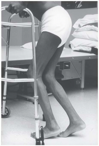
The loss of strength associated with disuse atrophy is more prominent in the lower limbs than in the upper limbs. Loss of muscle power during immobilization reaches -20% to -44% in knee flexors and extensors, comparing to an insignificant loss in the upper limbs of -5%. The decrease in peak muscle tension of knee flexors and extensor ranges from -19% to -26%, far more than the reduction in cross-sectional area of these muscles (-7%), indicating that loss of strength and power is proportionally greater than reduction in size of the respective muscles (23). The major contributors to the loss of strength and endurance in persons with disuse atrophy are the reduced number of myofibrils per fiber volume, reduction in size and number of mitochondria, muscle fiber nuclei, and reduction of sarcomeres in parallel (10).

Another aspect of disuse weakness is reduction of maximal instantaneous muscular power. It can be reduced to a greater extent than the peak muscle strength of one repetition maximum in bed rest subjects. The loss of strength has been well documented in immobility; however, the loss of explosive muscle power as measured by the maximal jump with both feet on force plate was only recently studied. After 45 days of bed rest, instantaneous muscular power was reduced 24%, and recovery required one and half months of remobilization (24). These findings indicate that specific training for instantaneous power should be considered during space flight or prolonged bed rest to preserve functions such as standing up, climbing, and performing transfer functions.

Decline in muscle twitch and tetanic tension parallels the decline in muscle strength. In rats, maximal tension obtained by electrical stimulation in the soleus muscle declined significantly after 6 weeks of immobilization (25). Changes in contractile forces of immobilized muscle are the result of diminished levels of myofibrillar proteins and also due to a reduction of sarcoplasmic reticulum calcium ion uptake, but not the rate of its release (26).


Loss of Endurance

Multiple studies have demonstrated that prolonged inactivity causes a significant and progressive reduction in muscle endurance. Unexercised muscle demonstrates a reduction of adenosine triphosphate (ATP) and glycogen storage sites and rapid depletion of them after resumption of activity. Reduction of muscle protein synthesis and oxidative enzyme function, and premature anaerobic energy production with rapid accumulation of lactic acid, are important factors leading to fatigability and reduced endurance (27).

Metabolic and enzymatic alterations in unexercised muscle result from reduced demand for oxygen and reduction in the blood supply. Initially after bed rest, succinate dehydrogenase enzyme activity per muscle fiber increases, but later, the overall amount is reduced (28). Oxidative enzyme activity and content as well as the number and size of mitochondria are all reduced during immobility (29). Unexercised muscle also shows a decreased ability to utilize fatty acids when compared with trained subjects.

Ferretti et al. studied peripheral and central factors that contribute to the decline of VO2max after 42 days of bed rest. VO2max was found reduced by 16%, cardiac output by 30%, and oxygen delivery by 40%, which parallels the reduction in muscle cross-sectional area of 17%, volume density of mitochondria by 16%, and total mitochondria volume in the magnitude of 28%. Oxidative enzyme activity falls by 11%. These decrements indicate a significant contribution of both peripheral and central factors in causing the decline in VO2max after immobility, confirming a close interrelationship between the muscular and the cardiovascular system (27).

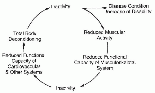
The following sequence of events transpires in the deconditioning process. Prolonged reduction of muscle repetitive contractions below 50% of maximum alters muscle protein synthesis and decreases glycogen and ATP storage, causing a reduction of oxidative enzymes, mitochondrial function, and microvascular circulation, impacting muscle metabolic activity. As a result, the oxygen supply is attenuated, and the extraction of oxygen from blood is diminished, further negatively affecting VO2max and cardiovascular reserve. The loss of muscle mass leads to reduction of muscle strength and endurance, reducing muscle blood flow, red blood cell delivery, oxidative enzyme activity, and oxygen utilization in the muscle precipitating a further loss of musculoskeletal and cardiovascular functional reserve to low or dangerous levels. In this cascade of events, specific muscle gene activation and expression are altered as well. Physical inactivity causes change in muscle fiber type composition and decreases formation of oxidative muscle fiber types I and IIa, the main factors in reduction of endurance and fitness (30).


Functional Impairments of Disused Muscle


Mobility and ADL

The progressive weakness and reduced stamina resulting from inactivity negatively impact the ability to perform basic mobility and ADLs. In the lower limbs, type I muscle fibers, which are active during standing and slow ambulation, are especially affected with a rapid reduction in endurance. If the quadriceps muscle is immobilized in an extended position, the deep layer of vastus intermedius, which has predominantly type I and type IIa fibers, shows the greatest histochemical changes in contrast to the rest of the muscle (31). Such accelerated rate of atrophy and weakness was also noted in hip and back extensors, hip abductors, and ankle plantar flexors and dorsiflexors, impacting the ability to walk (5). Disuse weakness and loss of endurance will result in impaired ability to perform ADLs and ambulate safely, reducing personal independence (Fig. 48-2).


Muscle Pain and Stiffness

Patients frequently complain of back pain during bed rest. The cause of this pain is still not fully understood. Recent studies in which spine length and degree of movement were measured by miniature ultrasound transmitters have demonstrated that back pain is more prominent when trunk movements are limited in a supine position. It is speculated that localized, prolonged, low-intensity isometric muscle contractions may cause this pain. Back pain can be averted by stretching exercises and walking (32).

Limb muscle pain and stiffness occur after generalized immobility or focal limb immobilization, especially in the presence of limb swelling. However, gentle static or intermittent stretching can alleviate this problem and prevent the reduction in number of sarcomeres in series (33). It has been shown that the position in which a joint is immobilized has a significant
influence on the number of sarcomeres present in a single muscle fiber (33). When immobilized in a shortened position (extensors in a fully extended position or flexors in a fully flexed position), a muscle can lose 40% of its original number of sarcomeres, contributing significantly to weakness and muscle stiffness (34).


FIGURE 48-2. A sequence of contracture development occurred from hip down to knee in a patient with traumatic hip fracture treated operatively with the pins. As a result of hip-flexion contracture and immobility, the hamstring and eventually posterior capsule with neurovascular soft tissue of the knee became tight and contracted, causing knee-flexion contracture. With these contractures, a person must walk on the toes, which increases energy expenditure.


Disuse Weakness, Deconditioning, and Cardiovascular Disease

A lack of adequate muscle activity adversely affects the cardiovascular and related systems. As noted in detail above, chronic inactivity impairs and reduces maximal oxygen consumption (VO2max), cardiovascular reserve, and fitness (35). Individuals with an inactive lifestyle and low level of fitness are more prone to develop coronary artery disease (CAD) and have greater odds of suffering myocardial infarction and death. A number of epidemiologic studies have demonstrated an inverse dose-response relationship between physical activity and mortality from CAD (36). The mechanism of this interaction is not known, but the current belief is that regular physical activity prevents or suppresses the chronic inflammation associated with atherosclerosis, CAD, and the metabolic syndrome (37, 38, 39).

A daily regimen of 30 to 45 minutes of endurance or aerobic activity will reduce the risk of CAD, non-insulindependent diabetes, hypertension, and breast and colon cancers. Encouraging lifestyle changes and promoting fitness and leisure physical activity for such patients are important aspects of prevention and treatment of CAD (40).

It is well documented that chronic exercises have antioxidant effect on several levels. Exercise training prevents or reduces low-density lipoprotein oxidation (formation of a powerful free radical, oxLDL), which has been implicated as a cause of endothelial cell damage and subsequent inflammation and development of atherosclerosis. Furthermore, exercise training can promote and improve endothelial cell function and release of nitric acid (41). As noted, inflammation plays a key role in CAD. Inflammatory markers such as C-reactive protein are reduced with exercise training, which may explain the beneficial effects of exercise on reducing mortality and morbidity associated with CAD (41). A study by Ambramson and Caccarino revealed that physical activity is independently associated with lower levels of inflammatory markers even in healthy subjects with no signs of CAD or other inflammatory diseases (37).

When physical inactivity is associated with other conditions causing muscle atrophy and weakness, the catabolic changes in muscle can be aggravated. For example, the elderly with age-related decreases in VO2max from baseline are particularly affected by prolonged bed rest. Similarly, trauma combined with strict bed rest can accelerate the loss of muscle strength and protein breakdown. This effect is maybe due to hypercortisolemia associated with acute trauma, resulting in muscle catabolism. In one study, intravenous hydrocortisone given to immobilized persons resulted in three times the rate of catabolism seen at baseline (42).


Prevention and Treatment of Muscle Weakness


Principles


  • Identify clinical and subclinical changes in strength, endurance, and physical function.


  • Determine whether additional conditions exist that may exacerbate atrophy and weakness caused by inactivity, such as acute trauma or chronic disease.


  • Ascertain the necessity for continued bed rest or immobility.


Strategies


  • Prevent muscle weakness by prescribing progressive resistive exercise, stretching, and aerobic exercise.


  • In most cases, use a combination of specific exercises to address all aspects of muscle weakness, including exercise for flexibility, strength, endurance, and fitness.


  • Remobilize the patient as quickly as possible; provide progressive mobility training.


  • Encourage 30 minutes of walking and leisure activities for at least 3 days a week for the general population.


Flexibility Exercise

Stretching to maintain optimal muscle resting length as well as viscoelastic properties is important for maintaining normal muscle function. Several animal studies indicate that passive stretching of striated muscle is associated with muscle hypertrophy, increase in muscle fiber area, increase in number of sarcomeres, and muscle fiber proliferation. In doing so, undifferentiated, quiescent myoblasts residing on the sarcolemma of muscle fibers (satellite cells) are activated and believed to be responsible for this stretch-induced muscle hypertrophy. Passive stretch is also a potent mechanical stimulus to influence gene expression and muscle proliferation. For example, both myogenin mRNA per microgram RNA and muscle cross-sectional area were significantly increased after 3, 6, 14, and 21 days of stretch (overload) in avian latissimus dorsi (43).

Muscle stiffness due to lack of stretch occurs through the reduction of the elongation properties of elastic and collagen fibers and myofibrils. It is also a result of structural changes in the muscle, such as muscle fiber angulations, reduction of sarcomeres in 18 series, and rearrangement of collagen fibers and their cross-links. Even a relative increase in muscle connective tissue may lead to muscle stiffness and reduction of joint motion. Muscles that cross two joints such as the hamstrings, gastrocnemius, and long back extensors are particularly prone to stiffness, even in healthy subjects with limited physical activity. In immobilized and inactive persons, however, this process is accelerated in the absence of activity-induced stretch. Stiffness and subsequent muscle belly shortening of two-jointed muscles can interfere with functional walking. Hip-flexion contracture at 35 degrees as a result of iliopsoas muscle tightness causes a 60% increase in energy consumption per unit distance during ambulation (5). Thus, daily stretching of a muscle for a half hour can prevent the loss of sarcomeres in series of the immobilized muscle and maintain elongational properties of muscle fibers and surrounding connective tissue, maintaining full ROM of joints.


Strengthening (Resistance) Exercises

To prevent disuse weakness and deconditioning, daily physical activity should be encouraged, and progressive resistance exercise (PRE) using isokinetic or isotonic exercises done on a regular basis. A minimum of once a day muscle contraction at 30% to 50% of maximal strength for 3 to 5 minutes, three times a week for a single muscle group, may suffice to prevent muscle loss and weakness in otherwise mobile, active individuals. Strengthening exercise prevents muscle atrophy and improves strength and endurance by mediating the activation of muscle genes, enhancing mitochondrial function, and improving protein synthesis, causing muscle fiber phenotypic changes and muscle angiogenesis (Table 48-2).

Muscle strength can be increased by concentric or eccentric contractions performed isokinetically or isotonically. To improve or maintain strength, there should be 8 to 15 repetitions for each muscle group done twice with brief pauses between sets. There are many studies that indicate that strenuous eccentric exercises may produce muscle damage since CK may increase 50-fold and cytokine and interleukin-6 (IL-6) fivefold. Furthermore, high-intensity eccentric exercises in untrained individuals frequently produce elevation of myofibrillar enzymes in plasma, ultrastructural damage of muscle fibers, infiltration of mononuclear blood cells, and acute inflammation (44). Therefore, it is important that eccentric exercises are started with lower intensity, fewer repetitions, and slower progression of applied resistance or loads.


TABLE 48.2 Resistance/Strengthening Exercises Guide


Establish one repetition maximum for each muscle group.


Select initial and subsequent intensity at 50%-80% of the maximum.


Repetition is performed 8-15 times, twice per session for each muscle group, three times per week or more.


Progressive increase in resistance (i.e., weights) and time until goal is achieved.


Include sequentially all muscle groups of the lower and upper limbs, back and abdominal muscles.


Focus on antigravity muscles, agonists, antagonists, and functional training.


Exercise should become a habit and part of a person’s wellness program.


Muscle weakness can also be prevented by the use of electrical stimulation. For example, applying local stimulation to the quadriceps while in a long leg cast may help preserve muscle bulk and strength and also may shorten rehabilitation time, a factor that may be particularly important in an athlete (45). For astronauts exposed to prolonged periods of microgravity with resulting muscle atrophy, functional electrical stimulation (FES) has prevented atrophy when applied 6 hours/day, with 1 second on and 2 seconds off stimulation at 20% to 30% of maximum tetanic force applied to the two pair of agonist-antagonist muscles in the legs (46).

For chronic disuse atrophy with weakness and stiffness, strengthening and stretching exercises may be required for several months, and, even then, return to normal strength and ROM may not be complete. For example, returning to normal functional capacity and fatigue resistance after 8 weeks of immobilization in persons with ankle fractures required 10 weeks of supervised physical therapy, demonstrating that more time is needed to restore muscle capacity than to develop muscle weakness (47).


Exercises for Endurance and Fitness

Aerobic exercise should be prescribed for physically inactive individuals with history of prolonged bed rest, immobility, or limited physical activity regardless of age and sex and for those with high risk for CAD and metabolic syndrome. Covertino (48) and others have reported that decline in VO2max is progressive and parallels the duration of bed rest and it occurs independently of age, sex, and the presence of any other disease and it is considered a major factor for the loss of cardiovascular fitness. Daily endurance exercises at 60% to 80% of VO2max or at the target heart rate are required to maintain or improve aerobic capacity (VO2max) in persons with deconditioning
or for those who desire to improve their fitness. If resistance exercises are performed on a regular basis for 8 weeks or more, improvements in endurance, VO2max, and cardiovascular fitness can also be expected (35). Daily or three to four times per week resistance or aerobic exercise to the muscles of the lower and upper limbs should be prescribed for 2 to 3 months to restore and increase endurance and fitness (49).

Short-time exercises can also prevent, attenuate, or reverse the process of deconditioning and functional decline during the period of bed rest. For example, high-intensity and short-duration isotonic ergometer exercises can maintain work capacity, plasma and red-cell volume, reverse negative water and electrolyte balance, and decrease the quality of sleep and concentration when compared with a no-exercise group during bed rest. However, isokinetic exercises, high-intensity short-duration, can only slightly attenuate the decrease in peak VO2max and the loss of red-cell volume but not loss of plasma volume, cannot reverse negative water balance, and have no effect on quality of sleep and concentration (50). These studies have pointed out that different training protocols are required to reverse or prevent the adverse effects of inactivity.

Regular physical activity and exercises do provide anti-inflammatory effect in conditions with low-grade chronic inflammation such as CAD or type II diabetes resulting in the reduction of inflammatory cytokines and increased production of anti-inflammatory cytokines (51). Regular endurance exercise also activates a number of genes, enhancing their expression and leading to the production of oxidative muscle fibers (type I and type IIa) and phenotypic changes of muscle fibers.

Changes in motor unit recruitment and force of contraction further contribute to the loss of endurance and easy fatigability of immobilized subjects (52). Current international guidelines suggest that 30 minutes/day or 3 to 4 hours/week of aerobic exercise should be enough to improve fitness and prevent cardiovascular deconditioning. One to two hours per week of this activity needs to be of higher intensity (40).


TABLE 48.3 Anatomical Classification of Contractures


Type


Primary Cause


Secondary Cause


Arthrogenic


Cartilage damage, congenital deformities, infection, trauma, degenerative joint disease


Immobility


Synovial and fibrofatty tissue proliferation (e.g., inflammation, effusion)


Immobility


Capsular fibrosis (e.g., trauma, inflammation)


Lack of ROM


Immobilization as primary cause


Mechanical position


Soft and dense tissue


Periarticular soft tissue (e.g., trauma, inflammation)


Immobility


Skin, subcutaneous tissue (e.g., trauma, burns, infection, systemic sclerosis)


Tendons and ligaments (e.g., tendinitis, bursitis, ligamentous tear, and fibrosis)


Immobility


Myogenic


Intrinsic (structural)


Traumatic (e.g., bleeding, edema)


Inflammatory (e.g., myositis, polymyositis)


Immobility


Fibrosis


Degenerative (e.g., muscular dystrophy)


Ischemic (e.g., diabetes, peripheral vascular disease, compartment syndrome)


Immobility


Extrinsic


Spasticity (e.g., strokes, multiple sclerosis, spinal cord injuries), hypertonicity


Lack of stretch


Flaccid paralysis (e.g., muscle imbalance)


Faulty joint position


Mechanical (e.g., faulty position in bed or chair)


Immobility


Immobilization as primary cause


Lack of stretch


Mixed


Combined arthrogenic, soft-tissue and muscle contractures noted in a single joint


JOINT CONTRACTURE

While joint contractures can be associated with a primary joint pathology, they more frequently result from a combination of contributory conditions coupled with a lack of joint motion. Changes resulting in limited joint motion may stem from joint pain, arthropathies, paralysis, capsular or periarticular tissue fibrosis, or muscle belly tendon changes. However, the single most common factor that contributes to the occurrence of fixed contractures is a lack of joint mobilization throughout the full allowable range. For instance, prolonged elbow immobilization in a flexed position will cause reduction of resting muscle length of flexors and capsular or soft-tissue tightness with resultant fixed joint contracture (53).

Many factors, such as limb position, duration of immobilization, and preexisting joint pathology, affect the rate of contracture development. Edema, ischemia, bleeding, and other alterations to the microenvironment of muscle and periarticular tissue can precipitate the development of fibrosis. Advanced age is also a factor since both muscle fiber loss and a relative increase in the proportion of connective tissue occur in the elderly (54). Microvascular changes and relative ischemia as found in diabetes mellitus can predispose to contractures, especially of the hand. Contractures that are precipitated by pathologic changes in the joints or muscles may be classified into three groups (Table 48-3): arthrogenic, myogenic, and soft tissue related. It is important to remember that all tissues surrounding a joint are likely to eventually become affected in joint contracture regardless of the initiating process.


Mechanical Properties of Connective Tissues

Connective tissue can be subdivided into five major groups: (a) loose connective tissue, (b) dense connective tissue (i.e., ligaments), (c) cartilage, (d) bone, and (e) blood vessels. Loose and dense connective tissues are complex, dynamic structures that are important for structural support, stabilization, and movement. It is not always well appreciated that these are living, changeable tissues that can adapt their structure and composition in response to a change in environment, particularly in response to mechanical stresses. An appreciation of the anatomic design is important to fully understand the mechanical properties of the loose and dense connective tissues and their relationship to passive stretch. Both types of connective tissues are composed of cells (fibroblasts) and intercellular macromolecules surrounded by polysaccharide gel, also called extracellular matrix. The intercellular substances, or collagen, impact the mechanical properties of the tissue, whereas the cells are important for homeostasis, adaptation, and repair functions (55).


Collagen

There are two types of intercellular substances in connective tissues: collagen fibers and proteoglycans. Fibers in tendons, ligaments, joint capsules, and muscle endomysium and perimysium are predominantly of the collagen type, although there is a significant population of elastic fibers in tendons. This is consistent with function in that tendons have great tensile strength and some elasticity that allows a joint complex to move through all stages of muscle contraction and relaxation. Ligaments, on the other hand, are relatively inelastic and are composed primarily of collagen fibers. Collagen is the most abundant protein in the body and accounts for more than 20% of total body mass. At least 12 different collagen types have been well-characterized. More than 30 different genes produce different aggregations of specific polypeptides, resulting in the different collagen types.

The terminology used in describing the organization and aggregation of collagen molecules is inconsistent and confusing. All collagen molecules have a unique protein conformation known as the triple helix, a result of three constituent polypeptide chains of the collagen molecule coiled together. The synthesis of these chains from amino acids, known as pro-chains, occurs in the rough endoplasmic reticulum of the fibroblast. The precise amino acid sequence differs between the different types of collagens and accounts for the tissuespecific properties. When the collagen molecules (monomers) are subsequently secreted from the cell, enzymatic cleavage of the end-part of the molecule occurs, and the molecules aggregate in a systematic manner to form fibrils in the extracellular space (56). Collagen fibrils, visible with the electron microscope, are grouped into fibers that are visible with the light microscope. Cross-linking between collagen fibrils is another important structural feature that varies with location and function. The type and location of collagen cross-linking are the key to tensile strength and can be altered, depending on the direction and magnitude of applied mechanical loads. The fibers aggregate into fiber bundles that are grouped together into fascicles. A large number of fascicles form the whole tendon or ligament (57). In striated muscle, collagen fibers form endomysium around the muscle bundles and perimysium around the muscle fascicles. These are covered with thin films of loose or dense connective tissues surrounding collagen fiber bundles of tendon or ligament (endotendon or endoligament), fascicles (peritendon or periligament), and the whole tendon or ligament (epitendon or epiligament). The epitendon and epiligament, as well as endomysium, are thought to be critically important in responding to mechanical loads and injury, and all play a part in the onset of myogenic, arthrogenic, or soft-tissue contractures (58).

In tendons and ligaments, type I collagen predominates, although types III, IV, and VI are also present. Important variations in collagen diameter have been found in association with site, age, activity level, and repair. Investigations in several animal and human models alike have demonstrated that changes in collagen diameter, density, and orientation follow Wolff’s law; that is, connective tissues orient themselves in form and mass to best resist extrinsic forces. This has been established in response to physiologic conditions (e.g., immobilization or exercise) as well as in response to injury. Changes in collagen are mediated by fibroblasts that are sensitive to mechanical stimuli, enzymes (collagenase and tissue inhibitor of metalloproteinases), and growth factors. These factors shift the dynamic equilibrium toward synthesis or degradation, depending on environmental factors (59). If extrinsic factors, such as stretch or weight bearing, are limited, or if a joint is immobilized in a foreshortened position, then collagen fiber density and mass will be readjusted to the new positions or to new loads, reducing ROM of a joint and breaking point of ligaments and tendons (60).


Proteoglycans

Although proteoglycans make up only about 1% of the dry weight of ligaments and tendons, their functions of lubrication, spacing, and gliding are essential (61). Proteoglycans also impact the viscoelastic properties of dense connective tissues. There are several different types of proteoglycans (e.g., hyaluronic acid, chondroitin sulfate, decorin, aggrecan, biglycan) that are specific to site and function. An examination of different regions of a tendon as it traverses around a bony pulley is an excellent example of adaptation of proteoglycans by dense connective tissues. The proximal region of the tendon (at a distance from the bony pulley) is only exposed to tensional forces and contains a scant amount of decorin providing some lubrication to the surrounding collagen fibers. By contrast, the region of tendon that is in contact with bone (and subjected to compression, gliding, and tension) contains approximately 10-fold more proteoglycans, most of which is chondroitin sulfate. In other words, the tissue is more like fibrocartilage in the area of compression to withstand the mechanical forces in that region. Work in animal models has demonstrated that these proteoglycan levels may adjust to environmental factors and be reduced by immobility (61).


Disuse Changes in Connective Tissue

After trauma or inflammation of connective tissue, undifferentiated mesenchymal cells start to migrate to the site of injury and gradually change into mature fibroblasts. The fibroblasts travel along fibrin layers, multiply, and develop collagen-producing organelles (56). These new collagen fibers are either arranged randomly in the loose connective tissue or packed and oriented in the direction of force and stretch in dense connective tissue. Hence, the mechanical property of newly formed connective tissue is the result of the type and amount of collagen produced, as well as bonding and orientation of the collagen fibers and the intensity and duration of loading and stretching (53,57).

The balance between synthesis and degradation is disturbed by physical factors, such as the lack of static stretch that is seen in prolonged immobilization or immobility. Trauma with bleeding into the soft tissue and muscle, inflammation, degeneration, or ischemia could all trigger an increased synthesis of collagen. In these conditions, additional lack of stretch and mobility may cause the collagen fibers to become more tightly packed and randomly arranged (57).

Only gold members can continue reading. Log In or Register to continue

Stay updated, free articles. Join our Telegram channel

May 25, 2016 | Posted by in PHYSICAL MEDICINE & REHABILITATION | Comments Off on Physical Inactivity: Physiological and Functional Impairments and Their Treatment

Full access? Get Clinical Tree

Get Clinical Tree app for offline access