Perioperative Pain Management


Perioperative Pain Management


Andrew J. Meyr

John S. Steinberg


Since the beginnings of recorded history, the management of pain has served as the focus and foundation of the science of medicine. In fact, the term patient is derived from the Latin word patior, translating “to endure suffering” (1). Pain is not only the primary reason that people seek medical care but is also an expected sequela of surgical intervention (2,3,4 and 5). It is the responsibility of all surgeons to appreciate the management of this most basic of human complaints and to understand how to implement appropriate interventions for the benefit of their patients.


HISTORY

The act of surgery provides unique perspective on how modern medicine approaches concepts of pain. Despite pain relief acting as the driving force of medical interventions for thousands of years, perioperative anesthetic techniques were not introduced until the middle of the 19th century. Classic physicians such as Hippocrates (460 to 377 BC) and Ambrose Paré (1520 to 1590) were known to use carotid compression in the perioperative setting, but it was a dentist in 1846 who is recognized with pioneering modern anesthesia. William Morton (1819 to 1868) led a surgical demonstration of a tumor excision from the jaw of a patient preoperatively anesthetized with ether before an astonished crowd at the Massachusetts General Hospital in Boston (6,7,8 and 9). Incredibly, the medical community and general public needed to be convinced that this form of intervention to prevent pain during surgery was necessary or even socially acceptable. Generations of public conception viewing pain as its Latin root poena, or “punishment,” spurned medical advances to treat and prevent pain in elective situations (1). It was not until the later part of the century when Queen Victoria was anesthetized during labor, and Pope Pius XII declared approval for anesthetic techniques, that the public came to accept it (1,6,7). Physicians were also initially skeptical. Surgeons often took pride in the ability to perform painful surgery on sensate patients, and some even considered it a rite of passage for younger surgeons (8,9 and 10). This history of surgery from a hindsight perspective reveals the almost barbaric nature of the practice.


LITERATURE REVIEW

Although it is now considered unacceptable to operate on sensate patients, we continue to allow unnecessary pain in the postoperative recovery period. Estimates provide that 26% to 69% of patients experience moderate to severe pain within the first 48 hours of outpatient ambulatory surgery, with only 25% reporting adequate pain relief throughout the perioperative period (11,12,13,14,15,16,17,18 and 19). In studies specifically examining lower extremity orthopaedic procedures, the majority of patients admitted episodes of moderate to severe pain that resulted in the loss of sleep and impairment of activities of daily living (13,15).

Additionally, pain as a surgical sequela contributes to other short-and long-term complications. Uncontrolled postoperative pain leads to increased levels of patient dissatisfaction, longer hospital stays, and higher health care costs (20,21,22,23,24,25,26,27 and 28). Acute postoperative pain activates the postinjury stress response and systemic effects extending beyond the operative site (21). Increased circulating catecholamines, vasoconstriction, platelet aggregation, peripheral thrombosis, hyperglycemia, and a decreased immune response with increased infection rates are all associated with uncontrolled postoperative pain (see Table 8.1) (21,29,30 and 31).

John Bonica (1917-1994), the founder of the modern multidisciplinary, multimodal approach to pain management, explains why current attitudes may exist with regard to acute pain. He notes that the diagnosis or source of the pain is usually easy to deduce, the conditions are often self-limiting, and treatments are generally very effective when properly utilized (32). Further, the lack of formal education and training with regard to pain management in medical schools and residency programs leads to physicians who are uncomfortable with treating pain and who have unfounded misconceptions about certain pain behaviors (33,34,35,36,37,38,39,40 and 41). These factors contribute to the medical and surgical communities taking a passive approach to postoperative pain management. Recognition of significant findings and aggressive interventions are deferred until critical situations are encountered. But just as pain during the intraoperative setting is no longer accepted, unnecessary pain throughout the recovery period can be controlled through active physician intervention.


CLINICAL POINTS OF EMPHASIS

All surgeons must take an active approach to postoperative pain management. This chapter highlights how the known physiologic and pathophysiologic mechanisms contributing to operative pain can be used in treatment interventions throughout the perioperative period. Specific consideration is given to preemptive analgesia, a multimodal analgesic approach, and the perioperative management of the chronic pain patient.


PHYSIOLOGY OF OPERATIVE PAIN


“ATTACK POINTS” OF PAIN PROCESSING

An evidence-based approach requires that medical intervention is based on known mechanisms (42,43). A thorough understanding of the underlying physiology allows the clinician to actively attack the source of a patient complaint, instead of passively covering up the symptoms. The majority of basic science with regard to the topic of pain physiology has been established
over the last 25 years. Particularly in the acute pain setting, these mechanisms are now understood and medical interventions that directly interrupt pain processing pathways are available.


TABLE 8.1 Systemic Effects of Acute Pain and the Postinjury Stress Response


Sympathetic nervous system


Increased sympathetic tone and circulating catecholamines.


Metabolic system


Increased systemic catabolism and hyperglycemia.


Cardiovascular system


Increased work rate, heart rate, blood pressure, vasoconstriction, and incidence of myocardial infarction.


Pulmonary system


Decreased ventilation.


Increased atelectasis and infection.


Coagulation system


Increased platelet aggregation, peripheral thrombosis, and pulmonary thrombosis.


Renal system


Increased urinary retention.


Gastrointestinal system


Decreased motility.


Increased nausea/vomiting and ileus.


Immunologic system


Decreased immune response.


Muscular system


Increased weakness, fatigue, atrophy, and spasms.


Psychological effects


Decreased overall patient satisfaction.


Increased anxiety, fear, anger, and suffering.


The effects of uncontrolled postoperative pain extend beyond the surgical site. Short and long-term complications can result from the undertreatment of this form of acute pain. Adapted from Hallivis R, Derksen TA, Meyr AJ. Peri-operative pain management. Clin Podiatr Med Surg 2008;25(3):443-463.


In many ways, acute pain can be viewed as a “healthy” or “normal” physiologic response. Following the initiation of a noxious stimulus in the form of tissue damage, the body activates pathways alerting the organism that homeostasis has been disrupted and that a change in behavior is necessary to prevent further injury. In fact, the spinal reflexes in response to acute pain form the most basic components of the peripheral and central nervous systems (32,44,45 and 46). While operative pain is best described by the mechanism of acute peripheral pain physiology, it serves no biologic function. The “normal” physiology of operative pain can only lead to complications (29).

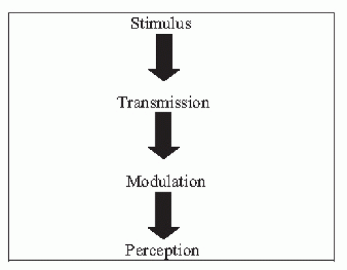
For a basic understanding of acute pain physiology, the process is viewed as a serial bottom-up system in which an external stimulus leads to a staged series of reactions within the peripheral and central nervous systems (47). The following model emphasizes clinical diagnosis and treatment presented in terms of four “attack points” where active intervention is possible to treat the source and interrupt the physiology of pain (48).


ATTACK POINT: STIMULUS

The initiation of the acute pain pathway comes in the form of a noxious stimulus. Interestingly, there is no quantitative or objective definition that separates a noxious stimulus from a nonnoxious stimulus. Put most simply, a stimulus is considered noxious if it leads to transduction, or activation of specialized peripheral nociceptors (47,48,49 and 50). Three general forms of peripheral nociceptors are likely to be activated during a surgical procedure: chemical, mechanical, and thermal (47,51,52).


Chemical Nociceptors

Chemical nociceptors undergo transduction when the free nerve endings are exposed to specific ions and cytokines within the surrounding extracellular matrix. These substances are released either directly as a result of cellular damage or indirectly through the normal inflammatory processes that accompany tissue damage and wound healing. Additionally, these substrates can either directly activate the nociceptor or sensitize the nerve endings making transduction more likely to occur. This process is summarized in Table 8.2, and is dependent on an intricate physiologic balance (44,47,48,49,50,51,52,53 and 54).

Activation of chemical nociceptors can develop at multiple stages of an operative procedure. Direct cellular damage occurs with the injection of local anesthetic, during dissection to the surgical target tissue and during the definitive procedure as intracellular components are spilled into the extracellular environment. The degree of tissue damage will determine the quantity and quality of the postoperative inflammatory response, particularly in the superficial layers (55). Uncontrolled hemostasis can also lead to direct and indirect activation of peripheral chemical nociceptors through the action of platelets (56). And although damage to any type of cell will result in the release of inflammatory cytokines, direct nerve damage additionally amplifies the noxious signal through the processes of ectopic discharge, peripheral sensitization, and central sensitization (47,57,58,59,60,61 and 62).


Mechanical Nociceptors

Transduction of mechanical nociceptors occurs when a physical stretch deforms unique transmembrane channels on free nerve endings. The resultant distortion of the channel allows for the influx of extracellular ions (Na+, K+, and Cl), and nociceptor depolarization occurs (47). Most mechanical noxious stimuli result in plastic deformation of the channel, but excessive stimuli can result in permanent damage (44). Activation of mechanical nociceptors is likely during an elective procedure. Both retraction (63,64,65,66 and 67) and tourniquet use (68,69,70 and 71) have been shown to contribute to postoperative pain through the mechanisms of mechanical nociceptors.


TABLE 8.2 The Mechanisms of Chemical Nociceptor Transduction at the Stimulus Attack Point


Cytokine Origin


Direct Cellular Damage


Indirect Inflammatory Response


H+


Mast cells: Histamine, Serotonin


K+


Platelets: Histamine, Serotonin


Reactive oxygen species


Basophils: Histamine


Histamine Serotonin


Plasma: Kinins, Interleukins, Thrombin, Trypsin


Kinins


Nerve Terminals: Substance P, CGRP


Arachodonic Acid: Prostaglandins, Leukotrienes


Cytokine Activation


Direct Nociceptor Activation


Indirect Nociceptor Sensitization


H+


Histamine


K+


Serotonin


Reactive oxygen species


Bradykinin


PGE2


Kallidin


Histamine


Prostaglandins


Serotonin


Leukotrienes


Kinins


Thromboxane


Thromboxane


Hydroxyacids


SRS-A


Interleukins


Thrombin


Substance P


Trypsin


CGRP


Peripheral chemical nociceptors can be activated by either direct or indirect means. The quantity and quality of the extracellular balance of these inflammatory cytokines at the surgical site will determine whether there is a physiologic inflammatory response associated with wound healing or a pathophysiologic reaction associated with peripheral sensitization. Adapted from Meyr AJ, Steinberg JS. The physiology of the acute pain pathway. Clin Podiatr Med Surg 2008;25(3):305-326.


Thermal Nociceptors

Thermal nociceptors are activated by both heat and cold stimuli, although most in the lower extremity are activated by heat between 42°C and 45°C (53,72). These temperatures are routinely generated during orthopaedic procedures, with the secondary sequela of thermal necrosis, from the use of power instrumentation and electrocautery devices (73,74,75,76,77,78,79 and 80).


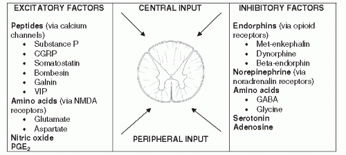
Figure 8.1 The modulation attack point. The influence of a noxious stimulus transmitted to the dorsal horn of the spinal cord is dependent on the complex interaction of central and peripheral, excitatory and inhibitory factors. (Adapted from Meyr AJ, Steinberg JS. The physiology of the acute pain pathway. Clin Podiatr Med Surg 2008;25():305-326.)


ATTACK POINT: TRANSMISSION

The transmission phase of the acute pain pathway involves peripheral afferent nerve fibers carrying the action potential generated by the noxious stimulus from the local site of tissue injury to the dorsal horn of the spinal cord (47,48,49 and 50). Two unique types of afferent nerve fibers are responsible for this conduction in the acute operative pain setting: the A-δ fiber and the C fiber.

A-δ fibers are myelinated nociceptors that are activated by both mechanical and thermal, but rarely chemical, stimulation. Some texts describe the A-δ fibers as giving rise to first pain because of the rapid nature of their transmission, nearly 10 to 25 times the speed of the unmyelinated C fibers (29). This pain is described as brief, localized, and sharp in nature. It is theorized that it provides the central nervous system with rapid information regarding the exact location of the noxious stimulus (21,29,43,47,49).

C fibers are unmyelinated nociceptors that are activated by chemical, mechanical, and thermal stimulation. They are believed to play the significant role in the development of pain associated with the inflammatory process because of the ability to respond to chemical stimulation. These unmyelinated fibers are intrinsically slower, giving rise to second pain. This is described as burning, throbbing, and aching in nature, and may provide the central nervous system with information regarding the extent or severity of the injury (21,29,48,49).


ATTACK POINT: MODULATION

At the most basic level, the modulation attack point represents the synapse between the primary sensory afferent of the periphery and the second-order neuron of the central nervous system. This site however, at the dorsal horn of the spinal cord, is actually a complex interaction of excitatory and inhibitory signals from both ascending and descending pathways (see Fig. 8.1).

A simplified explanation that provides a basic visual model was proposed by Wall and Melzack (81) in the middle of the 20th century. The gate control theory depicts the dorsal horn of the spinal cord as a single gate that is either opened (leading to propagation of the noxious stimuli into the central nervous system) or closed (blocking the peripheral noxious stimuli from being processed by the central nervous system). The status of the “gate” is determined by the interaction of various inhibitory and excitatory signals from peripheral and central sources in a constant balance. If the cumulative inhibitory signal is stronger
than the net excitatory signal, then the gate remains closed. If the cumulative excitatory signal is stronger than the net inhibitory signal, then the gate is opened and the signal is transmitted on (48,50,81,82,83,84 and 85).

While it is now known that the mechanisms of modulation are more complex than the status of a single gate either being opened or closed, the fundamental principles of the gate control theory remain true. The term modulation was specifically chosen to describe this stage because of the modification of the peripheral noxious signal that occurs. For each A-δ and C fiber that has been stimulated and is transmitting a noxious signal, there are thousands of peripheral nociceptors in the extremity that are not. In the same way, there are descending pathways from higher brain centers sending a constant stream of excitatory and inhibitory signals to the dorsal horn. Through these mechanisms, the dorsal horn synapse represents a balance of excitatory and inhibitory signals that has been triggered by the initial noxious stimulus (48).


ATTACK POINT: PERCEPTION

In many ways, perception represents the qualitative and psychological aspects of pain physiology. The previous attack points have emphasized concrete physiologic mechanisms and changes that occur in response to a noxious stimulus. While perception does involve the paleospinothalamic and neospinothalamic tracts terminating the noxious signal in the higher brain centers of the limbic system, frontal cortex, and somatosensory cortex (29,49,50), pain cannot be described in terms of a physiologic mechanism. Pain is not tissue damage, it is not an action potential, and it is not the influx of calcium ions within synapses of the spinal cord. It is a biologic and psychologic experience influenced by previous history, culture, religious beliefs, mood, and even the time of day or number of people in the room (86,87,88,89,90,91,92,93,94 and 95). Perception is the process of transforming an objective physiologic mechanism into a subjective patient complaint.


PATHOPHYSIOLOGY OF


OPERATIVE PAIN

The physiology of acute operative pain can be thought of as a distinct pathway beginning with the noxious stimulus and ending with perception (see Fig. 8.2). In the absence of sustained stimulation, the consequent physiology leading to perception does not occur. In more basic terms, the pain stops when the stimulus is removed. However, through both peripheral and central sensitization, this linear pathway can be transformed into a chronic cycle without a beginning or endpoint. In this situation the perception of pain continues in the absence of noxious stimulation. Although many of the molecules, synapses, and mechanisms used in these processes are the same, they are functioning in different ways. It is the pathophysiology of these systems that initiates chronic pain (96,97,98,99 and 100).


Figure 8.2 The clinical “attack points” of the acute pain pathway. The diagnosis and treatment of pain from the perspective of the four “attack points” of acute pain physiology allow the clinician to take an active approach to perioperative pain management.


PERIPHERAL SENSITIZATION

Localized, peripheral inflammation is a normal consequence of tissue damage and represents the important initial phase of the healing process (47). However, peripheral sensitization is an imbalance of this inflammation leading to the transmission of painful signals to the central nervous system out of proportion to, or in the absence of a noxious stimulus. It is a pathophysiologic process using the physiology of stimulus and transmission attack points (96).

Inflammatory cytokines can either directly activate chemical nociceptors or sensitize nociceptors making them more susceptible to transduction. Nociceptor sensitization is a unique concept in human physiology in that constant stimulation leads to a greater reaction, and not functional adaptation with a decreased response (29,47,56). In the presence of peripheral sensitization, quantitatively more nociceptors are available for activation, and transduction occurs at lower thresholds (44,45,50,52,85,96,97,98 and 99,101,102,103,104,105,106,107,108,109,110,111,112 and 113). Amplification of the noxious signal occurs in a sensitized state because a given stimulus leads to a relatively greater degree of transduction and transmission.


CENTRAL SENSITIZATION

In the same way, central sensitization can be thought of as an imbalance of the modulation attack point. There is a disproportionate excitatory signal from peripheral and central sources “opening the gate” and leading to noxious processing. Centrally at the dorsal horn, excitatory signals from C fibers displace magnesium ions from N-methyl D-aspartate (NMDA) receptors and open calcium channels at other excitatory synapses. A “wind-up” phase results in the postsynaptic neurons as excitatory neuropeptides and amino acids accumulate within the synapse (44,47,48,96,98,108,114,115,116,117,118,119,120,121 and 122).

Central sensitization is driven by peripheral excitatory signals (particularly from the C fibers), and it cycles to increase peripheral effects. Axonal reflexes lead to a secondary hyperalgesia where the perceived area of pain increases. A reflex arc develops within the activated peripheral nerve and spinal cord to cause activation and sensitization along the entire distal extent of that peripheral nerve, not just the branches of the injured tissue. Through this mechanism, nociceptors in the surrounding area of the injured tissue become activated (44,47,119,123,124,125,126 and 127). Allodynia also develops in the periphery when nonnoxious stimuli are perceived to be painful. In the setting of central sensitization and displaced magnesium ions, A-β peripheral afferents that are activated by light touch are registered as a noxious signal by NMDA receptors at the dorsal horn (29,47,98,114,128,129,130,131,132 and 133).

Peripheral and central sensitization have been described as a more efficient pain signal where amplification of the
noxious stimulus creates a cycle of inflammation and further sensitization. If noxious stimuli or peripheral sensitization are maintained, then central sensitization can be sustained indefinitely. In contrast, when the peripheral signal is removed, there is evidence that central sensitization resolves within minutes (44,47,98).

The noxious stimuli produced by the physical act of surgery are sufficient enough to generate peripheral and central sensitization pathophysiology in addition to the physiology of the acute pain pathway (134). An active approach to perioperative pain management takes into account and intervenes into both the physiology and pathophysiology of these mechanisms.


MULTIMODAL MANAGEMENT OF PERIOPERATIVE PAIN

The goals of pain management in the perioperative setting can be viewed in several ways. Physiologically, interventions should interrupt the known acute and chronic pain mechanisms to limit, in an attempt to prevent, peripheral sensitization, central sensitization, and the perception of pain. Put more broadly, the physiologic goal is to reach a resolution of the acute pain pathway without the development of a pathophysiologic chronic pain cycle. Objectively, the goals of pain management include quantifiable outcome measures such as increased time to the first perception of pain and a decreased total narcotic usage, total duration of pain, need for rescue analgesia, average pain intensity score, maximum pain intensity score, length of hospital stay, and pain/medication side effects. Finally, subjectively high levels of patient satisfaction with regard to pain management are sought. Although multifactorial, patient satisfaction involves an effective physician-patient relationship with respect to communication and the development of expected outcomes. These goals are best achieved with a multimodal approach (22,23,24,25,26,27,28,29 and 30,99,134,135 and 136). Niv and Devor (134) have compared interventional postoperative pain management to fighting a multiheaded Hydra, as can be illustrated by the work of John Singer Sargent’s depiction of the mythical hero Hercules (see Fig. 8.3). The mechanisms of physiology and pathophysiology at each of the attack points individually represent one of the Hydra’s heads. If physician intervention is unimodal and focuses on only one of the heads, then the rest are unobstructed. Perioperative pain management should instead intervene into multiple physiologic mechanisms at multiple attack points covering all phases of the perioperative period.

Only gold members can continue reading. Log In or Register to continue

Stay updated, free articles. Join our Telegram channel

Jul 26, 2016 | Posted by in MUSCULOSKELETAL MEDICINE | Comments Off on Perioperative Pain Management

Full access? Get Clinical Tree

Get Clinical Tree app for offline access