Other orthopaedic complications
Acute compartment syndrome in the elderly
Complex regional pain syndrome in the elderly
ACUTE COMPARTMENT SYNDROME IN THE ELDERLY
MARGARET M. McQUEEN
Introduction
Acute compartment syndrome (ACS) occurs when the pressure inside a confined compartment rises to a level and for a duration beyond which there is a critical reduction in blood flow to the tissues contained within. Without urgent decompression tissue ischaemia, necrosis, functional impairment and sometimes loss of limb will result. This is one of the few true emergency situations in orthopaedic trauma practice.
It is important to differentiate ACS from other related conditions. Exertional compartment syndrome is raised intracompartmental pressure (ICP) during exercise, causing ischaemia, pain and rarely neurological symptoms and signs. It normally resolves with rest but may proceed to ACS if exercise continues. Volkmann’s ischaemic contracture is the end stage of neglected ACS with irreversible muscle necrosis leading to contractures. The crush syndrome is the systemic result of muscle necrosis commonly caused by prolonged external compression of an extremity. In the presence of established muscle necrosis, ICP may rise as a result of intracellular and intracompartmental oedema, causing a superimposed ACS.
Epidemiology – who is at risk?
The incidence of ACS in a westernized population is 3.1 per 100,000 of the population per annum.1 The annual incidence for males is 7.3 per 100,000 compared with 0.7 per 100,000 for females, a 10-fold increase in risk for males. The age and gender-specific incidences are illustrated in Figure 6.1, showing a type B pattern2 or the L-shaped pattern described by Buhr and Cooke.3 The mean age is 32 years; the median age for males is 30 years and for females 44 years. It is clear therefore that older patients are at less risk of ACS. This is thought to be due to a combination of smaller muscle bulk allowing more room for swelling within a compartment and a possible protective effect of hypertension. The only exception to youth being a risk factor in ACS is in cases with soft tissue injury only. These patients have an average age of 36 years and are significantly older than those with a fracture.4
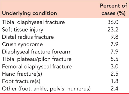
The most common underlying condition causing ACS is a fracture in 69% of cases (Table 6.1). Tibial diaphyseal fracture is the most frequent, occurring in 36% of cases of ACS and with a prevalence of ACS being reported in up to 14% of cases of tibial fracture.1,5,6 The second most common cause is soft tissue injury which accounts for almost one quarter of cases of ACS. The second most common fracture causing ACS is distal radius fracture. Other causes of ACS are listed in Table 6.2. Although most of these are uncommon, several are relevant to the older patient who may have comorbidities. The crush syndrome is probably more frequent than realized in the elderly who may lie in an obtunded state for many hours after a fall. In these circumstances creatinine kinase levels should be monitored. Comorbidities such as diabetes and treatment with anticoagulants are frequent in the older patient and treating surgeons should be aware that they may increase the risk of ACS developing.
Although there is a persistent belief that high energy injury is a risk factor for the development of ACS, there is no evidence to support this. In tibial diaphyseal fracture in adults complicated by ACS, the proportion of high compared with low energy injury shows a slight preponderance of low energy injury (59%).1 In an analysis of 1,403 tibial diaphyseal fractures presenting to the Edinburgh Orthopaedic Trauma Unit over the period from 1995 to 2007 there was an increased risk of ACS in closed compared with open fractures (P<0.05). This suggests ACS may be more prevalent after low energy injury, possibly because in low energy injury the compartment boundaries are less likely to be disrupted and an ‘autodecompression’ effect is avoided.
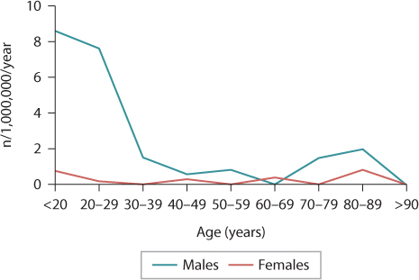
Figure 6.1 The age and gender related incidence of acute compartment syndrome showing a reduction in both genders with age.
Table 6.1 The underlying causes of acute compartment syndrome presenting to an orthopaedic trauma unit
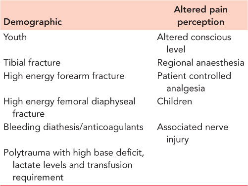
Risk factors for the development or late diagnosis of ACS are listed in Table 6.3. As well as demographic risk factors, altered pain perception can delay diagnosis. This can occur if the patient has an altered conscious state, has cognitive difficulties or with the use of spinal or epidural anaesthesia or patient controlled analgesia. Comorbidities such as diabetes and treatment with anticoagulants are frequent in the older patient and treating surgeons should be aware that they may increase the risk of ACS developing.
Diagnosis
Prompt diagnosis of ACS is the key to a successful outcome. Delay in diagnosis has long been recognized as the only cause of failure of the treatment of ACS. Delay in diagnosis may be due to inexperience and lack of awareness of the possibility of ACS, an indefinite and confusing clinical presentation, or anaesthetic or analgesic techniques that mask the clinical signs.
Table 6.2 Conditions causing acute compartment syndrome
Conditions increasing the volume of compartment contents |
Fracture |
Soft tissue injury |
Crush syndrome (including use of the lithotomy position) |
Revascularization |
Exercise |
Bleeding diathesis/anticoagulants |
Fluid infusion (including arthroscopy) |
Arterial puncture |
Ruptured ganglia/cysts |
Osteotomy |
Snake bite |
Nephrotic syndrome |
Leukaemic infiltration |
Viral myositis |
Acute haematogenous osteomyelitis |
Conditions reducing compartment volume |
Burns |
Repair of muscle hernia |
Medical comorbidity |
Diabetes |
Hypothyroidism |
Delay in treatment of ACS can be catastrophic, leading to serious complications such as permanent sensory and motor deficits, contractures, infection and, at times, amputation of the limb. In severe cases there may be systemic injury from the reperfusion syndrome. To avoid such complications it is essential for the treating surgeon to have a clear understanding of the clinical techniques necessary to make an early diagnosis. Early recognition and treatment of ACS also result in decreased indemnity risk in potential malpractice claims.7
CLINICAL DIAGNOSIS
Pain is considered to be the first symptom of ACS. The pain experienced is ischaemic and usually severe and out of proportion to the clinical situation. However, pain can be an unreliable indication of the presence of ACS because it can be variable in its intensity, may be absent in ACS associated with nerve injury or minimal in the deep posterior compartment syndrome. It cannot be elicited in the unconscious patient or where regional anaesthesia is used.8 Elderly patients with dementia may not be able to express the severity of their pain, so restlessness, agitation and anxiety with increasing analgesic requirements should raise the suspicion of the presence of an ACS.
Pain has been shown to have a sensitivity of only 19% and a specificity of 97% in the diagnosis of ACS (i.e. a high proportion of false negative or missed cases but a low proportion of false positive cases).9 There is general agreement, however, that pain if present is a relatively early symptom of ACS in the awake alert patient.9 Pain with passive stretch of the muscles involved is recognized as a symptom of ACS but is no more reliable than rest pain because the reasons for unreliability quoted above apply equally to pain on passive stretch. The sensitivity and specificity of pain on passive stretch are similar to those for rest pain.9
Table 6.3 Risk factors for the development or delayed diagnosis of acute compartment syndrome
Paraesthesia and hypoesthesia in the territory of the nerves within the affected compartment are frequently the first signs of nerve ischaemia, although sensory abnormality may be the result of concomitant nerve injury. Ulmer reports a sensitivity of 13% and specificity of 98% for the clinical finding of paraesthesia in ACS, a false negative rate that precludes this symptom from being a useful diagnostic tool.9 Paralysis of muscle groups affected by the ACS is recognized as being a late sign.9 This sign has equally low sensitivity as others in predicting the presence of ACS, probably because of the difficulty of interpreting the underlying cause of the weakness, which could be inhibition by pain, direct injury to muscle or associated nerve injury. If a motor deficit develops, full recovery is unlikely10,11 with full recovery in one series in only 13% of patients.10
Swelling is cited as a sign of ACS but the degree of swelling is difficult to assess accurately, making this sign very subjective. Casts or dressings often obscure compartments at risk8 and some compartments such as the deep posterior compartment of the leg are difficult to access.
Peripheral pulses and capillary return are always intact in ACS unless there is major arterial injury or disease or in the very late stages of ACS when amputation is inevitable. If ACS is suspected and pulses are absent, then arteriography is indicated. Conversely, it is dangerous to exclude the diagnosis of ACS because distal pulses are present.
Using a combination of clinical symptoms and signs increases their sensitivity as diagnostic tools.9 To achieve a probability of over 90% of ACS being present, however, three clinical findings must be noted. The third clinical finding is paresis, so to achieve an accurate clinical diagnosis of ACS the condition must be allowed to progress until a late stage. This is clearly unacceptable and has led to a search for earlier, more reliable methods of diagnosis.
COMPARTMENT PRESSURE MONITORING
Measurement of ICP was developed when it was established that ACS was caused by increased tissue pressure. Because raised tissue pressure is the primary event in ACS, changes in ICP precede the clinical symptoms and signs.12 Most methods use a slit or wick catheter inserted into the relevant compartment connected to a pressure transducer through a column of saline. Patency can be confirmed by gentle pressure over the catheter tip; an immediate rise in the pressure should be seen. Care must be taken to avoid the presence of air bubbles in the system as this can result in falsely low readings. The slit and wick catheters are equally accurate.13 ICP can also be measured by introducing a transducer into the compartment either by siting it in the lumen of the catheter or placing it at the tip. For continuous pressure measurement the latter requires an infusion of saline with a risk of increasing ICP; the former is expensive and difficult to resterilize. These methods all measure ICP which is an indirect measurement of muscle blood flow and oxygenation. Near infrared spectroscopy measures tissue oxygen saturation non-invasively by means of a probe placed on the skin and is a technique with promise but requires further validation in humans subjected to injury.
ICP is usually monitored in the anterior compartment in the tibia because this is most commonly involved in ACS and is easily accessible.5 There is a risk of missing an ACS in the deep posterior compartments but measuring two compartments is much more cumbersome. If the anterior compartment alone is monitored, the ICP in the deep posterior compartment must be measured if there are unexplained symptoms in the presence of normal anterior compartment pressures. It is important to measure the peak pressure within the limb, which usually occurs within 5 cm of the level of the fracture.14
Threshold for decompression
Much debate has occurred about the critical pressure threshold, beyond which decompression of ACS is required. Historically tissue pressure measurements of between 30 and 50 mmHg were used as the threshold for decompression, but it is now recognized that apparent variation between individuals in their tolerance of raised ICP is because of variations in systemic blood pressure. Whitesides and his co-authors were the first to suggest the importance of the difference between the diastolic blood pressure and tissue pressure, or ΔP.15 They stated that there is inadequate perfusion and relative ischaemia when the tissue pressure rises to within 10–30 mmHg of the diastolic pressure and this is supported by further evidence from experimental studies16,17 or the similar concept that the difference between mean arterial pressure and tissue pressure should not be less than 30 mmHg in normal muscle or 40 mmHg in muscle subject to trauma18 or antecedent ischaemia.19 A clinical study of 116 patients with tibial diaphyseal fractures validated the experimental evidence. The authors concluded that a ΔP of 30 mmHg is a safe threshold for decompression in ACS.5
The use of ΔP as a threshold for fasciotomy has also been supported in other studies.20,21
The sensitivity and specificity of continuous compartment pressure monitoring has recently been reported.6 Using a pressure threshold of a ΔP of 30 mmHg for more than 2 hours as an indication for fasciotomy in 850 patients, there were 11 false positives and 9 false negatives giving a sensitivity of 94% and a specificity of 98.4%. The positive predictive value was 92.8% and the negative predictive value was 98.7%. To achieve similar accuracy with clinical symptoms and signs, three signs need to be present with the third being paralysis.9
To derive the full benefit of monitoring for ACS, the diagnosis should be made on the basis of sequential differential pressure measurements rather than awaiting the development of clinical symptoms and signs. It has been demonstrated that this approach reduces both the delay to fasciotomy and the development of sequelae22 and that the appearance of clinical symptoms and signs lags behind the pressure changes.5 It has been assumed that these pressure thresholds apply equally to anatomic areas other than the leg, although this has not been formally examined.
Timing
Time factors are also important in making the decision to proceed to fasciotomy. It is well established experimentally and clinically that both the duration and severity of the pressure elevation influence the development of muscle necrosis and sequelae.5,11,17,23 Continuous pressure monitoring allows a clear record of the trend of the tissue pressure measurements. In situations where the ΔP drops below 30 mmHg, if the ICP is dropping and the ΔP is rising, then it is safe to observe the patient in anticipation of the ΔP returning within a short time to safe levels. If the ICP is rising, the ΔP is dropping and less than 30 mmHg, and this trend has been consistent for a period of 2 hours, then fasciotomy should be performed. Fasciotomy should not be performed based on a single pressure reading except in extreme cases. Using this protocol, delay to fasciotomy and the sequelae of ACS are reduced without unnecessary fasciotomies being performed.5 For ICP monitoring to be most effective in reducing delay, it must be used as the primary indication for fasciotomy.
Treatment
The single most effective treatment for ACS is fasciotomy, which if delayed can cause devastating complications. Nevertheless, other preliminary measures should be taken in cases of impending ACS. External limiting envelopes such as dressings or plaster casts, including the padding under the cast, should be split and spread. The limb should not be elevated above the height of the heart as this reduces the arteriovenous gradient.24 Hypotension should be corrected, because this will reduce perfusion pressure. Oxygen therapy should be instituted to ensure maximum oxygen saturation.
FASCIOTOMY
The basic principle of fasciotomy of any compartment is full and adequate decompression. Skin incisions must be made along the full length of the affected compartment (Figure 6.2) and it is essential to visualize all contained muscles in their entirety. There is no place for limited or subcutaneous fasciotomy in ACS. Any muscle necrosis must be thoroughly debrided to avoid infection.
In the leg, all four compartments should be released using a double incision four-compartment fasciotomy. The anterior and lateral compartments are released through a lateral skin incision over the intermuscular septum between the compartments (Figure 6.2) and the two posterior compartments are accessed through a skin incision 2 cm from the medial edge of the tibia (Figure 6.3). It is important to allow a generous skin bridge between the two, especially in the elderly, when skin circulation may be compromised. Single incision fasciotomy of all four compartments has been described but double incision fasciotomy is faster and probably safer than single incision methods because the fascial incisions are all superficial. Using the single incision method, it can be difficult to visualize the full extent of the deep posterior compartment. Both methods seem to be equally effective at reducing ICP.
In other areas decompression is usually simple with incisions made directly over the affected compartments affording good exposure of the muscle groups. The exceptions are the foot and hand. In the foot there are a number of compartments to decompress, and a sound knowledge of the anatomy is essential. In most cases dorsal incisions are sufficient, but in hindfoot injury a separate medial incision may be required to decompress the calcaneal compartment. Decompression of the hand can usually be adequately achieved using two dorsal incisions that allow access to the interosseous compartments, but if there is clinical suspicion or raised ICP on measurement, then incisions may be made over the thenar and hypothenar eminences, allowing fasciotomy of these compartments.
Fasciotomy incisions must never be closed, primarily because this may result in persistent elevation of ICP. The wounds should be left open and dressed, and approximately 48 hours after fasciotomy a ‘second look’ procedure should be undertaken to ensure the viability of all muscle groups. Skin closure or cover should not be attempted unless all muscle groups are viable. Delayed primary closure should be used if possible but without tension on the skin edges. Commonly in the leg this technique is possible in the medial but not the lateral wound. Dermatotraction or gradual closure techniques should be used with caution as they may cause skin edge necrosis25 and require a prolonged time to achieve closure. Split skin grafting is required less in the older patient, possibly because of reduced muscle bulk.11 Although offering immediate skin cover this technique has the disadvantage of a high rate of long-term morbidity.26 The recent introduction of vacuum assisted closure (VAC) systems is likely to be a significant advantage in this area and may reduce the need for split skin grafting.
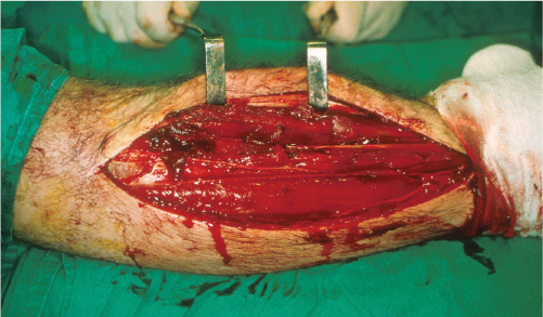
Figure 6.2 Fasciotomy of the anterior and lateral compartments of the leg using a lateral incision. Note the length of the incision which allows visualization of the full length of the compartments in order to ascertain the viability of the muscles.
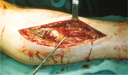
Figure 6.3 Fasciotomy of the superficial and deep posterior compartments of the leg through a medial incision. The superficial posterior compartment is being retracted to expose the deep posterior compartment. Care must be taken to ensure that there is a sufficiently wide skin bridge between the medial and lateral skin incisions.
MANAGEMENT OF ASSOCIATED FRACTURES
It is now generally accepted that fractures, especially of the long bones, should be stabilized in the presence of ACS treated by fasciotomy.27,28,29 and 30 In reality the treatment of the fracture should not be altered by the presence of an ACS, although cast management of a tibial fracture is contraindicated in the presence of ACS. Fasciotomy should be performed prior to fracture stabilization in order to eliminate any unnecessary delay in decompression. Stabilization of the fracture allows easy access to the soft tissues and protects the soft tissues, allowing them to heal.
Complications of ACS
Complications of ACS are unusual if the condition has been treated expeditiously. Delay to fasciotomy of more than 6 hours is likely to cause significant sequelae,31 including muscle contractures, muscle weakness, sensory loss, infection and non-union of fractures.5,32,33,34 and 35 In severe cases amputation may be necessary because of infection or lack of function.36
REFERENCES
1. McQueen MM, Gaston P, Court-Brown CM. Acute compartment syndrome. Who is at risk? J Bone Joint Surg Br, 2000;82:200–3.
2. Court-Brown CM. The epidemiology of fractures and dislocations. In: Court-Brown CM, Heckman JD, McQueen MM, Ricci WM, Tornetta P, III, McKee MD, editors. Rockwood and Green’s Fractures in Adults. 8th ed. Philadelphia, PA: Wolters Kluwer; 2015. pp. 59–108.
3. Buhr AJ, Cooke AM. Fracture patterns. Lancet, 1959;1:531–6.
4. Hope MJ, McQueen MM. Acute compartment syndrome in the absence of fracture. J Orthop Trauma, 2004;18:220–4.
5. McQueen MM, Court-Brown CM. Compartment monitoring in tibial fractures. The pressure threshold for decompression. J Bone Joint Surg Br, 1996;78:99–104.
6. McQueen MM, Duckworth AD, Aitken SA, et al. The estimated sensitivity and specificity of compartment pressure monitoring for acute compartment syndrome. J Bone Joint Surg Am, 2013;95:673–7.
7. Bhattacharyya T, Vrahas MS. The medical-legal aspects of compartment syndrome. J Bone Joint Surg Am, 2004;86-A:864–8.
8. Kosir R, Moore FA, Selby JH, et al. Acute lower extremity compartment syndrome (ALECS) screening protocol in critically ill trauma patients. J Trauma, 2007;63:268–75.
9. Ulmer T. The clinical diagnosis of compartment syndrome of the lower leg: Are clinical findings predictive of the disorder? J Orthop Trauma, 2002;16:572–7.
10. Bradley EL, III. The anterior tibial compartment syndrome. Surg Gynecol Obstet, 1973;136:289–97.
11. Duckworth AD, Mitchell SE, Molyneux SG, et al. Acute compartment syndrome of the forearm. J Bone Joint Surg Am, 2012;94:e63.
12. McQueen MM, Christie J, Court-Brown CM. Compartment pressures after intramedullary nailing of the tibia. J Bone Joint Surg Br, 1990;72:395–7.
13. Shakespeare DT, Henderson NJ, Clough G. The slit catheter: A comparison with the wick catheter in the measurement of compartment pressure. Injury, 1982;13:404–8.
14. Heckman MM, Whitesides TE, Jr., Grewe SR, et al. Compartment pressure in association with closed tibial fractures. The relationship between tissue pressure, compartment, and the distance from the site of the fracture. J Bone Joint Surg Am, 1994;76:1285–92.
15. Whitesides TE, Haney TC, Morimoto K, et al. Tissue pressure measurements as a determinant for the need of fasciotomy. Clin Orthop Relat Res, 1975;(113):43–51.
16. Heckman MM, Whitesides TE, Jr., Grewe SR, et al. Histologic determination of the ischemic threshold of muscle in the canine compartment syndrome model. J Orthop Trauma, 1993;7:199–210.
17. Matava MJ, Whitesides TE, Jr., Seiler JG, III, et al. Determination of the compartment pressure threshold of muscle ischemia in a canine model. J Trauma, 1994;37:50–8.
18. Heppenstall RB, Sapega AA, Scott R, et al. The compartment syndrome. An experimental and clinical study of muscular energy metabolism using phosphorus nuclear magnetic resonance spectroscopy. Clin Orthop Relat Res, 1988;(226):138–55.
19. Bernot M, Gupta R, Dobrasz J, et al. The effect of antecedent ischemia on the tolerance of skeletal muscle to increased interstitial pressure. J Orthop Trauma, 1996;10:555–9.
20. Ovre S, Hvaal K, Holm I, et al. Compartment pressure in nailed tibial fractures. A threshold of 30 mmHg for decompression gives 29% fasciotomies. Arch Orthop Trauma Surg, 1998;118:29–31.
21. White TO, Howell GE, Will EM, et al. Elevated intramuscular compartment pressures do not influence outcome after tibial fracture. J Trauma, 2003;55:1133–8.
22. McQueen MM, Christie J, Court-Brown CM. Acute compartment syndrome in tibial diaphyseal fractures. J Bone Joint Surg Br, 1996;78:95–8.
23. Petrasek PF, Homer-Vanniasinkam S, Walker PM. Determinants of ischemic injury to skeletal muscle. J Vasc Surg, 1994;19:623–31.
24. Matsen FA, III, Wyss CR, Krugmire RB, Jr., et al. The effects of limb elevation and dependency on local arteriovenous gradients in normal human limbs with particular reference to limbs with increased tissue pressure. Clin Orthop Relat Res, 1980;(150):187–95.
25. Janzing HM, Broos PL. Dermatotraction: An effective technique for the closure of fasciotomy wounds: A preliminary report of fifteen patients. J Orthop Trauma, 2001;15:438–41.
26. Fitzgerald AM, Gaston P, Wilson Y, et al. Long-term sequelae of fasciotomy wounds. Br J Plast Surg, 2000;53:690–3.
27. Gelberman RH. Upper extremity compartment syndromes. In: Compartment Syndrome and Volkmann’s Contracture, Mubarak SJ, Hargens AR, editors. 1st ed. Philadelphia, PA: WB Saunders, 1981.
28. Gershuni DH, Mubarak SJ, Yaru NC, et al. Fracture of the tibia complicated by acute compartment syndrome. Clin Orthop Relat Res, 1987;(217):221–7.
29. Rorabeck CH. The treatment of compartment syndromes of the leg. J Bone Joint Surg Br, 1984;66:93–7.
30. Turen CH, Burgess AR, Vanco B. Skeletal stabilization for tibial fractures associated with acute compartment syndrome. Clin Orthop Relat Res, 1995;(315):163–8.
31. Rorabeck CH, Macnab L. Anterior tibial-compartment syndrome complicating fractures of the shaft of the tibia. J Bone Joint Surg Am, 1976;58:549–50.
32. Court-Brown, McQueen M. Compartment syndrome delays tibial union. Acta Orthop Scand, 1987;58:249–52.
33. Gelberman RH, Szabo RM, Williamson RV, et al. Tissue pressure threshold for peripheral nerve viability. Clin Orthop Relat Res, 1983;(178):285–91.
34. Hargens AR, Romine JS, Sipe JC, et al. Peripheral nerve-conduction block by high muscle-compartment pressure. J Bone Joint Surg Am, 1979;61:192–200.
35. Karlstrom G, Lonnerholm T, Olerud S. Cavus deformity of the foot after fracture of the tibial shaft. J Bone Joint Surg Am, 1975;57:893–900.
36. Finkelstein JA, Hunter GA, Hu RW. Lower limb compartment syndrome: Course after delayed fasciotomy. J Trauma, 1996;40:342–4.
COMPLEX REGIONAL PAIN SYNDROME IN THE ELDERLY
ROGER M. ATKINS
Introduction
Complex regional pain syndrome (CRPS) consists of abnormal pain, swelling, vasomotor and sudomotor dysfunction, contracture and osteoporosis. It used to be considered a rare complication of injury, caused by sympathetic nervous system abnormalities in psychologically abnormal patients. Modern research is altering this view radically. This review will examine CRPS within the context of orthopaedic trauma surgery in the elderly. Therefore, the emphasis, descriptions and concepts differ slightly from those found in publications from the International Association for the Study of Pain (IASP).
Some important definitions
A cardinal feature of CRPS is abnormalities of pain perception1 which are unfamiliar to orthopaedic surgeons.
Allodynia: painful perception of a stimulus which ought not to be painful, for example, gentle stroking of the affected part.
Hyperalgesia: increased sensitivity to pain. Gentle touching with a pin is unbearably painful.
Hyperpathia: temporal and spatial summation of an allodynia or hyperalgesia. Repetitive stimulus becomes increasingly unbearable. The pain continues and may be accentuated by stimuli such as sudden noise or a draft of cold air. These are genuine perceptions of pain. The patients are not malingering or mad.
Nociceptive pain arises from direct stimulation of peripheral pain receptors, for example following fracture.
Neuropathic pain arises from the nerves themselves without any precipitating stimulus. Spontaneous or burning pain, mechanical or thermal hyperalgesia, allodynia and hyperpathia are common.1
Sympathetically maintained pain (SMP) consists of pain, hyperpathia and allodynia relieved by selective sympathetic blockade.
Taxonomy
Historically CRPS had many synonyms (Table 6.4) with differing diagnostic criteria. To overcome the resulting confusion, IASP reclassified the condition and proposed the name ‘complex regional pain syndrome”’ (CRPS)2,3 with standardized diagnostic criteria.1
CRPS was divided into type 2 (CRPS 2) caused by nerve damage and type 1 (CRPS 1) without nerve damage. The clinical features differ.4 From a surgeon’s perspective, a diagnosis of CRPS 2 should indicate causative nerve damage susceptible to surgical intervention, for example, sural nerve entrapment following Achilles tendon surgery.
Table 6.4 Synonyms for complex regional pain syndrome
Complex regional pain syndrome |
Reflex sympathetic dystrophy |
Sudeck’s atrophy |
Causalgia |
Minor causalgia |
Mimo-causalgia |
Algodystrophy |
Algoneurodystrophy |
Post-traumatic pain syndrome |
Painful post-traumatic dystrophy |
Painful post-traumatic osteoporosis |
Transient migratory osteoporosis |
IASP’s classification remains controversial, with suggestions that it medicalizes pain5 and over diagnoses the condition.6
Clinical features
CRPS is a biphasic condition with early swelling and vasomotor instability (Figure 6.4) giving way over a variable timescale to late contracture and joint stiffness (Figures 6.5 and 6.6).7 The hand and foot are the most frequent sites, although CRPS in the knee is increasingly recognized.8,9 The elbow is rarely affected, whereas shoulder disease is common and some cases of frozen shoulder are probably CRPS.10
CRPS usually begins up to a month after a precipitating trauma but may be spontaneous.7 As the direct effects of injury subside, a new diffuse, unpleasant, neuropathic pain arises.11 Pain is unremitting (although sleep may be unaffected), worsening and radiating with time and may be increased by limb dependency, physical contact, emotional upset and by extraneous factors.
EARLY PHASE OF CRPS
Vasomotor instability (VMI) and oedema dominate the early phase, although this is less marked with more proximal CRPS. The classical description divides the early phase of CRPS into two stages depending on the type of VMI.7,12,13 and 14 These stages are rarely seen except in severe cases.7,14,15 Initially the limb is dry, hot and pink but after days or weeks, it becomes blue, cold and sweaty (Figure 6.4). Usually, however, VMI is accompanied by increased temperature sensitivity with variable abnormality of sweating.4,14,15
Oedema is marked, particularly where the distal part of the limb is affected and over time becomes more fixed and indurated with coalescence of tissue planes and structures.
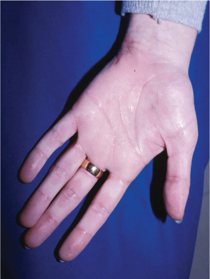
Figure 6.4 The hand in the early phase of CRPS. There is excessive sweating and the skin is bluish in colour.
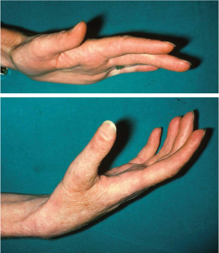
Figure 6.5 This patient’s right hand is in the late phase of CRPS. There is atrophy of the hand with digital spindling, extension contractures and loss of joint creases and subcutaneous fat.
Loss of joint mobility is due to swelling and pain combined with an inability to initiate movement.16,17 and 18 As the disease progresses, stiffness is increasingly due to contracture. Only if the disease can be halted before fixed contracture has occurred can complete resolution occur.
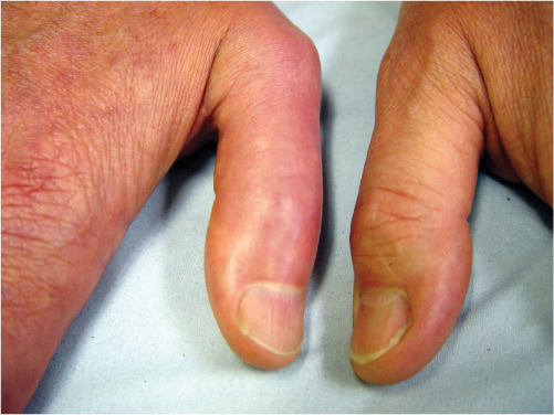
Figure 6.6 The thumbs of a patient with late CRPS 1 of the right hand. There is distal spindling of the affected thumb particularly distally with discolouration and ridging of the nail.
LATE PHASE OF CRPS
VMI recedes, oedema resolves and atrophy of the limb occurs affecting every tissue. The skin is thinned and joint creases and subcutaneous fat disappear (Figures 6.5 and 6.6). Hairs become fragile, thickened, uneven and curled, while nails are pitted, ridged, brittle and discoloured brown. Palmar and plantar fascias thicken, simulating Dupuytren’s disease.7,19 Tendon sheaths become constricted, causing triggering and increased resistance to movement which combined with muscle contracture and fibrosis leads to reduced tendon excursion. Joint capsules and collateral ligaments become shortened, thickened and adherent, causing joint contracture.
Within orthopaedic practice, the large majority of patients who demonstrate the features of early CRPS will not go on to develop severe contracture.
BONE CHANGES
Bone involvement is universal with increased uptake on the delayed bone scan in early CRPS throughout the affected region.20 Later, this returns to normal and there are radiographic features of rapid bone loss: patchy, subchondral or sub-periosteal osteoporosis, metaphyseal banding and severe osteoporosis (Figure 6.7). Despite the osteoporosis, fracture is uncommon, presumably because the patients protect the painful limb very effectively.
INCIDENCE
Severe CRPS with major contracture is uncommon with a prevalence of less than 2% in retrospective series.21,22 Population-based studies show 55 cases per 100,000 person-years in the United States23 and 262 per 100,000 person-years in the Netherlands.24
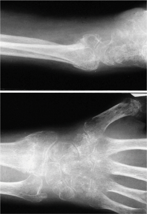
Figure 6.7 The radiographic appearance of a distal radius fracture complicated by the late phase of CRPS. There is profound osteoporosis.
Related posts:

Stay updated, free articles. Join our Telegram channel

Full access? Get Clinical Tree


