Osteoarthritis


Osteoarthritis


Todd P. Stitik

Jong-Hyun Kim

Doreen Stiskal

Patrick Foye

Robert Nadler

James Wyss

Sausheen Heslop


INTRODUCTION

Physiatrists, particularly those in the outpatient setting, will likely increasingly take care of patients with osteoarthritis (OA). This is due to both the growing prevalence of OA within the general population and the recognition by physiatrists, other physicians, and the general public that physiatrists possess the knowledge base and skill set to manage patients with various musculoskeletal conditions. Traditional physiatric training emphasizes function, therapeutic exercise, orthoses and assistive devices, and coordination of care with other health care professionals, especially physical therapists, occupational therapists, and social workers. Training nowadays often also includes instruction in musculoskeletal injection procedures and pain management pharmacotherapy. This combination of traditional physical medicine and rehabilitation along with interventional and pharmacologic pain management techniques places the physiatrist in a position of being able to provide the full spectrum of nonsurgical patient care.

Although the field of PM&R has not yet drafted specific management guidelines analogous to those published by the American College of Rheumatology (ACR) (1), the ACR guidelines center around many basic nonpharmacologic principles that are integral to physiatric training. Therefore, part of the nonpharmacologic treatment section of this chapter is loosely structured based upon these management strategies. The pharmacologic treatment section of this chapter is also based upon the ACR guidelines as medication recommendations were derived in part from this evidence-based medical research. Of note, the ACR guidelines are scheduled to be undergoing revision under the guidance of a subcommittee that now includes a physiatrist. These revised guidelines are scheduled to be completed during 2010.

Although physiatrists are also frequently involved in the postsurgical rehabilitation of patients who have undergone arthroplasty, this topic is covered elsewhere in this textbook.


PATHOLOGY AND PATHOPHYSIOLOGY

OA is really a group of disorders with different etiologies but similar pathologic changes. In primary OA, it is believed that excessive loads cause failure of an otherwise normal joint (2). The changes eventually involve all of the tissues including the articular cartilage, subchondral bone, synovial tissue, joint capsule, ligaments, and muscles that act on the joint. Histologically, small tears known as fibrillations and larger tears known as clefts both develop. These defects begin in the superficial zone of cartilage, extend into the transitional zone, and are also propagated by enzymatic breakdown of cartilage (3, 4). Eventually, large areas of cartilage loss occur, thus essentially exposing the underlying subchondral bone. Other changes within the cartilage include an eventual decline in the ability of the chondrocytes to replicate, an initial increase in the water content of cartilage, a significant reduction in proteoglycan content, a reduction in the size of type II collagen fibers, shortening of the glycosaminoglycan chains, a diminution of the keratin sulfate concentration, and an increase in the proportion of chondroitin-4-sulfate consistent with immature cartilage being produced in an attempt to regenerate the lost cartilage (5). Cartilage matrix changes lead to increased matrix permeability and decreased matrix stiffness, both of which may increase tissue vulnerability to additional mechanical damage. Although the chondrocytes within the osteoarthritic joint acquire the ability to replicate and produce new chondrocytes that are very metabolically active, they produce collagen, proteoglycan, and hyaluronan (HA) that do not aggregate well and are not adequately stabilized in the extracellular matrix. Eventually, this chondrocytic repair response declines, although the exact reasons for this are poorly understood. Theories, however, include failure of the matrix to stabilize and protect the chondrocytes and a down-regulation of the chondrocytic response to anabolic cytokines.

The order of appearance of the changes in the cartilage and underlying subchondral bone is debatable. Specifically, one theory is that the subchondral bone undergoes remodeling in response to the external load applied to it due to cartilage breakdown (6). In contrast, Radin hypothesized that subchondral bone stiffening associated with remodeling in response to externally applied loads precedes and subsequently promotes cartilage loss (7). Fibrous, cartilaginous, and osseous prominences known as osteophytes eventually develop around the periphery of the joints (marginal osteophytes) but can also develop along joint capsule insertions (capsular osteophytes) or protrude from the degenerating joint surfaces (central osteophytes). Cystlike bone cavities containing myxoid, fibrous, or cartilaginous tissue eventually form within the bone. Regardless of the exact sequence of events, both cartilage degeneration and subchondral bone remodeling are found by the time the patient is symptomatic (8). Changes also occur within the synovial membrane and the synovial fluid.
Specifically, the synovial membrane may have a mild to moderate inflammatory reaction and may contain fragments of articular cartilage (9). Within the synovial fluid, pathological changes include significant alteration in synovial fluid HA, including a decrease in the concentration of normal molecular weight hyaluronate and the production of abnormal hyaluronate. The resultant decreased hyaluronate concentration is a result of both defective hyaluronate production and increased hyaluronate breakdown. In addition, there is increased water content and an increased concentration of inflammatory mediators (10). These pathological changes result in defective synovial fluid viscosity, elasticity, barrier exclusion and shielding. Exposure of synovial nociceptors perhaps explains in part the pain associated with the osteoarthritic joint.


EPIDEMIOLOGY

More than 21% of US adults (46.4 million persons) have been reported as having “self-reported doctor-diagnosed arthritis”—for example, the patients themselves reported that a physician had previously diagnosed them with arthritis. Based on cognitive and validation studies, “self-reported doctor-diagnosed arthritis” is thought to provide the most credible estimate of overall arthritis prevalence, with acceptable sensitivity and specificity for surveillance purposes (11). As the average age of the US population increases, an estimated 67 million Americans will acquire some form of arthritis by the year 2030 (12). Among various types of arthritis, OA is the most common and affects 27 million people in the United States (13). Approximately 10% to 30% of those affected with OA have significant pain and disability (10). OA is second only to ischemic heart disease as a cause of work disability in men over age 50 (14).

Although incidence and prevalence may vary widely according to the type of epidemiologic studies, as well as by whether clinical or radiological definitions are used, the overall epidemiology of OA has been described. At least 37% (and up to 68% in some studies) of persons 60 years and older have radiographic evidence of knee OA. By contrast, 27.2% of adults 26 years and older have radiographic evidence of hand OA. Clinically, 12.1% of adults aged 60 or older have symptomatic knee OA. The prevalence of symptomatic hand OA was higher in women (9.2%) than in men (3.8%) (10, 15, 16).

The causes of OA are multifactorial and the risk factors may differ for each joint. Given the anticipated increase in OA prevalence, understanding the risk factors associated with OA can aid in further clarifying the disease process as well as helping to potentially delay OA progression.


RISK FACTORS


Genetic Factors

Multiple genes appear to confer an increased risk for OA, and the contribution of heritability and possibly genetics to the occurrence of OA varies by joint. The genetic predisposition to OA seems to be based on a variety of physiologic vulnerabilities including changes in collagen and enzymes within cartilage, variations in cytokines or growth factor profiles in cartilage, and genes that dictate joint shape and structure (16, 17).


Age and Gender

The prevalence and incidence of OA correlates markedly with age; however, age alone does not appear to cause OA. Rather, the increase in OA with age is likely a consequence of biologic alterations that occur with the aging process. These include decreased strength, slower neurologic responses with a decline in proprioception, a decreased response of chondrocytes by growth factors, and age-related accumulation of glycation end products, which is a haphazard process impairing the function of the cartilage (18).

Younger adults also develop OA secondarily from injury and loss of biomechanical integrity.

Data show that a greater number of men present with OA before age 50; while after age 50, the condition is more common in women (19). Women experience greater severity of symptoms and report more problems with morning stiffness, joint swelling, and night pain. Women also have a higher incidence of Heberden’s nodes (swelling of the distal interphalangeal [DIP] joint of the hands), a phenomenon with a familial tendency.


Ethnicity

African Americans demonstrate a greater severity rating of knee OA than Caucasians (20). The prevalence of hip OA is higher in European Caucasians than in African-Americans. Hip OA is rare in China and in those of Chinese descent in the United States (21, 22). Anatomic abnormalities that are prevalent in the United States are rare in the hips of persons of Chinese descent, which may indicate that genetic predisposition to developmental abnormalities is a factor in this ethnic variation (23). However, it may also be related to other variables including variations in body mass index (BMI), nutritional factors, and the impact of lifestyle differences and health care disparities between populations.


Obesity

Obesity is a risk factor for the development of OA. The reason why increased body weight is associated with OA remains somewhat uncertain but may involve cartilage degeneration via excessive load bearing (24). Another mechanical factor of knee OA in overweight individuals appears to be knee alignment. In patients with varus knees, BMI appears to be related to OA severity, especially medial tibiofemoral OA (25).

In the Framingham cohort study, the BMI measured at entry into the study predicted the presence of radiographic knee OA 36 years later. It also found there was strong association between body weight and knee OA, especially in women; for women in the most overweight, relative risk was 2.07, and for men, it was 1.51 (26).

In contrast to the knee, a more modest association between body weight and bilateral hip OA has been described (27).
Various factors including abnormalities that are unrelated to obesity; adiposity lying below the hip joint, which does not contribute to loading; and distribution of body weight load more broadly may be an explanation of less clear relationship between obesity and hip OA (16).

Some studies have shown that there is an association between obesity and hand OA, with BMI being directly proportional to carpometacarpal (CMC) OA in both genders, suggesting that obesity may predispose to OA, perhaps via an inflammatory or metabolic intermediary that has not yet been identified (28, 29, 30).


Occupation

Occupations characterized by repetitive joint loading (overuse) or high physical demands may increase the risk of developing OA. For example, farmers are at high risk for hip OA, jackhammer operators for shoulder and elbow OA, miners for knee and spine OA, and dockyard workers for knee and finger OA (16). These associations especially correlate in the setting of injury.

A systematic international review and synthesis (OASIS) states with a high degree of scientific evidence that sports and recreational activities are a risk factor for knee and hip OA and that the risk correlates with intensity and duration of exposure. But the risk is lesser than that associated with a history of trauma and overweight (31). Early diagnosis and treatment of sports-related injuries, with a goal of maintaining joint-surface integrity, should help decrease the subsequent risk of developing OA at the injured joint (32).


Muscle Weakness

Poor quadriceps strength may predispose individuals to the development of OA (33). In a longitudinal study, women who had no radiographic evidence of OA initially but who had knee-extensor weakness were more likely to develop OA than women with no initial weakness (34). On the contrary, the progression of knee OA is more likely associated with the factors such as level of hyaluronic acid and generalized OA rather than quadriceps weakness (35, 36).


JOINT BIOMECHANICS

A deviation from normal joint biomechanics amplifies joint vulnerability leading to OA. Biomechanical alteration may include disruption or incongruity of the articular surface, dysplasia, malalignment, instability, disturbance of innervation of the joint, ligament and muscles, and inadequate muscle strength or endurance.

Knee OA with varus alignment increases the risk of medialjoint disease progression, and knee OA with valgus alignment increases the risk of lateral-joint space progression (37). In a study of varus-aligned knees, a thrust was associated with a threefold increase in the likelihood of OA progression (38).

A higher incidence of varus-valgus laxity is seen bilaterally in the knees of those with OA (both the involved and the nonarthritic knees), suggesting that laxity may predate disease development and contribute to the disease process (39). Impaired proprioception has been seen in patients with OA compared with age-matched controls, which may also indicate that loss of proprioception precedes disease development (40).

The role of mechanical loading in the development of OA is clearer. Moderate, intermittent cyclic joint loading has been shown to be beneficial and essential to healthy joint function, but continuous compression of the cartilage suppresses metabolic activity including collagen and proteoglycan synthesis and causes tissue damage. Joint immobilization has also been shown to be detrimental, reducing cartilage thickness and proteoglycan content. In addition, intense exercise or a sudden increase in exercise, particularly in older persons, produces catabolic changes in cartilage (41, 42).


SYNOVIAL JOINTS


Normal Anatomy/Physiology

The normal synovial joint consists of articular cartilage, subchondral bone, the synovial membrane, synovial fluid, and the joint capsule (Table 31-1). Some synovial joints also have labral tissue, interosseous ligaments, menisci, and fat pads. Most synovial joints are supported by the periarticular muscles, tendons, and ligaments. These structures are all important in ensuring proper joint function.

Normal cartilage has a surface zone, a middle zone, a deep zone, and then a zone of calcified cartilage, where the cartilage attaches to the underlying subchondral bone. In the surface zone, the collagen content is highest and the collagen fibers are oriented parallel to the joint surface. In the middle zone, collagen fibers are oriented in multiple directions, and proteoglycan content is increased. In the deep zone, collagen fibers are oriented perpendicular to the articular surface. Collagen fibers attach to the subchondral bone after a transition through the zone of calcified cartilage (43).

The articular cartilage has several functions including friction reduction, shock absorption, and the spread and transmission of weight loads to the underlying bone. Articular cartilage is composed of an extracellular matrix and chondrocytes. Under normal circumstances, there is a balance of repair and degradation of the cartilage. In OA, this balance is disrupted resulting in altered matrix and chondrocyte. Cartilage nutrition is maintained by diffusion of nutrients from the synovial fluid and facilitated imbibition (the process by which cartilage absorbs synovial fluid in response to sequential loading and unloading).

The subchondral bone also plays a role in normal joint protection as a shock absorber. The interdigitation of the cartilage and bone between the deepest layer of cartilage and the subchondral bone plate (cortical end plate) serves to transform shear forces into tensile and compressive stresses. Subchondral bone can attenuate about 30% of the loads through the joint, whereas articular cartilage attenuates only 1% to 3% of load forces (32, 44). In addition to its shock-absorbing function, the subchondral bone plays a supportive role in maintaining
the joint environment. The subchondral bone contains not only bone marrow and trabecular bone but also end arteries and veins. The subchondral bone has marked porosity, with vessels penetrating the calcified cartilage zone. These help provide nutrients to the cartilage and facilitate the removal of metabolic waste products. The perfusion of these vessels accounts for more than 50% of the glucose, oxygen, and water requirements of the cartilage (44). The underlying bone also undergoes turnover as per Wolff’s law that states that the bone remodels in response to externally applied forces. Thus, loads applied to the joint also help the subchondral bone to remain metabolically active.


TABLE 31.1 Components of Normal Synovial Joints


Articular cartilage (aka hyaline cartilage)


Chondrocytes


Extracellular matrix


Water


Collagen


Glycosaminoglycans


Hyaluronate (hyaluronic acid)


Subchondral bone


Synovial membrane


Synoviocytes


Synovial capillary plexus


Small unmyelinated nociceptive C fibers


Lymphatic channels


Synovial fluid


Hyaluronate (hyaluronic acid)


Lubricin


Plasma exudate


Water


Plasma solutes


Proteins


Glycosaminoglycans


Proteolytic enzymes (e.g., proteases, alkaline phosphatase)


Mononuclear cells (lymphocytes, monocytes, macrophages)


Joint capsule


Collagen bundles


Elastic tissue


Arterioles


Venules


Lymphatic channels


Large myelinated proprioceptive feedback nerve fibers


Small unmyelinated nociceptive C fibers


Periarticular muscles, tendons, and ligaments


The synovial membrane is another structure that provides joint protection. The synovial membrane is normally 1- to 2-cell layers thick. The cells themselves are known as synoviocytes and are responsible for the formation of synovial fluid via plasma ultrafiltration and production of hyaluronate specifically by type B synoviocytes. The synovial fluid undergoes the normal process of turnover, as it is removed from the joint via diffusion into the surrounding lymphatics and capillaries. Synovial fluid provides nutrient support to the periarticular cartilage and acts as either a “joint lubricant” or a “shock absorber” depending upon the load imposed upon the joint. This differential action is known as viscoelasticity and can be understood by the rate of change of an externally applied load. Specifically, under conditions of a slowly changing load, such as slow range of motion (ROM) of the joint, the synovial fluid molecules line up so as to act as a viscous liquid (aka joint lubricant) and dissipate the externally applied energy as heat. By contrast, under high load conditions, such as stepping off of a curb, the molecules within the synovial fluid act as an elastic liquid by absorbing the externally applied energy. Another function of synovial fluid is referred to as barrier exclusion by which the movement of inflammatory cells (e.g., neutrophils) and molecular debris (e.g., fragmented cartilage know as “joint mice”) within the joint is limited due to the overlap of the large molecular weight hyaluronate molecules. Finally, shielding effects refer to the coating of articular nociceptors, thereby helping to prevent pain receptor binding by inflammatory mediators (e.g., prostaglandins and bradykinins), and the coating of synoviocytes, thereby decreasing synovial membrane permeability.

The surrounding joint capsule is composed of dense connective tissue containing collagen bundles and some elastic fibers. The capsule encloses the joint and provides stability and limits ROM. The periarticular structures include the surrounding muscles and their tendinous insertions onto the joint. The muscles and tendons function to move the joint and are also theorized to protect the joint by providing support.


Overall, the joint is protected by several different tissues that serve as shock absorbers including articular cartilage, subchondral bone, synovial fluid, and the periarticular muscles/tendinous insertions. Since articular cartilage is only 1 to 2 mm thick in most regions, it is too thin to serve as the sole shock absorber. A defect, therefore, in any of the adjunctive shock absorbers contributes to degeneration of the joint, as is discussed below.


INTRODUCTION TO NONPHARMACOLOGIC TREATMENT

A significant amount of research has been performed on nonpharmacologic treatment interventions for OA. Several of these have been validated by prospective studies and are part of the updated 2000 ACR guidelines for the medical management of hip and knee OA (1). The following 12 key nonpharmacological modalities appear in the guidelines: patient education, self-management, social support, weight loss, aerobic exercise, physical therapy (PT) ROM exercises, muscle-strengthening exercises, assistive devices for ambulation, patellar taping, appropriate footwear, lateral wedged insoles, and occupational therapy (OT) (Table 31-2).

There is relatively good evidence to support the efficacy of nonpharmacologic interventions. Unfortunately, compliance with one of the most effective interventions, therapeutic exercise is often poor as has been demonstrated in the elderly (45). Other patients might not be eligible to be enrolled in a structured therapeutic exercise program due for instance to insurance-related restrictions. In other cases, a patient might not be willing to undergo therapeutic exercise due to a previous unsuccessful attempt at PT. In these circumstances, it becomes important for the physiatrist to distinguish a true nonresponder from a patient who did not actually receive a valid attempt at PT.


TABLE 31.2 Nonpharmacologic Guidelines for Hip and Knee OA


General Guideline


Example of a Specific Intervention


Patient education


  • Self-management programs (e.g., Arthritis Foundation Self-Help course)


  • Arthritis Foundation publications


  • Education of the patient’s family, friends, or other caregivers


Health professional social support


  • Telephone contact


  • Direct health professional contact


Weight loss (if overweight)


  • Exercise


  • Dietary changes


Physical therapy


  • ROM exercises


  • Strengthening (especially quadriceps strengthening for knee OA)


  • Pain-relieving modalities


  • Assistive devices for ambulation


  • Specific interventions for some knee OA patients


○ Patellar taping


Orthotic intervention


  • Lateral-wedged insoles if genu varum deformity


  • Shock-absorbing insoles if no genu varum deformity


  • Knee bracing


Occupational therapy


  • Joint protection


  • Energy conservation


  • ADL training


The following sections on nonpharmacologic treatment should help convince the reader that the physiatrist is in an excellent position to direct this aspect of management as it incorporates many of the educational, exercise, orthotic, and functional aspects of physiatric training.


Patient Education and the Arthritis Foundation


Overview

Despite the link between OA and high disability rates in the United States, the cost-effective interventions such as patient education to reduce the burden of OA are currently underused and often go unnoticed. Thankfully, one national voluntary health agency champions the cause for this population. The Arthritis Foundation is the largest private, not-for-profit contributor to arthritis research in the world, funding more than $380 million in research grants since 1948. The Arthritis Foundation’s overall mission is to improve lives through leadership in the prevention, control, and cure of arthritis and related diseases. In this capacity, it has been particularly active in the area of patient education and in the delivery of community-based exercise programs (46). During its 60-year history, the foundation has grown into over 150 chapters and service points nationwide to provide community-based programs, to fundraise, and to act as the primary vehicle to inform and empower the millions of Americans with arthritis. The foundation emphasizes education for the public, patients, and their caregivers. By focusing on how individuals may
take control over the debilitating aspects of the disease, this volunteer-driven organization supplemented by dedicated staff to provide professional expertise and support for volunteer efforts helps individuals with arthritis improve quality of life through its many community-based, educational programs and activities. Outreach is provided through telephone and e-mail information services as well as the foundation’s internet site, www.arthritis.org, which serves as an outstanding resource for online information and support. Professional education directives also benefit health care providers to better diagnose and treat patients with arthritis. Publications, for both professional and consumer groups, continue to provide accurate and sound health information to record numbers.


Rationale for Educational Interventions

OA often produces pain, weakness, and decreased physical function, thus negatively influencing quality of life. Psychological and socioeconomic variables influence the pain experience of OA; for example, depression may affect a patient’s belief about his or her ability to cope with OA pain. High levels of anxiety, manifested by persistent attempts to avoid knee pain, may lead to loss of muscle bulk, generalized deconditioning, and subsequently a loss of confidence. With avoidance of movement, a chronic cycle of inactivity, muscle atrophy, and weakness produces further pain and inactivity. Eventually, a vicious cycle develops and perpetuates, often with a concomitant decline in function.

Since psychosocial and physical factors explain some of the pain and functional variability of persons with OA, interventions must address either or both of these domains. In a study of 69 hospital-based outpatients, pain severity, obesity, and helplessness were the most important determinants of disability (47). Patient education is one of the interventions that has demonstrated a reduction in pain and enhanced function despite continued structural pathology.

A tenet of all health care management is that the client/patient is an integral member of the team. By acquiring knowledge about the disease and the options available, an individual can best manage the implications of an arthritis diagnosis. Patient education as a planned, organized set of learning experiences designed to facilitate voluntary adoption of behaviors or beliefs conducive to health empowers the patient with new information, skills, beliefs, and attitudes to best influence health status, quality of life, and possibly health care utilization. Self-management strategies and self-efficacy, the belief that one is capable of successfully executing the behavior required to produce a certain outcome, intermingle into a framework for the adoption of the new knowledge and behaviors and strengthen the individual’s confidence that he or she can perform these new actions.


Educational Interventions

To affect behavior changes, self-help educational programs use five major strategies: contracting, feedback, modeling, reinterpreting physiological symptoms, and persuasion. The Arthritis Self-Help Program developed at the Stanford Arthritis Center incorporates these educational principles to instruct course members about pain, fatigue, and stress management; development of an individualized exercise program; purposes and effective use of medications; identification of coping strategies; and forming a partnership with the health care team. Past participants have experienced such benefits as increased knowledge about their arthritis, increased frequency of exercise and relaxation, increased self-confidence, decreased depression, decreased pain, and decreased physician visits.

Studies have also looked at the effects of patient education combined with strengthening exercises. Persons with knee OA, who participated in an 8-week walking and education program, reported less pain and medications than controls (48). In a similar study, after the 8-week intervention, persons with knee OA had an increase of 15% in distance walked during the timed 6-minute walk test, with a concomitant increase in stride lengths at both normal and fast speeds (49). The Arthritis Foundation’s research-based Walk with Ease program motivates individuals to increase their exercise levels through development of a solo or group walking programs in a safe and effective manner. Participants report such benefits as increased physical activity, increased walking distance and speed, decreased pain, and decreased depression. This program is beneficial for the participant with OA who is apprehensive about performing exercise. The curriculum empowers the individual to take the first step toward a regular physical activity program and the adoption of a healthy behavior.

For those wishing to engage in moderate physical activity, they may join one of the foundation’s three Life Improvement Series Exercise programs. These evidence-based programs include the Arthritis Foundation Aquatic Program (AFAP) (to be discussed later in this chapter), the Arthritis Foundation Exercise Program (AFEP), and the new Arthritis Foundation Tai Chi Program. The latter two programs can be performed either in sitting or in standing and offer adults with arthritis options to increase physical activity while minimizing the stresses to their joints. The AFEP, formally known as people with arthritis can exercise (PACE), has been given at community centers around the country for over 20 years. This program consists of patient education infused with gentle exercise and activities that help to improve flexibility, strength, endurance, and function. Evidence shows that regular participation in these classes helps participants to better control the symptoms of the disease and the friendly and supportive atmosphere that can lead to improved mood and self-esteem. In a randomized control trial with 346 adults with arthritis, Callahan et al. found that if participants attend a majority of AFEP classes of an 8-week series of sessions twice a week, they may expect improvements in symptoms, self-efficacy for arthritis management, and upper and lower extremity function (50). They also noted that sustained improvement may require continued participation in the program.

In addition to flexibility and strength deficits with this disease, many adults with arthritis report difficulties with balance and walking. Tai Chi is an ancient, gentle form of exercise that emphasizes breathing and mental focus during the
performance of slow, continuous movements and postures. These techniques are thought to integrate the mind and the body resulting in a harmonious inner and outer self. Known benefits include increasing flexibility, muscle strength, fitness, balance, and cardiovascular (CV) health. The Sun Style of Tai Chi is especially suited for persons living with arthritis. This style utilizes forward or backward stepping motions, which is believed to improve mobility. When one foot moves forward or backward, the other foot follows. The higher stance places less stress on lower extremity joints. The coordinated breathing tied to motion promotes symptom relief and relaxation. Thus, the target audience for the Arthritis Foundation Tai Chi program is individuals with arthritis or related conditions characterized by diffuse muscle pain, decreased muscle strength, and fatigue. Preliminary outcome data suggest that participation results in improved lower extremity strength, balance, and reach.


Weight Loss and OA


Overview

Obesity is a growing epidemic in the United States and worldwide. It is estimated that currently 33.2% of women and 27.6% of men in the United States are overweight or obese (51). These statistics in conjunction with the apparent correlation between obesity and OA have prompted a number of studies. Multiple population-based studies have shown that increased body weight is associated with OA, especially of the knee, and that being overweight actually precedes the development of knee OA, rather than vice versa (52, 53). In addition, it has been demonstrated that risk ratios for radiographic knee OA increase with weight, even in normal BMI range, and that weight loss protects against knee OA development (54).

A prospective study of 1,180 male medical students followed them over a median duration of 36 years. For each 8 kg (18 lb) heavier that men were during ages 20 to 29 years, the incidence of subsequent knee OA was substantially increased (relative risk =1.7) but without an increased risk of hip OA. In addition, BMI at ages 20 to 29 years was more predictive of future knee OA than BMI at ages 30 to 39 or 40 to 49 years, indicating that duration of obesity may play a cumulative role (55).

Human obesity is associated with an increased risk of knee OA, specifically at the patellofemoral compartment and the medial tibiofemoral compartment, and most notably with simultaneous OA at both of these sites and in the knees bilaterally (52, 56). Conversely, knee injury predisposes to knee OA that is usually unilateral, rather than bilateral (57). Body weight has a weaker association with hip OA than with knee OA (58). Interestingly, overweight patients may also be at increased risk for OA of the hand, thus affecting joints other than those intuitively considered to be weight bearing (58).

Women are two to three times more likely to develop knee OA in comparison with men and are twice as likely to suffer from knee OA bilaterally (59). A cross-sectional survey of 6,987 males and 7,689 females found that, without exception, for any given category of body weight, the risk of OA was higher in women than in men within the same BMI category (60). It is believed that gender difference in reference to gait may play a role in this finding. In the McKean study done on 42 healthy subjects (18 men and 24 women) and 39 individuals (24 men and 15 women) afflicted by OA (59), it was found that OA women generated less torque at the knee and ankle and had decreased ROM at the knee. Meanwhile, OA men had results that were comparable to healthy men.


Effect of Weight Loss on OA

In keeping with the theory that increased weight causes a proportionate increased risk in development of OA, studies were done to determine if losing the weight could lessen the risk. The Framingham Study showed that women with weight loss of approximately 11 lb during the 10-year period preceding the study decreased their risk of symptomatic knee OA by over 50%. Conversely, previous weight gain was associated with a slightly increased risk of subsequent knee OA (61). Thus, it appears that weight loss can be effective as a primary means of prevention against the onset of symptomatic knee OA.

Weight reduction seems to not only help prevent onset of OA, but it may also alleviate symptoms when the diagnosis of OA has already been made, especially for OA of weightbearing joints (62, 63). In one study where the participants lost an average of 100 lb over a 12- to 18-month period through gastric bypass surgery, there was a considerable improvement in pain, function, and knee ROM during stance and swing phase of walking (54). Research appears to indicate that even small amounts of weight loss may be beneficial for OA, especially for knee OA. For example, a study done in 2005 on elderly women in Denmark found that reducing BMI greater than two units over a 10-year period was associated with a 50% decreased risk in developing OA (64). Meanwhile, phentermine use as an anorectic agent in obese women with knee OA has been shown to be more effective than placebo at achieving 12 lb of weight loss and improving OA symptoms at the hips and especially at the knees (65).

While an ideal weight loss program would typically start with an appropriate exercise program and dietary modification as a first-line approach, the various health risks of obesity may justify the more aggressive treatment, for example, also using anorexic agents and surgical approaches, under close medical guidance (66). The potential short-term and long-term benefits of weight loss might outweigh the potential side effects (67). Thus, many clinicians and researchers now feel that pharmacotherapy may also be an appropriate and effective supplementary treatment, in addition to diet and exercise (68, 69).

It has been demonstrated that the most effective nonpharmacologic weight loss interventions combine fat and caloric restriction, increased physical activity, behavioral reinforcement, and an extended weight maintenance program, with support from the physician and weight-loss support groups (68). These measures require a considerable commitment by the patient, and substantial weight reduction is often difficult to achieve and maintain (70).


The overall goals of weight loss include benefits not only to the patient’s OA but also to the patient’s general health and functional status (71). For example, in patients with knee OA, obesity is an independent risk factor for disability (72). An extensive review of clinical studies has shown that moderate weight loss (5% to 10% of baseline body weight) has clear benefits in terms of overall health (73).

There are still questions to be answered, but it appears as though weight loss can have a resounding beneficial effect only on the likelihood of developing OA, the symptoms of OA, and on the likelihood of progression of OA. This can be accomplished by a carefully monitored, comprehensive weight loss program including dietary modifications, appropriate exercise, and possibly pharmaceutical and surgical interventions.


Physical Therapy and OA


Background

While medications, injections, and orthotic interventions are valuable treatments discussed elsewhere within this chapter, it is important for rehabilitation specialists to also have specialized knowledge of the role that PT, particularly with respect to therapeutic exercise, can play in patients with OA. Many medical treatments are more successful at decreasing pain than they are at decreasing disability. But exercise often can target factors that lead to disability (74), and long-term benefits may include improving fitness and encouraging participation in physical activity programs that may help to reduce the risk of various comorbid conditions associated with obesity and a sedentary lifestyle (75). Such exercise programs may include stretching, strengthening, aerobic conditioning, aquatic programs, proprioceptive training, and use of joint-protection techniques. Until fairly recently, many physicians recommended against most exercises in OA patients, but the benefits of a well-planned exercise program have become increasingly clear within the medical literature (76, 77, 78). In 2005, the Ottawa Panel evaluated the strength of the scientific evidence on the efficacy of therapeutic exercise (79). They found 26 randomized clinical trials and controlled clinical trials for analysis. This resulted in 16 positive recommendations for therapeutic exercise and physical activity for pain management and improvement of physical function in this population. Exercise is now considered to be an integral component of primary prevention (preventing the occurrence of OA), secondary prevention (before OA has caused significant clinical symptoms), and tertiary prevention (after the diagnosis has been established) (80).

While exercise is believed to be essential for improving patient outcomes, unfortunately, definitive recommendations cannot be made regarding optimal exercise programs for any joint region with OA since with only a select number of high-level randomized clinical trials available. The inability to make judgments about the best possible exercise program is further compounded by the wide range of exercise interventions that appear in the literature. Clinical and research designs have incorporated programs that focus on simple local joint exercises to complex, multijoint exercise plans that also include other modalities (81). Furthermore, contemporary research studies utilize different modes of exercise delivery, such that the exercises are taught to one individual or in a group setting programs, in a supervised clinical setting, community center, or on a home basis. The advantage of this array allows for the health care practitioner to select exercise regimens that may maximize a patient’s adherence.


Flexibility

ROM deficits are well-known sequelae of OA (76). Hip OA typically is accompanied by deficits in internal rotation, followed later by restricted abduction and flexion. Knee OA classically involves extension lag, but flexion may also be limited. The pathophysiology of ROM deficits is probably multifactorial, including articular changes within the joint as well as shortening of myotendinous structures in areas of pain and/or weakness. Joint stiffness is a common complaint of OA patients. Decreased ROM is often found not only at the OA joint but also at other joints within the same lower limb and even in the contralateral lower limb (76). When muscles are shorter than their ideal length, they are at a biomechanical disadvantage when they are required to generate force. Thus, a stretching program to address inflexibilities should probably be incorporated early in an exercise program for OA patients. ROM is also obviously important for functional activities. For example, various degrees of knee flexion are needed for mobility, for example, requiring at least 70 degrees for walking on level surfaces, 83 degrees for climbing stairs, and 93 degrees to get up from a chair. Neutral knee extension is required for all of these activities (76). Upper limb ROM deficits in OA patients are often seen in the fingers, for example, at Heberden’s node at the DIP joints, with functional implications for ADLs and fine motor skills.

A flexibility program often begins with patients gently moving their joints through the readily available ROM, to prevent further loss of range. Next, stretching should be added to reverse some of the previously accrued ROM deficits. OA patients should be taught how to perform the stretching program properly, generally with slow, gentle, and sustained stretching. Sustained stretching generally involves holding the stretch for at least 20 to 40 seconds, and perhaps longer, before relaxing and then repeating the stretch. Sudden, jerky or ballistic stretching should be avoided since it may cause exacerbation of the OA.

The rehabilitation specialist should provide the OA patient with guidance as to which muscle groups should be targeted by the stretching program. For patients with OA of the hip or knee, stretching of the quadriceps, hip flexors, and hamstrings muscles is crucial (76). Of course, any specific ROM deficits that have been identified as problematic within a specific patient should be addressed when formulating the stretching program. While some patients may be able to begin a flexibility program with little hands-on instruction, many will benefit from learning proper techniques from a qualified therapist and then transitioning to a home exercise program.


Strengthening

Muscle weakness has long been considered to be the best correlate of functional limitations in individuals with OA, especially knee OA (74). Some studies of patients with knee OA have shown that quadriceps weakness, in particular, is the single most important predictor of lower limb functional limitations. Quadriceps weakness was a stronger predictor of disability than was pain, radiographic findings, or other factors (82).

There are many potential reasons why the quadriceps muscles are important in knee OA. It was previously theorized that quadriceps weakness in patients with knee OA was due to deconditioning from disuse, perhaps secondary to the pain of the OA. But more recent studies have shown that quadriceps weakness actually seems to precede the development of knee OA, that is, apparently serving as a causative factor for knee OA rather than resultant sequelae. Explanations for this include the role of the quadriceps muscles in knee joint protection. Presumably, repeated lower limb impulse loading may contribute to the onset or progression of knee OA. Strong knee extensors can decrease the impulse loading of the lower limb by slowing the deceleration phase before heel strike (74).

Given that the quadriceps are the muscles most often associated with knee OA, it is not surprising that they are also the most well-studied muscle group within therapeutic strengthening programs for patients with knee OA (45). A recent review by Baker and McAlindon showed that despite many differences in research designs and strengthening programs, overall the vast majority of studies assessing quadriceps strengthening have found positive outcomes in reducing pain and/or disability (74).

Strengthening exercises are often classified as closed kinetic chain (where the distal aspect of the limb is against a fixed source of resistance, e.g., the foot on the floor during squatting) or open kinetic chain (where the distal part of the limb is free in space, e.g., knee extension while seated or recumbent). Open kinetic chain exercises may allow the patient to more specifically strengthen just one movement at one joint (e.g., isolated knee extension), but these exercises increase shear forces at joints, which may exacerbate OA. Conversely, closed chain exercises seem preferable because they cause less shear forces and may more closely mimic the many synergistic movements and firing patterns required for everyday functional tasks (76).

Strengthening exercises are often classified as isometric, isotonic, and isokinetic. Isometric exercises involve contraction of muscles while they are at a fixed length, such that little or no joint motion is taking place. Isometric exercises are a reasonable initial approach for strengthening in patients who cannot tolerate repetitive joint motion, for example, with painful, inflamed joints (76). Isometric contractions held for 6 seconds, performed at 70% of the maximal voluntary contraction (MVC) and repeated 5 to 10 times daily, have been shown to increase strength, but contractions greater than 50% of MVC can produce postexercise soreness (83). While isometric contractions can be learned quickly and can result in rapid strength gains, functional benefits from isometric contraction exercises might be limited to a small range around the joint angle of training (74, 76). Therefore, these are suggested to be used prior to or in conjunction with other forms of exercises (83). In a recent study that compared the functional benefits of either a dynamic exercise program or an isometric resistive program to a control group, subjects who engaged in either exercise program increased physical function and decreased knee pain more than those participants who did not exercise (84).

Isotonic exercises are often used to maximize strength (76). By definition, isotonic refers to joint movement through its ROM, against a constant weight or resistance. Isotonic work consists of concentric and eccentric contractions, depending on whether the muscle length is shortening or elongating during the contractions, respectively. Eccentric contraction is more stressful than concentric but also results in greater strength gains. For the patient with OA, strengthening exercises should be performed within a pain-free range and progress toward the outer ROM (83).

Isokinetic exercises involve constant speed of motion throughout the joint range during muscle contraction, while the amount of resistance may vary throughout the range. Isokinetic exercises are infrequently used, due to equipment requirements and uncertain correlation to functional activities. Instead, functional training has replaced isokinetics as a component of a comprehensive exercise regime.

In summary, isotonic and closed chain exercises are probably the most beneficial therapeutic exercises in OA patients, but individuals who find these too stressful may perform isometric exercises. The goal should be to progress to isotonic and functional exercises that include a combination of open and closed chain activities (74, 81).


Balance and Proprioceptive Training

If lower extremity proprioception is suboptimal, the force of impact transmitted up to the hip and knee will be increased during weight-bearing activities (76). Repetitively, such forces may promote progression of OA and the associated symptoms. Similar to strengthening exercise prescription, the best method of optimizing balance in OA patients is not fully clear. Some older studies have shown that postural stability in OA patients is improved by strength training or especially by aerobic walking programs, while other studies have found, ironically, that exercises specifically designed to improve balance had no such effect (85). More recently, Fitzgerald investigated the use of agility training and balance/perturbation training techniques for a 73-year-old woman with complaints of instability with her bilateral knee OA. After 12 sessions, the subject reported no further incidents of instability and returned to recreational walking, tennis, and golf (86).


Joint Protection

Joint protection is one goal of exercise in a patient with OA. Flexibility, strength, and proprioception are optimized in hopes of reducing joint stresses, decreasing shock impacts to the joint, and maximizing joint movement and alignment (76).
Recently, the uses of patellofemoral taping techniques have been applied to relieve knee pain and improve reported physical function. While the taping group reported significantly less pain and improved function than those with no tape, the placebo tape group also reported benefit (86). Energy conservation techniques may help in minimizing unnecessary activities and joint stressors, so that the OA patient will be able to use his or her sometimes limited abilities to gain the most functional independence possible.


Modalities

Various passive modalities have been used in OA patients, with varying degrees of support within the medical literature. Application of therapeutic heat, through use of hydrotherapy, hydrocollator packs, or paraffin, seems to be effective at improving myotendinous flexibility, allowing enhanced subsequent stretching or promoting relaxation leading to pain relief. Similarly, therapeutic cold is well-tolerated and highly effective analgesic modality. A recent review of the evidence indicates that two randomized studies found that ice massage or ice packs applied to the knee improved muscle strength and ROM or decreased swelling, respectively (87). Some higher-level studies support the benefits of repeated use (4 weeks or more) of high-intensity burst modes and acupuncture-like transcutaneous electrical nerve stimulation (TENS) in relieving pain and improving function in OA patients (76, 86). In a meta-analysis that specifically addressed the use of TENS for knee OA, the authors concluded that the data supported its use and reported a moderate effect size of 0.45 (81). Subsequently, TENS has been recommended as the only electrophysical agent by the American Physical Therapy Association in its recent clinical practice guidelines for patients with knee pain (88). Conversely, the medical literature contains little or no support for the routine use of electrical stimulation, iontophoresis, or ultrasound in OA patients (76, 89). Possible reasons for the ineffectiveness include the limited treatment length, inconsistent dosage, and uncontrolled treatment area size and mechanical frequencies (81). In general, modalities are probably most appropriate when used as a means of facilitating more active components of the exercise program, rather than as treatments given in isolation. They may also be used for short-term relief of acute exacerbations.

Manual therapy is another intervention that physical therapists employ in conjunction with exercise to treat patients with arthritis. It is a general term used to describe passive movements applied by the physical therapist and may include passive ROM, passive accessory joint motions, soft-tissue mobilization, or massage techniques (88). These techniques aim to increase joint motion or reduce joint stiffness. Deyle et al. found that subjects who received manual therapy to the hip, knee, or ankle in addition to exercise reported significant improvements in pain, 6-minute walk test, and self-reported function when compared with a control group (90).


Compliance

Obviously, no exercise program can be expected to be beneficial if the patient is not compliant with performing the therapeutic activities. Health care workers treating OA patients should encourage patients to be actively engaged in performing their therapeutic exercises. Barriers to compliance should be actively elicited so that obstacles can be addressed and surmounted. Some clinicians advocate patient diaries to track compliance with recommended exercises.

A recent study on compliance in OA patients found that demographic, fitness, psychosocial, and disability-related measures did not predict compliance with any consistency. The researchers found that activity during the first few months of an exercise program was the strongest predictor of longer-term exercise compliance. Results also suggested that exercise should be prescribed at least three times each week, for approximately 35-minute sessions (91). To retain these treatment benefits longer than 6 months, the findings of a systematic review indicate that moderate evidence exists for when exercise programs contain booster sessions (92). Community-based exercise classes serve as excellent adjuncts for the long-term maintenance of the benefits obtained from the therapeutic program (81).


Lower Limb Orthotic Intervention


Background

Pain relief and joint protection via structural support and realignment are principle benefits that can be realized by the OA patient from the use of orthotic devices. The institution of a lower limb orthosis is often a team effort, involving the physician, physical therapist, and the orthotist. While the physician can direct care and prescribe bracing intervention, physical therapists can also recommend bracing during the course of treatment, and the orthotist will subsequently evaluate the patient for appropriate device function, fit, and usage. After the orthosis has been issued, the physical therapist can continue to work with the patient to help ensure proper usage and short-term compliance while the physician can monitor for long-term compliance.

Lower limb OA braces are prescribed to inhibit multiplane joint mobility so as to relieve pain associated with weight bearing. As will be discussed, pain reduction is achieved by supporting the affected joint, reducing the muscular force needed to stabilize the joint, and redirecting axial loads, which lead to intra-articular bone-on-bone forces. The clinician working with the OA patient should provide proper education on brace function and application. In particular, the prescribing physician should set proper outcomes expectations by explaining to the patient that the brace will not correct current “damage to the joint,” rather it will provide support, potential pain relief, and hopefully prevent further joint injury.

Clinically, the chief complaint of a knee OA patient is often pain within the knee and/or the surrounding tissues. Unicompartmental gonarthrosis of the medial compartment is most often seen and can be attributed to the forces present during the varus moment that is present during mid-stance of normal human gait. In order to be most effective, a knee OA brace must act to decrease this varus-related compressive force.

The early history of research as it relates to knee OA and the application of an orthotic appliance to alter forces and produce
pain relief includes two main studies. Smith et al. described the emphasis of their studies as follows: “It was often possible during weight bearing for the physician to control instability of the knee by applying forces with his own hands, and in so doing relieve the associated pain. This relief can occur even though the disease is still present, a fact emphasizing the role of mechanical stress in pain production. If pain can be reduced by the proper application of forces manually, it may also be possible to relieve it with a properly designed brace” (93). In 1975, the Canadian Arthritis Research Symposium—University of British Columbia knee orthosis (CARS UBC), as described by Cousins and Foort, was designed specifically for the treatment of knee OA (94). The brace was comprised of plastic thigh and calf shells, utilized universal hinges and a telescoping tube assembly, and required a waistband for suspension.

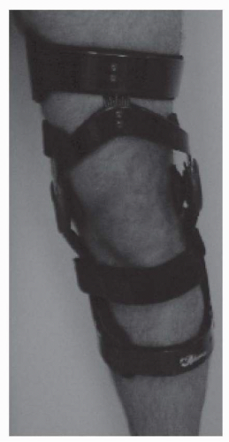
Knee bracing for medial compartment gonarthrosis currently involves application of a three-point force system across the coronal plane of the knee joint, that is, valgus bracing that “unloads” the medial compartment. Current bracing terminology refers to this type of orthosis as an “unloading brace” (Fig. 31-1). Manufacturing trends have focused on single and double upright brace designs that are fabricated from high strength, very light weight materials.

A more current study showed that the external varus moment at the knee was reduced during initial contact and loading response phases of the gait cycle (95). The interval between 10% and 15%, or loading response, is the point of peak varus loading; therefore, a reduction in the external varus moment about the knee during this interval is felt to be important for diminishing load and subsequent pain reduction. There was, in fact, improvement in pain and function with use of the brace as measured by a visual analog scale (VAS). A second study showed that the mean femorotibial angle decreased from 185.1 degrees to 183.7 degrees. In addition, isokinetic quadriceps muscle strength increased from an average of 36.8 to 42.8 Nm (96). Another study was performed during which patients were asked to walk with and without a brace, and condylar separation angles were measured under fluoroscopic guidance. Twelve out of fifteen patients demonstrated an average condylar separation angle of 2.2 degrees, while the three subjects without separation were obese (97). Lindenfeld et al. evaluated the biomechanics of an unloader brace on patients with medial compartment involvement and found that the adduction moment at the knee was altered when the brace was worn (98). This change may reduce the load transmitted through the medial compartment, resulting in less pain. Lastly, the clinical knee score and bone mineral density of the proximal tibia were assessed in an attempt to evaluate the efficacy of valgus knee bracing. The knee score improved after 3 months, and increases in bone mineral density were seen more in the lateral tibial condyle than in the medial, suggesting that the brace transmits forces across the knee joint from the medial to the lateral side (99).


FIGURE 31-1. Typical knee OA unloading brace.

A clinician should consider using a simple knee sleeve for those patients who present with mild OA-related knee pain but without any significant angular deformity. Although there is still no scientific evidence from clinical studies that highlight positive results using only a sleeve, there are many anecdotal patient accounts. Since the nonjointed sleeve provides no significant structural support to the knee, patient-reported feelings of improved stability and reduced pain are likely due to an improvement in proprioception that theoretically occurs with use of the sleeve. In 1999, a study was performed in order to measure the efficacy of a sleeve along with pharmacological treatment versus an unloader brace and medication as compared to the control group of patients who were given medication only. A significant improvement in function, in addition to disease-specific quality of life (p = 0.001), was found in both the neoprene-sleeve and unloader brace groups as compared to the control group. The unloader brace was on average more effective than the neoprene sleeve (100).

Wedged shoe soles/insoles are additional orthotic devices that can possibly be used to counter OA-related knee joint compressive forces. Patients have reported pain relief in the medial compartment after applying a lateral wedge under the calcaneus. Sasaki and Yasuda examined the effects of using lateral heel wedges with load transducers and roentgenograms to observe the biomechanical effects (101). The variables that were under study included the ground reaction force (GRF), the tibiofemoral angle, and the tibiocalcaneal angle. Although the GRF and the tibiofemoral angle did not exhibit a change, the lower limb mechanical axis, however, approached an upright position, and the calcaneal angle corrected in the valgus direction with reference to the subtalar joint. The observed changes were believed to reduce the axial load bearing on the medial joint surface. The use of the wedged insole at a certain angle serves as a “stimulant” to the patient, because it causes an inclination of the floor to which the patient must compensate.
Patients must adapt to the wedge via changes in muscle activity and posture, ultimately modifying their standing and walking mechanics. In addition to their effects on knee pain and forces acting at the knee, other investigators have examined the effects of lateral wedged insoles on knee kinetics. In this study, three-dimensional analysis was performed on 17 subjects with and without them wearing a 5-degree lateral wedged insole. They found no significant differences in temporal and spatial parameters; joint angles at the hip, knee, and ankle; or kinetics at the hip and ankle. The external varus moment and estimated medial compartment load at the knee were reduced significantly (102). One can interpret the results to indicate that lateral wedged insoles are a potential effective component of a conservative treatment regimen for patients with mild medial compartment OA.

Viscoelastic shoe inserts presumably provide shock absorption at the knee and as such are another intervention at the foot and ankle for the treatment of painful knee OA. Walking has been linked to the generation of impulsive loading on the human locomotor system. Impulsive loading from normal gait has been associated with “damage” to some elements of the locomotor system. Specifically, boney vibratory forces during normal gait have been detected at 25 to 100 cycles/second at initial contact and are believed to lead to articular cartilage deterioration (103). The feet, menisci of the knees, intervertebral discs, and vertebral bodies are the so-called “natural shock absorbers,” as one of their functions is to absorb and dissipate the energy that acts upon the body during heel strike. Depending on gait velocity and the foot’s viscoelastic properties, the dynamic forces to which the foot is subjected during gait may be considerably higher than the body weight. Under normal physiologic conditions, the intermittent and continuous shock and load experienced during gait tends to cause a slowly progressive weakening of the natural shock absorbers and may later lead to articular cartilage degeneration and OA. Insufficient damping of incoming shock waves can overload the proximal joint and contribute to the process of joint degeneration (103, 104).

Voloshin and Wosk clearly showed that viscoelastic inserts reduce the amplitude of incoming shock waves by 42%. They, therefore, can dampen overloading and in a sense possibly prolong the life of articular cartilage. Conservative treatment, using viscoelastic shoe inserts, for patients with clinical symptoms of knee OA too yielded an improvement in symptoms by 78% (103).

After medial knee compartment involvement, patellofemoral compartment involvement is the second most common site of involvement in knee OA. The principal biomechanical factor noted to cause patellofemoral joint pain is felt to be patellar maltracking. Specifically, it is believed that there is abnormal lateral patellar tracking, thus increasing lateral patellar facet contact forces (105). The purpose of orthotic intervention is to help achieve and maintain optimal patellar tracking in order to prevent abnormal compression forces and degenerative changes. Knee braces used for patellofemoral compartment OA are specifically designed to reduce patellar compression as well as to prevent excessive lateral shifting. Criteria evaluated at time of prescription are patient vocation/avocation, likely compliance, and cost. Patellar-stabilizing braces, infra-patellar straps, and patellar taping are some of the current orthotic treatment options.

The neoprene-sleeve patellar stabilizing brace consists of a patellar cutout and force inducing buttress pads around the inferior and lateral aspects of the patella. The brace has two circumferentially wrapped rubber straps that apply dynamic tension to a crescent-shaped lateral patellar pad. Palumbo found this sleeve useful in the treatment of patients with patellofemoral arthritis (106). Cushnaghan et al. demonstrated that medial taping of the patella resulted in a 25% reduction in knee pain compared to lateral or neutral taping (107).

Although hip OA is also extremely common, there is a much smaller body of literature on orthotic intervention for this body region. This is not surprising given the practical difficulties encountered with bracing at the hip joint. Ankle OA, by contrast, is much less common than hip and knee OA. Literature pertaining to orthotic intervention for ankle OA is quite limited and largely anecdotal.


Hand and Occupational Therapy for OA patients

In addition to the many potential benefits from PT interventions, OT is generally believed but not proven to be of benefit in select patients (1).


OT for Upper Limb OA

OA can affect a variety of upper limb structures including the shoulder girdle region, the elbow, the wrist, and the hand. OA in the shoulder region may involve the glenohumeral joint, the acromioclavicular joint, and rarely the sternoclavicular joint and nonarticular locations such as the subacromial space due to impingement of the rotator cuff tendons and/or the subacromial bursa by acromion bone spurs. Elbow region OA may be secondary to prior trauma or as a result of inflammatory joint disease or may be primary due to repetitive overuse. OA of the wrist region can occur due to prior trauma, inflammatory joint disease, prior infection (especially prior gonococcal or Lyme infection), or perhaps due to overuse. OA of the hand region
is very common and especially affects the first CMC joint, the proximal interphalangeal (PIP) joints, and the DIP joints.

Hand OA can be associated with significant pain and disability, especially in women (108). Upper limb weakness due to deconditioning from pain-related disuse and stiffness is common in OA patients. The Arthritis Hand Function Test can provide reliable, standardized measurements of strength and dexterity in adults with OA (109).

An OT program can be targeted toward the associated pain, disability, and weakness (110). Modalities, including therapeutic heat or cold, may provide pain relief, especially during an exacerbation. Adaptive equipment may include devices that empower the patient to independently perform activities such as opening jars, buttoning clothing, or holding utensils. Occupational therapists often prescribe assistive devices to assist with ADLs and train patients in their use. Although it is believed that patients with OA are frequent users of such assistive devices, there is a scarcity of scientific evidence for the prescription, provision, and use of these in OA patients (111).

Only gold members can continue reading. Log In or Register to continue

Stay updated, free articles. Join our Telegram channel

May 25, 2016 | Posted by in PHYSICAL MEDICINE & REHABILITATION | Comments Off on Osteoarthritis

Full access? Get Clinical Tree

Get Clinical Tree app for offline access