Chapter 28 Orthoses in total joint replacement
Review of the literature
Several authors indicate that a hip dislocation in the early postoperative period in a hip with proper orientation and restored mechanics can be treated successfully with patient education and adjunctive bracing.1,2,9,12 This group included a retrospective study of 80 patients managed with a hip orthosis to prevent dislocation following hip revision surgery.7 The primary shortcoming of such studies is the lack of control groups, statistical analysis, and long-term outcomes. DeWal et al.4 retrospectively reviewed 91 patients who dislocated for the first time and 58 patients who had a history of recurrent dislocation to determine if hip abduction orthoses were effective in reducing redislocations after a closed reduction. They found that patients fit with a hip abduction orthosis redislocated at the same rate as those who did not receive an orthosis.4 The study had several limitations: no orthotic protocols were provided, the report did not state whether the patients who received hip orthoses had injuries of similar severity, and the report did not indicate whether the patients were wearing the orthosis at the time of the dislocation.2 Differences in surgical procedures, prosthetic components, and types of hip orthoses used all can impact the dislocation rate and subsequent management with a hip orthosis. Clearly, long-term, controlled studies are needed to determine which patients may benefit from orthotic management following dislocation or as a means of preventing first-time dislocations in complicated cases.
New surgical procedures may have an impact on the dislocation rate. Constrained acetabular liners have significantly reduced the incidence of dislocation but are subject to premature wear and disruption. Many surgeons now use prosthetic components with larger articulating bearing surfaces to help prevent dislocation.2 Surgical procedures now are being performed with smaller incisions, leaving the abductor muscles more intact and more able to provide hip stability. In addition, new hip resurfacing procedures are emerging, which replace the acetabular component and cap the head of the femur, leaving most of the head intact. This procedure, performed on younger patients, retains more bone and may help to prevent dislocation. Although early reports are promising, long-term study is needed to evaluate the impact of these new procedures on component longevity and resistance to dislocation.
Dislocation following hip surgery
Dislocation of a total hip prosthesis is a difficult clinical problem. The incidence of dislocation varies, depending on whether the procedure is a primary (1.7,5 3.9%,13. 7.2%2) or a revision arthroplasty (11.2%2–14.4%13). The rate of dislocation for these procedures is also linked to factors such as component selection,2 prosthetic component orientation,4,7,9 and surgical technique.6,9,11 Dislocation rates were higher for patients with a history of dislocation, those with poor abductors or adductor spasticity, patients with anterior wall weakness or global instability, those with acetabular transplants, and patients with two or more surgeries on the affected side.7
It is important to understand the cause and direction of instability in order to provide appropriate orthotic management. Patients who dislocate with satisfactory radiographic positioning of the prosthetic components often can be managed with a closed reduction of the hip and a hip orthosis to discourage motion that can lead to dislocation.9 If the referring physician has not indicated the direction of dislocation, the orthotist should review the patient’s activity at the time of the dislocation and clarify the instability with the physician.7 Posterior dislocations occur 85% of the time and usually involve hip flexion, adduction, and internal rotation. Activities associated with posterior dislocation include sitting and reaching toward the uninvolved side, exiting a vehicle, reaching for an object on the floor, leaning over to apply shoes, or rising from a low chair, toilet seat, or soft cushion. Anterior dislocation is linked with external rotation and extension and often occurs during activities such as reaching up on a high shelf, extending hips and trunk to move back into bed, reaching behind the body while standing to put on a coat, or lying in bed with hips extended.7 Anterior dislocations are also seen in patients diagnosed with hip dysplasia, who have excessive femoral anteversion.
Often physicians are able to reduce a dislocated hip with a closed reduction performed under local anesthesia. In cases where the pain is too severe or when efforts to reduce the hip are unsuccessful while the patient is awake, a closed reduction can be performed under general anesthesia. Once reduced, a total hip can be stabilized with an orthosis until the compromised soft tissue heals and creates scarring around the joint. This usually prevents further dislocations if the femoral and acetabular components are well positioned. In the past, hip spica casts have been shown to be useful for this purpose. Currently, orthoses offer several advantages over casts. Orthoses weigh less, which makes them easier to tolerate during ambulation. An orthosis can be carefully removed, providing access for wound care and hygiene. In addition, hip orthoses are widely available in adjustable, prefabricated models, facilitating immediate stabilization of the patient, limited in-patient hospitalization, and earlier return to activities (Fig. 28-1).1,7
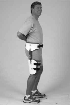
Management of posterior dislocations
The hip orthosis used to treat a hip that dislocates in a posterior direction is generally proximal to the knee. A snugly fit pelvic band suspends the orthosis and provides an attachment point for the hip joint. A laterally placed, adjustable range of motion hip joint capable of controlling flexion, extension, abduction, and adduction attaches to a snug-fitting thigh cuff that holds the hip in 10 to 20 degrees of abduction and allows 0 to 70 degrees of flexion. This joint position combined with properly fitting pelvic and thigh components provides a kinesthetic reminder against excessive flexion, adduction, and internal rotation. Another study recommends that the hip be held in 0 to 10 degrees of flexion, externally rotated, and abducted 15 to 20 degrees for posterior dislocations.9 Once properly fit, most patients ambulate with minimal support and can perform most activities of daily living while wearing the orthosis. The orthosis is worn under clothing to ensure protection in all positions, including toileting. Innovations include joints that abduct as the hip moves into greater amounts of flexion and rotational control incorporated into the hip joint mechanism. Internal and external continue to be the most difficult motions to control in an orthosis that ends proximal to the knee. When used to prevent another dislocation, the orthosis is worn at all times.2,7,9,12 If radiographs demonstrate malposition or instability of either the femoral or acetabular prosthetic component, if an orthosis is not effective, and if the patient is a good candidate, surgery is generally performed to stabilize the hip.9
Management of anterior dislocations
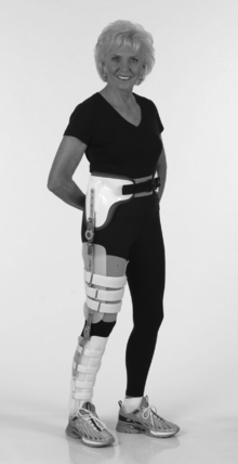
When patients have anterior wall weakness or global instability, extension, rotation (usually external rotation), and abduction usually is the mechanism of dislocation. Extension range is blocked at −40 degrees, and flexion range is generally safe up to 70 degrees, unlike posterior dislocations where flexion is one component of dislocation.7 Patients who dislocate in an anterior direction often demonstrate global instability due to acetabular insufficiency. To provide rotational control, a knee–ankle–foot orthosis (KAFO) rather than a simple thigh cuff is suspended from the pelvic band. Although this adds considerable bulk to the orthosis, it also provides increased directional control and stability. Patients who wear a hip–knee–ankle–foot orthosis (HKAFO) often need additional assistance to perform activities of daily living because the orthosis adds considerable bulk and motion restriction. Patients generally wear the orthosis at all times for 3 to 6 months. Often, fluoroscopy is used to determine the exact mechanism of dislocation. In conjunction with the physician, the orthotist should adjust the range of motion settings to address the patient’s specific instability.7
In another study highlighting orthotic management of anterior dislocations, the author recommends setting the hip joint to allow flexion between 20 and 30 degrees and provide 0 to 10 degrees of internal rotation and 20 degrees of abduction.9 For the most severe revision surgeries and patients with compromised bone, a custom lumbosacral orthosis and molded thigh cuff without a hinge on the affected side with thigh cuff and hinge on the contralateral side can be used in place of a prefabricated hip orthosis. Because anterior dislocations occur only 15% of the time and precautions are very different than those for posterior dislocations, the orthotist should educate other staff members about the positions that can place the patient at risk for dislocation in an anterior direction (Fig. 28-2).
< div class='tao-gold-member'>
Related posts:
 Appropriate technologies for assistive devices in low-income countries
 Orthoses for the muscle disease patient
 Upper limb orthoses for the person with spinal cord injury
 Sports adaptations for the physically challenged athlete
 Protective equipment to the upper limb in sport
Stay updated, free articles. Join our Telegram channel

Full access? Get Clinical Tree


