Fig. 13.1
Normal whole-body bone scintigraphy
Bone pain is a severe debilitating condition which frequently impairs the quality of life. Patients often receive analgesics, bisphosphonates, and external beam radiotherapy to provide symptomatic relief. However, each therapy has its side effect and not so effective in patients with widespread metastasis. Bone-seeking radiopharmaceuticals are one of the therapeutic options available for palliation of bone pain and should be used in conjunction with other therapy modalities in osteoblastic metastasis detected by bone scintigraphy. Approximately 60–80 % of these patients benefit from these therapy with mild transient hematological toxicity [96]. Although previous radiotracer like 89Sr, 153Sm-lexidronam, or 186Re-etidronate only provided bone pain palliation, recently introduced radium-223 chloride, an alpha-emitting radioisotope, was found to provide additional survival benefit in a phase 3 trial [97]. Ongoing studies with bone-seeking radiopharmaceuticals combined with other cytotoxic therapies will further clarify the potential patient benefit.
A-Bone Scintigraphy (BS)
Plain radiographs provide assessment of anatomic structure of bone, whereas BS gives us the ability to detect bone’s blood flow and metabolism. In addition to its high sensitivity, the visualization of the entire skeletal system in a single examination is another advantage of BS. However, the specificity of BS is low and needs to be correlated with other anatomic imaging modalities. In recent years, widespread use of SPECT-CT scanner allowed us simultaneous acquisition of 3D scintigraphic and CT data from the same region, and specific diagnosis can be reached in a single study.
Standard BS was performed as whole body for the investigation of metastatic disease. The three-phase BS consists of blood flow, blood pool, and delayed phases that shows blood flow and bone metabolism. Three-phase BS is used for localized involvement of bone lesions like osteomyelitis, tumors, or fractures [1, 2].
Indications of BS
1.
Staging and follow-up of primary/metastatic bone tumors
2.
Etiology bone pain that cannot be identified by radiography
3.
Evaluation of patients with suspicion for fractures
4.
Evaluation of bone and soft tissue infections.
5.
Evaluation of joint involvement in patients with arthritis
6.
Detection of prosthetic complications like aseptic loosening and infection
7.
Assesment of bone graft viability
8.
Investigation of maturation of soft tissue calcifications before surgery
9.
Diagnosis of avascular necrosis (AVN)
10.
Metabolic bone diseases
11.
Reflex sympathetic dystrophy
12.
Diagnosis and follow-up of Paget’s disease
13.
Evaluation of chronic back pain
14.
Suspicion of child abuse
Radiopharmaceuticals Used and Uptake Mechanisms
Tc-99m-labeled diphosphonates (e.g., methylene diphosphonate (MDP)) are used in bone scintigraphy [1–3]. After intravenous injection, radiopharmaceutical is biodistributed to the whole body via blood and passes to extravascular area by passive diffusion. Tc-99m-labeled phosphates are absorbed by chemical bonds to hydroxyapatite crystal found on bone surface. About 40–50 % of these compounds will be affixed to the bone by 4 h after injection. Excretion is primarily renal [4] and 70 % of the administered dose is eliminated by 6 h. The recommended dose for an adult patient is 20 mCi (740 MBq). The whole-body radiation dose from a Tc-99m MDP bone scan is approximately 0.01 rad/mCi (4.2 mSv); the urinary bladder is the critical organ because of renal excretion, receiving about 0.1–0.2 rad/mCi [3, 4].
The uptake of radiopharmaceutical in bone depends on blood flow and osteoblastic activity. There is a normal physiological activity in areas where bone turnover is increased like in joints, muscle insertion areas, and excretion pathways (mainly kidney and bladder and small amount by intestines). In areas where only osteoclastic activity is seen or in metabolically inactive sclerotic bone islands, normal radiopharmaceutical biodistribution is seen. Radiotracer uptake is increased in conditions where blood flow or bone metabolism is increased. Primary or secondary bone tumors, fractures, trauma, arthritis, degenerative joint changes, neuropathic joint pathologies, and pathological fractures due to osteoporosis were the conditions that osteoblastic activity could be seen [1–3].
Imaging Technique
Standard BS
Whole-body images of skeleton were obtained 2–6 h after injection of radiotracer (Fig. 13.2).
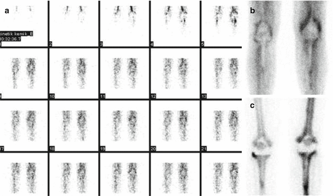
Fig. 13.2
Three-phase BS in a patient with knee prosthesis, (a) blood flow, (b) blood pool, (c) delayed phase
Three-Phase BS
The area of interest was focused by gamma camera, and by using dynamic acquisition, blood flow (obtained as the tracer is injected) and blood pool phase that reflects soft tissue involvement (acquired immediately after the flow portion of the study and completed within 10 min of injection of the tracer) were acquired (Fig. 13.3). After 2–5 h whole-body images were taken as late images.

Fig. 13.3
Patient suffering from prostate cancer. Whole-body BS shows widespread bone metastases showing increased radiotracer uptake, PSA level: 2300 ng/ml
Patient Preparation
BS does not require specific preparation. Oral or i.v. hydration may increase the image quality by decreasing the background. Although radiation dose is lower than abdominal or thoracic CT, patient could further decrease this dose by excessive fluid intake and micturition. Patient should be asked for possibility of pregnancy. The main source of radiation to the fetus comes from the radioactivity from the bladder (17 mrad/mCi (4.6 μGy/MBq) at 8th week, 9.7 mrad/mCi (2.6 μGy/MBq) at 18th week). This risk can be decreased with hydration and frequent micturition [3, 5]. Although BS is not recommended for pregnant patient, if necessary one should know that radiation dose to fetus is low enough for BS to be used safely.
Interpretation of the Study Report
In normal BS, in addition to whole skeleton, normal mid radiotracer activity at soft tissue, kidney, bladder, and thyroid cartilage can be seen [1–3]. The areas that show high radiotracer activity than normal bone are called as hyperactive-hot focus. This finding can be seen in fractures, osteomyelitis, neoplastic diseases, and arthritis. Areas that show less uptake than normal are called hypoactive and cold focus. These can be seen in lytic lesions, tumor necrosis, early osteonecrosis, metallic foreign bodies, and after radiotherapy.
Another scintigraphic pattern is called superscan. This pattern is characterized by diffuse increased uptake in the whole skeleton and decreased activity in kidneys. It is commonly seen in patients with widespread bone metastases or hyperparathyroidism [3].
Clinical Applications
Detection and Follow-Up of Bone Metastases
BS is readily available, highly sensitive imaging modality for screening of bone metastases with no absolute contraindications. It is 50–70 % more sensitive than radiographs in detecting skeletal metastases. This is because about 30–70 % of the bone mineral content must be lost before a metastasis is detectable on a radiograph [5, 6]. Contrary, 5–10 % change in the ratio of the lesion to normal bone is required to detect an abnormality on BS [6]. Also lesions in regions like sternum and scapula are also difficult to be detected by direct graphy due to overlapping bone structures. In general the sensitivity for bone scan in detecting bone metastases is between 62 and 100 % [6]. The success of BS is high in the detection of bone metastases from cancers like prostate which makes osteoblastic metastases or the metastases that lead to osteoblastic reaction. Asymmetric increased uptake throughout the skeleton is characteristic of bone metastases [6]. Other conditions like fractures, degenerative disease, or other benign conditions that may lead to increased uptake may confuse the diagnosis, so correlation with other imaging modalities may help in the differential diagnosis. BS is probably not the procedure of choice for screening skeletal involvement in patients with multiple myeloma or lymphoma which usually make bone marrow or osteolytic metastases. However, combination of planar BS images with SPECT-CT can be used to improve the detection of lytic bone lesions.
SPECT imaging provides 3D images of a given lesion and results in overall improved sensitivity with the detection of 20–50 % more lesions in the spine when compared to planar images [6]. SPECT-CT can further improve accuracy by improving anatomic localization and by better evaluation of underlying bone pathology through CT data [7].
Whole-body imaging by BS is advantageous because about 90 % of patients with skeletal metastases present with multiple lesions. Detection of all metastatic sites could possibly effect patient management especially when local therapy is considered. Most of the metastatic lesions are in the axial skeleton. In patients with a known malignancy, 60–70 % of axial lesions are due to metastases, whereas about 30 % of lesions in the extremities or skull are due to metastases [1, 2]. In the evaluation of vertebral lesions, there are certain patterns of the radioactivity distribution which may help in the differential diagnosis, particularly when SPECT images are performed. Increased uptake beyond the vertebral body especially in facet joints is common due to degenerative changes [5]. Tracer uptake involving the vertebral body and extending to pedicles is usually indicative of metastatic disease [5, 6]. After chemotherapy for bone metastases, radiotracer uptake may increase in metastatic areas within 3 months after therapy. This is a sign of bone healing process after the death of tumor cells and called “flare phenomenon” [5]. It is commonly seen in breast or prostate cancer after chemo-/hormonotherapy and characterized by increase in the number and size of the pretherapy BS lesions. This phenomenon may lead to confusion in the differential diagnosis of progression or good therapy response. BS must be repeated 6 months after the end of therapy for differential diagnosis. Decrease in the number and intensity of lesions in the repeated scintigraphy is seen in good response, whereas progressive disease will show further progression [5].
BS in Prostate Cancer
Thirty-five percent of patients with prostate cancer have bone metastases in the first clinical presentation (Fig. 13.4) [8, 9]. Presence of bone metastases positively correlate with PSA (prostate-specific antigen) and Gleason score. Rate of bone metastases were 2.3 % for patients with PSA less than or equal to 10 ng/mL, 5.3 % for patients with PSA between 10.1 and 19.9 ng/mL, and 16.2 % for patients with PSA levels between 20 and 49.9 [6]. The rate of bone metastases increases to about 50 % when the PSA level is greater than 50 ng/mL [8–10]. Based upon the Gleason score, detection rates are 5.6 % of patients with a score of less than or equal to 7 and 29.9 % of patients with a score of 8 or greater [7–9]. Rate of bone metastases is low when PSA is below 10–20 ng/ml. BS should be limited to patients for initial staging with elevated PSA (greater than 10 ng/mL), high Gleason scores (8–10), locally advanced disease, elevated alkaline phosphatase, or bone pain symptoms. Patients who are receiving hormonal therapy (antiandrogen) may have bone metastases, even when their PSA is normal [11].
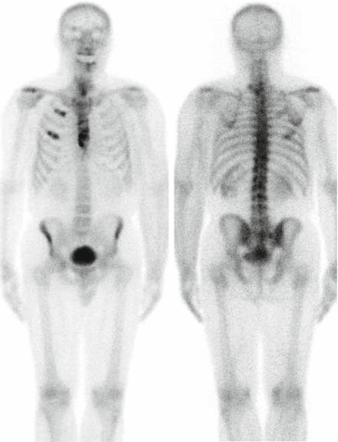
Fig. 13.4
Patient with rib metastases. Linear uptake in ribs is a sign of bone metastases
BS in Breast Cancer
Bone metastases occur in 70 % of patients during the course of disease in locally advanced breast cancers [12]. BS could detect metastases 2–12 months before the direct graphy (Fig. 13.5). However, as in other cancers specificity of the technique is low. The rate of positive BS changes with the stage of patient (2.5 % for stage 1, 7 % for stage 2, 16 % for stage 3, and may be as high as 14–28 % in stage IV disease [6, 12]). BS must be performed if the patient complains about bone pain, has elevated tumor markers, and has a stage of 3 or 4.
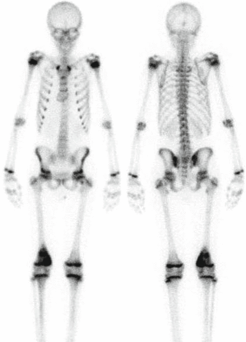
Fig. 13.5
Osteosarcoma in the right femur distal metadiaphyseal region
Recurrence was seen in 36 % of patients with locally advanced breast cancer during 4 years of follow-up and most of these were seen at bone. The rate of recurrence is higher in patients with lymph node metastases at the primary diagnosis. Although bone pain is a common symptom in these patients, up to 30–50 % of these patients can be asymptomatic. Increased radiotracer uptake in sternum is important for a patient with breast cancer as it has a 75 % likelihood of being a metastasis. There is also a strong correlation between the side of the sternal met, the side of the primary breast lesion, and the presence of internal mammary lymph node involvement.
BS also can be used in the therapy response evaluation of treatments like chemotherapy. However, low specificity of the BS due to benign changes and flare phenomenon limits its utilization. Due to its higher specificity, FDG (F-18-fluorodeoxyglucose)-PET-CT replaces BS in therapy response evaluation. However, BS is still used due to its readily availability and low cost. BS provides advantage in osteoblastic metastases where FDG shows lower sensitivity [5].
BS in Lung Cancer
Advanced lung cancers often metastasize to bone and BS is a routine imaging method for staging [1, 2]. BS is especially useful if curative surgery is planned. However, the utilization of BS is decreased with the increased availability of FDG-PET-CT in the staging of lung cancer. FDG-PET-CT not only detects early bone lesions (bone marrow, small sized cortical lesions etc) but also gives useful information about the lymph node and distant metastases [13, 14].
BS is not indicated in tumors which cause osteolytic metastases or bone metastases like renal cell cancer and multiple myeloma. It can be useful if patient is symptomatic and used with SPECT-CT that can detect osteolytic lesion and complications due to metastases like pathological fractures. MRI and FDG-PET-CT are the methodologies of choice for the staging of these tumors.
BS can be used as complementary to MIBG (metaiodobenzylguanidine) in patients with neuroblastoma and pheochromocytoma. MIBG is superior in detection of early bone marrow metastases, but BS may help in the tumors with no MIBG uptake.
Solitary Lesion on BS
A solitary rib lesion has about a 10 % probability of representing a metastasis in a patient with a known malignancy [15]. The chance of solitary lesion detected on BS to be malignant depends on the patient history, localization and shape of the focus. For example, in patient with malignancy rate of solitary lesion in vertebra or pelvis to be malignant is as high as 60–70 %; however, this rate decreases if benign diseases like degenerative disease is detected by direct graphy. Similarly the rate of rib lesion to be malignant is low as 10 % especially in patient with trauma history. In a study held in Hacettepe University in patient with breast cancer patient, this rate was found to be as high as 26 % (Fig. 13.5) [16]. The percentage increased by the number of new lesions detected and reached 100 % when 5 new lesions were identified [17].
The Diagnosis, Staging, and Follow-Up of Primary Bone Tumors
MRI is the modality of choice in the preoperative detection of soft tissue involvement and lesion boundaries in primary bone tumors like osteosarcoma (OS) and Ewing’s sarcoma (ES). Three-phase BS is a sensitive method for the detection of bone tumors; however, due to increased local blood flow and osteoblastic activity in late images, BS may overestimate lesion size. Due to its high sensitivity, BS mainly used for screening of bone metastases [18]. The most common malignant bone tumor in childhood and adolescence is osteosarcoma. Primary tumor and metastases show bone formation that can readily be detected by BS (Fig. 13.6). About 80 % of cases have localized tumor at presentation, whereas the remainder present with metastases most commonly to lungs and bones. Although radiotracer uptake within a pulmonary lesion is very specific for a metastatic focus of osteosarcoma, BS is less sensitive than CT in the detection of pulmonary metastases especially when the metastases are small. Pulmonary metastases can be detected in 20 % (2D planar imaging BS) to 40 % (3D SPECT imaging) of patients [5]. BS could detect bone metastases with a high sensitivity of 80 %, but specificity may be low as 60 % because of benign changes that may mimic malignancy. Nonetheless, the presence of skeletal metastases is associated with a poor prognosis and will change treatment, and a baseline bone scan is generally recommended.
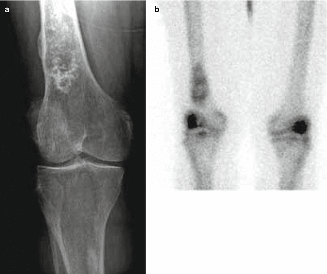
Fig. 13.6
Patient with an enchondroma in the right distal femur, (a) X-ray graphy, (b) bone scintigraphy
Ewing’s sarcoma is the second most common malignant bone tumor. BS is also used for staging purposes; however, ES has the tendency to have early bone marrow metastases which decrease the sensitivity. FDG-PET and MRI are the modalities that has widespread application for bone metastases. ES and metastases are typically “hot” on blood pool and delayed bone scan images. On follow-up, the skeleton is the first site of metastatic disease in about 25 % of patients. Baseline and follow-up bone scans are therefore very useful in these patients. FDG-PEt also shows increased uptake in Ewing’s sarcoma and is superior to bone scan for the detection of osseous metastases [19].
Early after chemoradiotherapy, benign reactive nonspecific radiotracer uptake on BS can be seen in the tumor bed. However, persistent uptake after 6–12 months raises the suspicion of local recurrence. CT and MRI could not be reliable in differentiation of necrosis/fibrosis from tumoral recurrence. Tl 201, Tc-99m sestamibi, and FDG-PET studies were useful in the evaluation of response [1–3]. There is a positive correlation between decrease in uptake and therapy response.
BS is not only used in bone tumors but also used in the evaluation of suspicious lesion detected on direct graphy. Although benign lesions show less and malignant lesion shows more uptakes, there is a significant overlap between benign-malignant lesions (Fig. 13.7). BS mainly gives information about lesion number and distribution.
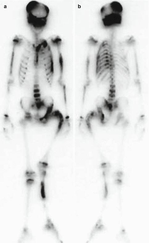
Fig. 13.7
A patient with fibrous dysplasia showing increased uptake on BS which produced left leg deformity
In benign tumors, BS may also help in the differential diagnosis by showing the bone metabolism. Aneurysmal bone cyst show increased activity on all three phases. About two-thirds of the lesions will be photopenic centrally, surrounded by a “ring” of increased activity [20].
Bone islands (enostosis) are often confused with sclerotic bone metastases. Most of them show no radiotracer uptake; however, about 30 % of large bone islands may demonstrate mild increased uptake. Enchondroma is a benign neoplasm of the medullary canal composed of mature hyaline cartilage; they can have a very variable appearance on bone scan demonstrating very mild to prominent uptake of tracer.
Langerhans’ cell histiocytosis is characterized by a varied and abnormal proliferation of histiocytes. The spectrum pathological entities included bone involvement (solitary and multifocal). Musculoskeletal manifestations occur 78 % of the time, and there is a predilection for marrow involvement with the flat bones of the skull and ileum being the most frequently involved.
On scintigraphy, up to 30–40 % of skeletal lesions may not be detected, and 10 % may appear cold [5]. Nonetheless, BS is considered to be complimentary to skeletal radiography in this disorder. Howarth et al. reported the sensitivity and specificity of the radiographic skeletal survey of 100 % and 61 %, respectively, compared to 91 % and 55 % for bone scintigraphy. For solitary lesions, radiographic sensitivity and specificity were 95 % and 73 %, respectively, compared with 88 % and 77 % for BS [21].
BS is most useful in two bone tumors. Among these osteoid osteoma is characterized as increased uptake in three phases. In the late phase, osteoid osteoma may be seen as more intense area in the middle as “double density” sign. BS provides whole-body screening and normal BS rules out osteoid osteoma diagnosis. BS is also used in other benign tumoral disorder like fibrous dysplasia. This entity may be 70–80 % monostatic and 20–30 % polyostotic. The diagnosis of monostotic form is late and most of these patients are asymptomatic (Fig. 13.8). Fibrous dysplasia is characterized by increased osteoblastic activity; however, these findings may be nonspecific. BS is helpful to screen polyostotic involvement and detection of biopsy site [1–3, 5, 6].
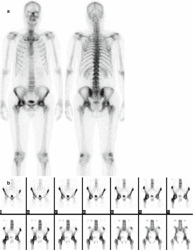
Fig. 13.8
Patient with avascular necrosis, (a) whole-body BS, (b) bilateral hypoactive areas in femoral heads in SPECT images
Bone Fracture
X-ray graphy is preferred for the initial diagnosis of acute trauma. It enables to see the anatomic fracture lines and help orthopedicians manage the patient with severe pain. However, it lacks enough sensitivity in small fractures and in bones with overlapping structures like scapula. BS is highly sensitive (>95 %) in detection of fractures [5]. About 80 % of bone scans will show increased activity at a site of fracture by 24 h and 95 % by 72 h, although patients over 75 years old and debilitated patients may not show activity for several days. A gradual decline in activity occurs over time with about 80 % of exams normalizing by 1 year and 90 % becoming normal by 2 years. The minimum time for normalization of activity is 6 months after properly fixed fracture [1, 2]. Nonunion occurs when there is failure of the fracture site to heal by 6–8 months following the injury. The tibia is the most frequent site of nonunion. Nonunion may occur secondary to an inadequate blood supply, osteoporosis, improper immobilization, or infection. Two types of nonunion have been described: reactive and atrophic. Reactive nonunion is characterized by persistent increased or normal uptake at the fracture site (80 %). This bone has potential for healing, and these fractures tend to show significant improvement following therapies like electrical stimulation. It is not possible to distinguish a reactive nonunion from a delayed union. Atrophic nonunion appears as a photon-deficient area between the fracture ends, which themselves may have increased activity. This group of fractures is usually not responsive to percutaneous electric stimulation. The etiology of the nonunion must be investigated with In-11-labeled leukocytes or Ga-67 to exclude infection.
Stress or Insufficiency Fracture or Shin Splints
Stress fractures occur due to repetitive trauma on normal bone. Bone adapts to stress with internal remodeling; however, if the trauma continues or high enough to exceed repair mechanisms, micro and gross fractures occur. Up to 40 % of patients with stress fractures can be asymptomatic [5]. Bone scan is much more sensitive than X-ray graphy for diagnosing stress fractures [5]. More than 80 % of stress fractures will not be evident on initial radiographs, while the sensitivity of bone scan for the diagnosis of stress fracture approaches 100 % [3]. On bone scan, stress fractures usually appear as solitary, focal, fusiform areas of increased tracer activity generally lasting for 10–12 weeks. SPECT imaging is much more sensitive than planar imaging for the detection of femoral neck stress fractures and can demonstrate fractures in patients with normal planar imaging [10].
Insufficiency fractures are common problem in elderly and debilitated patients. Most common cause of insufficiency fracture is osteoporosis. In these patients, proximal femur fracture is a diagnostic problem. Patient has history of pain and trauma history; however, X-ray graphy may be normal. These patients need urgent therapy before avascular necrosis or dislocation occurs. BS is preferred rather than X-ray graphies, and if the BS is not conclusive, MRI is the method of choice. In the sacrum, stress fractures are associated with osteoporosis or prior radiation therapy to the pelvis (an insufficiency fracture) [3]. The osteoporotic sacrum develops a characteristic “butterfly” or “Honda sign” fracture on BS. Plain films are typically normal but may show a sclerosis in sacral ala.
Shin splints (periosteal reaction) are due to abnormal movement of the either the soleus muscle tendon complex or posterior tibialis muscle which produces a periostitis-type lesion. They are characterized by abnormal linear (contrary to stress fracture not focal) uptake of tracer along the posteromedial tibial cortex. Shin splints have not been demonstrated to progress to stress fractures. Shin splints are mainly treated with anti-inflammatory drugs [22, 23]. However, stress fractures need longer immobilization.
Evaluation of Bone and Soft Tissue Infections
X-ray graphy is the first imaging modality in the suspicion of osteomyelitis. Most common problem is the early diagnosis. In the first 3 days, the radiological findings are nonspecific. Bone demineralization can be detected after 7 days, and after 14 days periosteal new bone formation can be seen. BS was shown to be positive 24–72 h after the beginning of infection [24]. Three-phase bone scintigraphy is performed in suspicion for osteomyelitis. BS could differentiate soft tissue infection vs osteomyelitis and acute vs chronic phase of new bone formation. Increased activity in blood flow and blood pool phases shows hyperemia and raises the suspicion of acute infection. Increased activity in late images show increased bone formation and osteoblastic activity. In patients with only soft tissue infections, there will be increased activity in the first two phases of three-phase BS, but late static images will be normal and no increased osteoblastic activity. In osteomyelitis all three phases will be positive, and three-phase BS helps in the differential diagnosis of OM, cellulitis, and noninflammatory reactive bone pathologies (degenerative joint disease, chronic osteomyelitis). However, it is difficult to differentiate acute vs chronic OM and trauma-postoperative changes. In these patients, infection-specific radiotracers must be used. In complicated OM (diabetic foot or patients with prosthesis), Ga-67 scintigraphy and BS combination is useful [1, 2, 24]. Ga-67 is more specific than MDP in detection of infection. However, it also accumulates in the healing bone, so Ga-67 must be evaluated with BS. Ga-67 activity higher or more extended than the activity seen on BS is indicative of infection.
Radiolabeled leukocyte studies (In-111 or Tc-99m HMPAO) are more preferred for the diagnosis of complicated OM than Ga-67. There will be no significant leukocyte accumulation in fractures and prosthesis without infection. There is also accumulation of radiolabeled leukocytes imaging in bone marrow so the specificity and accuracy of the radiotracer is higher in peripheral extremities where bone marrow is less. When there is a problem in the differential diagnosis, bone marrow scintigraphy with Tc-99m nanocolloid could help to prevent interpreting physiological bone marrow activity as infection. Due to this limitation, Ga-67 or FDG-PET-BT must be preferred in vertebral osteomyelitis. Although BS activity remains high after therapy, infection-specific radiotracers return to normal faster than BS so can be used in the follow-up of antibiotic therapy.
Identification of Prosthetic Complications Like Loosening and Infection
X-ray graphy is the first imaging choice in the evaluation of prosthetic complications. It can help in the diagnosis of fractures, loosening, and identification of heterotopic ossification. BS when combined with Ga-67 and leukocyte scintigraphy is useful in the evaluation of prosthesis (Fig. 13.3) [1–3]. Prosthesis complications change with prosthesis type, postoperative time, age of the patient, etc.; however, there are some scintigraphic patterns that may help in the differential diagnosis. There is a high increased osteoblastic activity around prosthesis for 9–12 weeks, and moderate activity remains usually for 1 year. Tracer activity at trochanter major in hip prosthesis is a benign sign of remodeling; additionally mild radiotracer activity in the tip of prosthesis due to mechanic effect on small surface is a normal finding. BS is more sensitive than direct graphy in differential diagnosis of infection and loosening; however, specificity is similar. The sign of loosening is increased activity (higher than iliac crest activity) in the lateral aspect or tip of the diaphyseal component of prosthesis with accompanying radiolucency in X-ray graphy. This finding may overlap with infection; however, the uptake in the infection is higher, and increased activity in blood flow and blood pool phases are not observed in loosening unlike seen with infection. If the suspicion is high for infection Ga-67 and leukocyte, scintigraphy can be used with BS.
Viability of Bone Graft
Three-phase BS may evaluate viability of bone graft noninvasively and more sensitive than X-ray graphy [1–3]. BS findings change according to the graft type. In microvascularized autografts, perfusion is intact so BS show uniform uptake in three phases in viable graft. Vascular patency to the graft is characterized by normal or diffusely increased tracer uptake throughout the graft. When the vascularization of the grafts is injured, they do not concentrate tracer and will appear as photon-deficient areas on late bone images with reduced flow on the perfusion study. BS is most useful for predicting vascular patency and osseous metabolic activity when the scan is performed within 1 week of surgery (typically between 2 and 11 days after surgery). Scans performed later may be incorrectly interpreted as showing graft viability because of “periosteal creep” which is a new bone deposition on a nonviable graft. Allografts are devascularized when they are implanted so they are seen as hypoactive; in the repeated studies, graft shows progressive activity and revascularization progresses to the center. BS is a sensitive method in the revascularization monitorization than X-ray graphy [25].
Soft Tissue Calcifications
Myositis ossificans (MO) is a benign process characterized by heterotopic ossification usually within soft tissue (connective tissue, muscle, etc.). Three-phase BS is used for the evaluation of MO maturation. This entity is characterized by increased blood flow and osteoblastic activity before calcification is seen on X-ray graphy. The recurrence rate is higher if excised before maturation. When the activity in MO decrease to level in adjacent bone, maturation is completed [26].
Avascular Necrosis (AVN)
MRI and BS are used in the evaluation of femur head avascular necrosis. Osteonecrosis can be seen on BS before radiography. This enables physicians to early intervention that could prevent complications. SPECT images further help in detection of AVN by providing 3D and high-quality images (Fig. 13.9) [27]. In early phases, AVN is seen as hypoactive areas; in the healing phases, there is an increased activity starting from the periphery to the center.
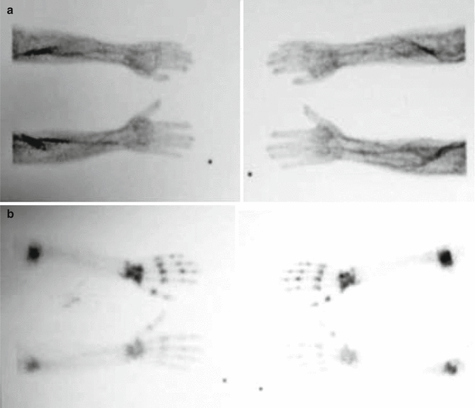
Fig. 13.9
Increased vascularity and periarticular uptake in a patient with reflex sympathetic dystrophy
In adults most of these patients referred to BS in the late phase and images show mainly increased uptake. Scintigraphy is sensitive but specificity must be increased with radiography by excluding activity due to nonspecific degenerative disease [28]. Patients with suspect of AVN should be evaluated by direct graphy, and if MRI is not available, BS with SPECT may be used. It must be remembered that BS may miss small osteonecrosis. If the clinical suspicion is high, patient must undergo MRI.
Metabolic Bone Diseases
Reflex Sympathetic Dystrophy (RSD), Complex Regional Pain Syndrome
Due to classic scintigraphic pattern, BS is diagnostic in RDS [1–3, 17]. In 0–6 months increased blood flow and increased periarticular uptake in delayed phase. Six months to 1 year perfusion turns to normal but late images are permanent (Fig. 13.10) [29]. After 1 year, late images also turn to normal. This pattern is defined for hand and not seen in foot and knees so specificity of the method decreases in extremities in late stages. Scintigraphy also can be used for the therapy response.
Fig. 13.10
Paget’s disease in skull
Paget’s Disease
Paget’s disease is mainly asymptomatic and mostly presents itself above 40 years old. As 85 % of this disease is polyostotic, BS is an ideal imaging method for whole-body screening [1, 2, 30]. It has higher sensitivity and specificity than X-ray graphy and may help in the therapy monitorization and detection of complications like sarcoma. The most commonly involved bones are the vertebral bodies (30–75 %), skull (25–65 %), pelvis (30–75 %), and proximal long bones (25–30 %) [1]. There are generally considered to be three phases: active lytic phase, mixed phase, and blastic phase; in early osteolytic active phase, BS shows high uptake that includes the whole bone, whereas in the late osteosclerotic phase, radiotracer activity decreases (Fig. 13.11). Sarcomatous degeneration occurs only rarely in pagetoid bone (about 1 % of patients). Increase in the activity of healed lesion may present a fracture or neoplastic transformation.
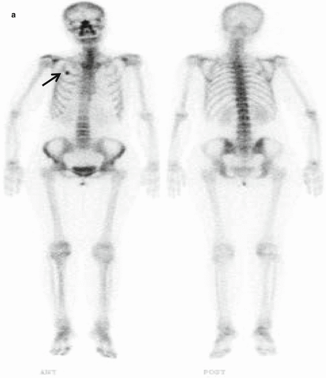
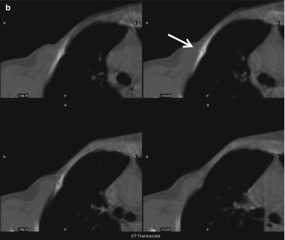
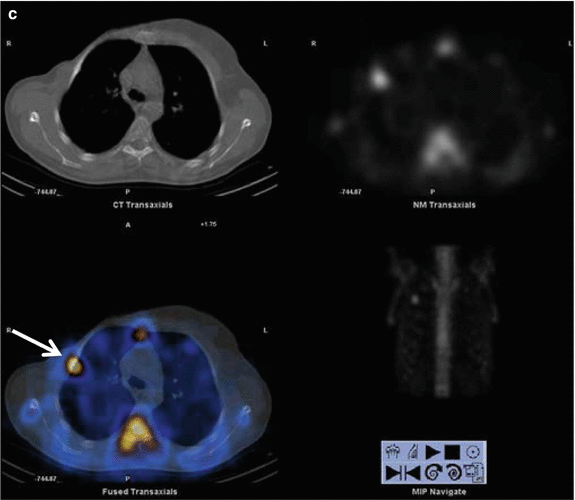
Fig. 13.11
A 40-year-old patient with breast cancer with mildly elevated tumor markers Ca 15-3: 27 U/mL (0–25). (a) Whole-body bone scintigraphy shows focal uptake in the right second rib (black arrow). (b, c) SPECT-CT images show benign fracture line that helped to exclude metastases (white arrows)
Detection of Compression Fractures and Evaluation of Back Pain
BS and MRI play a complementary role in chronic back pain in relation with patients’ age and clinical presentation. In older patients, disc pathologies are a common source of pain, so in these patients MRI is used. BS is preferred in younger patients with no localized chronic back pain. SPECT imaging is highly sensitive in spondylosis and osteoid osteoma that may cause bone pain in this patient group. For the evaluation of bone grafts and pseudoarthrosis, CT and BS are preferred for the evaluation of osteoblastic activity and fractures. Increased osteoblastic activity in the lumbar fusion area after 1 year is compatible with pseudoarthrosis. BS as with MRI could also detect sacroiliitis which may cause back pain. BS is positive especially in the early period, and radiotracer activity decreases in the late phases. Vertebral compression fracture is a common problem in elderly. Radiological images have difficulty in detection of acute fracture from the old ones. Most elderly patients will have a positive bone scan by 48 h; however, the exam may not become positive for 3–7 days in up to 5–10 % of patients. By 1 year, about 60 % of compression fractures will normalize and 95 % will become normal by 3 years. When multiple fractures are present in different stages of healing, osteoporosis is the most likely diagnosis.
Facet syndrome is a disorder related to the lumbar facet joints that can produce both local and radiating pain associated with poorly defined or absent neurological findings. Although degenerative changes of the facets are commonly seen in these patients, there is poor correlation of symptoms with X-ray graphy or CT findings. SPECT bone imaging will usually detect an abnormality (sensitivity 100 %), but the exam suffers from a lack of specificity (70 %). Nonetheless, a normal exam confirms the absence of facet syndrome, and the examination can also demonstrate other, non-facet-related lesions [2].
B-SPECT-CT in Bone Diseases
Although BS has high sensitivity, it lacks specificity. This problem is recently solved by hybrid SPECT-CT which provides morphological and functional imaging.
SPECT-BT Applications in Benign Diseases
Most common benign applications of SPECT-CT are bone infections. Addition of SPECT-CT to planar imaging helps in the identification of exact localization of radiotracer uptake and characterization. CT part of SPECT-CT identifies infection-related osteolysis and sequestration of a bone segment. It also increases specificity by identification of degenerative changes [31]. In a study by Horger et al., the sensitivity of specificity of Tc-99m MDP BS in detecting OM is found to be 78 % and 50 %, respectively. The values increase to 78 and 86 % by addition of SPECT-CT [32].
Radiolabeled leukocyte imaging is routinely used in bone infections with high sensitivity and specificity. However, planar 2D imaging and inherent lack of landmark in scintigraphic imaging create problem in localization of uptake and decrease the test’s accuracy. For the accurate localization, BS with Tc-99m MDP or anatomic imaging methods are needed for correlation. Addition of SPECT-CT increased the accuracy of radiolabeled leukocyte imaging by providing 3D imaging with fused anatomic localization at the same time.
Filippi et al. had found that SPECT-CT changed suspicious planar and SPECT interpretation of radiolabeled leukocyte imaging in 10/19 (52.6 %) areas in patients with diabetic foot. Addition of SPECT-CT ruled out OM in six patients, confirmed OM in one patient, and identified soft tissue infection in addition to bone infection in three patients [33]. The same authors in 28 patients (15 with suspicion of OM, 13 with orthopedic implant) found that SPECT-CT with Tc-99m HMPAO provided accurate anatomic localization of all positive foci and helped diagnosis in 10/28 (35.7 %) patients.
SPECT-BT helped in the differentiation of bone and soft tissue infection. It identifies structural changes after trauma. In patients with knee prosthesis, it could also identify synovial infection without prosthesis infection [34].
SPECT-CT has also additional value after spinal fixation. In a study by Damgaard et al., SPECT-CT was able to identify loosened screw in 6/9 patients suffering from pain after lumbar surgery [35]. Another application for SPECT-CT is patients suffering from pain of unclear etiology in extremities. Linke et al. evaluated contribution of SPECT-CT to three-phase BS in 34 patients evaluated as degenerative disease by planar and SPECT. SPECT-CT reclassified seven patients as having fracture and one as benign bone tumor. In 15 patients with a diagnosis of osteomyelitis, SPECT-CT reclassified 4 patient as osteoarthritis, 4 as fracture, and 1 as soft tissue infection. In eight patients with diagnosis of fracture, two evaluated as OM and two as osteoarthritis. Totally SPECT-CT changed diagnosis in 23/71 patients (p < 0.01) [36].
< div class='tao-gold-member'>
Only gold members can continue reading. Log In or Register to continue
Related posts:


Stay updated, free articles. Join our Telegram channel

Full access? Get Clinical Tree


