Fig. 44.1
Bone structures of knee joint
Tibial plateau: it consists of two parts, medial and lateral, and they have different size. The main load-bearing part is medial plateau.
Patella: It is the largest sesamoid bone of the body functioning in the extensor mechanism. Largest contact surface of patella appears when the knee is flexed 45° [2].
Intra-articular Structures
There are three main intra-articular structures. These are synovia, meniscuses, and ligaments.
Synovia provides lubrication to the knee joint. In some cases, its hypersecretion might cause effusion and pain (pigmented villonodular synovitis).
Meniscuses provide equal load distribution on the knee joint and provide shock absorption. It is divided into medial and lateral meniscus.
Medial meniscus: It has a semicircular shape, and its rear part is larger. Its middle one-third portion tightly holds MCL. Most of the injuries occur in the external rotation of tibia (Fig. 44.2).
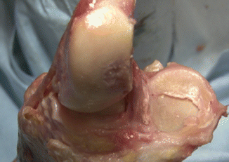
Fig. 44.2
Intra-articular structures of knee joint
Lateral meniscus: It has a circular shape compared to medial meniscus and covers a larger portion of the articular surface. It does not have connection to LCL, and therefore is more mobile. There are two ligaments which run to the medial femoral condyle through posterior horn of the lateral meniscus, and named in reference to their relations with PCL. The ligament located anterior to the PCL is called anterior meniscofemoral ligament (Humphrey ligament), while the one located posterior to PCL is called post meniscofemoral ligament (Wrisberg ligament). The injuries usually occur during knee flexion [3]. During medial and lateral meniscus examination, it is assessed through McMurray and Apley tests.
Anterior, Posterior Cruciate and Collateral Ligaments
Anterior cruciate ligament resists tibia’s displacement to the front. It is assessed by anterior drawer, Lachman, and pivot shift tests. It is composed of two bands named in reference to the place of their tibial attachment: AM (anteromedial) and PL (posterolateral). The positions of the bands are different during flexion and extension, and they cross one another during flexion [3]. AM flexion stability ensures PL extension stability. The injury there usually occurs when the tibia in internal rotation is forced to extension and when it is in hyperextension (Fig. 44.3).
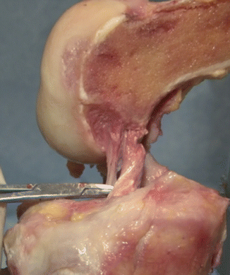
Fig. 44.3
Anterior and posterior cruciate ligaments
Posterior cruciate ligament resists tibia’s displacement to the back. It is assessed by posterior drawer test, and the symptom of posterior sag is checked. The injury there occurs following anterior tibial traumas and when the tibia in internal rotation is forced to hyperflexion.
Medial collateral ligament (MCL) has superficial and deep fibers. It resists against valgus straining and may rupture after valgus traumas. In gonarthrosis with severe varus alignment deformity, it typically loosens superficial fibers during TKR.
Lateral collateral ligament (LCL) resists against varus straining (Fig. 44.4).
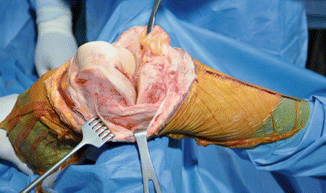
Fig. 44.4
Lateral collateral ligament
Medial collateral ligament is tighter compared to the lateral ligament; therefore, motion is less in the medial compartment.
Posteromedial and lateral capsule structures resist against rotation.
Extra-articular Structures
It consists of extra-articular muscles. Quadriceps is the strongest extensor of the knee. It is composed of four muscle groups. Extensor muscles are three times stronger than flexors.
Hamstring muscles are composed of biceps, sartorius, gracilis, and semitendinosus. Moreover, pes anserinus is composed of sartorius, gracilis, and semitendinosus.
Q Angle
It is the angle between the ASIS and the midpoint of the patella. It is approximately 12° in females while being 15° in males. Growing angles lead to anterior knee pain and patellar chondromalacia. Increased angle may develop patellar subluxation. If femoral anteversion is increased and tibial external rotation is available, then Q angle increases [4].
There are five main structures supporting the knee joint. These are as follows:
Anterior Support Complex: It is composed of quadriceps tendon, patellar tendon, medial and lateral retinacula, and infrapatellar fat pad.
Medial Support Complex: It is composed of medial head of gastrocnemius, hamstring muscles, and MCL. This resists against valgus traumas. If medial (valgus) instability exists, then
Abduction stress test is positive in flexion, and it becomes positive in PCL rupture in extension as well
ACL is typically ruptured
PCL is typically ruptured
Medial capsular ligament ruptured
MCL ligament may have ruptured
Lateral Support Complex: It is composed of lateral head of gastrocnemius, popliteus, tendon and LCL (Fig. 44.5). This structure protects against varus traumas. If it has lateral (valgus) instability, then
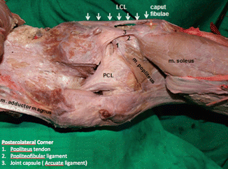
Fig. 44.5
Posterolateral corner of knee joint. Posterolateral corner: 1 popliteus tendon, 2 popliteofibular ligament, 3 joint capsule (arcuate ligament)
Abduction stress test is positive in flexion, and it becomes positive in PCL rupture in extension as well
ACL is typically ruptured
PCL is typically ruptured
Lateral capsular ligament ruptured
LCL ligament may have ruptured
Posteromedial Corner is supported by posteromedial capsule, semimembranosus tendon, and sheath. It is a significant structure in providing static stability (Fig. 44.6).
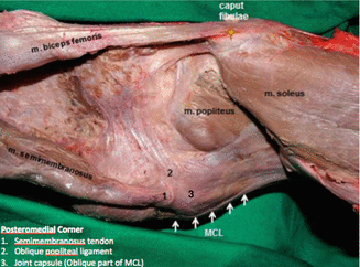
Fig. 44.6
Posteromedial corner of knee joint. Posteromedial corner: 1 semimembranosus tendon, 2 oblique popliteal ligament, 3 joint capsule (oblique part of MCL)
Posterior Support Complex is composed of oblique popliteal ligament, arcuate ligament, popliteus tendon, and gastrocnemius muscle.
Oblique ligament is the continuation of semimembranosus and provides significant stability in extension. Popliteus tendon on the other hand prevents compaction of lateral meniscus and plays a significant role in the stability of the posterolateral corner. Gastrocnemius muscle provides a dynamic contribution to the stability of the posterolateral corner.
Alignment Deformity
Genu varum → occurs when mechanical axis of lower extremity runs more than 15 mm medial to the alignment center in frontal plane
Genu valgum→ occurs when mechanical axis of lower extremity runs lateral to the alignment center in frontal plane
Genu recurvatum → occurs if the knee can be passively extended more than 5° in sagittal plane
Normal Knee Kinematics
Range of Joint Motion
The range of motion in knee joint ranges from −10° (recurvatum) to 130°. Functionally acceptable range of motion is between 0 and 90°. The rotation in knee joint varies depending on the flexion. When in full extension, there is little rotation, while in 90° flexion, 45° of external rotation and 30° of internal rotation are possible. During the motion of knee joint, displacement occurs in the medial and lateral meniscus [5].
Joint Motion
Femoral Rollback
It is the displacement/rollback to the posterior as the knee flexion increases. Between 0 and 90° of flexion motion, femorotibial contact point displaces 14 mm to the posterior.
Flexion and extension motion → It occurs between the femoral lower point and meniscus upper point.
Twisting motion → It occurs between the meniscus base and tibia [6].
Kinetics
Extension is performed by quadriceps mechanism with extension patellar system, and flexion is performed by hamstring muscles.
Knee Stabilizers
The actual stabilizers of the joint are muscles and ligaments around the knee. Anterior cruciate ligament is subjected to 170N and 500N load during normal walking and running, respectively. In a young person, it can bear maximum 1750N. Anterior cruciate can extend as far as 10–15 % at the most, beyond which tear begins to occur [4].
Joint Forces
Tibiofemoral joint
Surface forces on knee joints increases by three times of normal body weight while walking on a flat plane, and by four times when climbing up stairs. Meniscuses serve as transferring agents in load transfer. Their complete removal increases joint stress.
Patellofemoral joint
Patella helps in extension by extending support arm and providing stress distribution. It is the thickest cartilage in the body for it is the joint that bears the biggest load. While climbing down the stairs, compressive forces between patella and trochlea increase as far as two to three times of body weight. Following patellectomy, the length of the moment arm decreases as much as the thickness of the patella.
Following are the factors that improve patellar motion in TKR: external rotation of femoral component, placement of tibial and femoral component to lateral, placement of patellar component to medial, and avoiding placement of tibial component to internal rotation.
Axis of Lower Extremity
Mechanical axis: It runs from the femoral head center to the center of ankle.< div class='tao-gold-member'>Only gold members can continue reading. Log In or Register to continue
Related posts:

Stay updated, free articles. Join our Telegram channel

Full access? Get Clinical Tree


