Fig. 10.1
On multiplanar reformatted oblique coronal image (a), proximal ulnar (white arrow) and radial head fractures (black arrow) are readily seen. Three-dimensional reconstructed image in bone window (b) demonstrates the two fracture lines, prominent elbow dislocation, and the free bone fragment (arrowhead) in front of the humerus
Trauma remains the leading cause of death in people of age 45 and younger [1]. Multisystem trauma (polytrauma) is defined as injury to more than one body region such as the head, chest, abdomen, and extremities, which may be potentially fatal to the patient [2].
The introduction of MDCT led to further advantages in the setting of trauma. Scanning time was substantially reduced, and motion artifacts that were commonly observed with the slower scanners could be eliminated. Thinner slice collimation available allowed high-quality reformatted images in differently oriented planes whenever necessary [3].
In evaluating the multisystem trauma patients, time interval between the initial evaluation in the emergency room and the CT scanner becomes highly important. Dedicated trauma departments should have their own CT scanner available for the emergent multitrauma patients [4].
In the abovementioned case, after the initial evaluation and the stabilization of the patient in the emergency room, we were able to perform a multisystem CT examination in our dedicated CT scanner without any significant time loss. While lying in the CT gantry, owing to multitrauma and massive pain at his left elbow, a routine positioning to the patient could not be given. Nevertheless, although the patient was not in the proper position on the table, we were able to perform the CT examination and acquire qualified images without necessity of removing the splint.
2.
Is it possible to perform initial and control musculoskeletal CT examinations with reduced doses by using dose modulation strategies?
Since its introduction in the 1970s, besides in the trauma setting, CT has played a major role in the diagnosis of many musculoskeletal diseases. Even though image quality is altered by metallic artifacts, CT also found indications in postsurgical imaging [5]. However, CT is a technique working on the principle of X-ray radiation that is responsible for the majority of medical irradiations [6].
In the last few years, MDCT has benefited from technological innovations, thereby considerably reducing the dose delivered to the patient. With these technological innovations and better control of the image acquisition parameters, it is now possible to obtain CT images in lower doses [7].
There are some rules of thumb to reduce the radiation from CT in the evaluation of the diseases of the musculoskeletal system such as (1) comply with the diagnostic indications; (2) where possible, see if a non-radiating imaging method (i.e., ultrasound or MR imaging) can help to solve the clinical problem in question; (3) limit the CT coverage and number of acquisition phases during dynamic and perfusion examinations; (4) adopt the kilovoltage and milliampere according to the indications and morphology of each case; (5) reduce the kilovoltage during the exploration of the peripheral joints and arthro-CT; and (6) choose modern methods to reduce the dose (i.e., iterative reconstructions, automatic modulation of the milliamps, etc.) [7].
While evaluating the patients especially in pediatric age who are planned to perform initial or reexamination (postoperative or control) of CT scan, basic dose reduction strategies should always be in the clinicians’ minds. Without sacrificing the image quality requisite for a proper diagnosis, one should care ALARA principle which describes the diagnostic X-ray utilization as “As Low As Reasonably Achievable” [8].
3.
Compared to the standard axial images, are multiplanar reformatted and/or three-dimensional –reconstructed images more beneficial for orthopedists in guiding preoperative session?
Clinical applications of CT have increased extensively over the past decade and continue to widen. The introduction of three-dimensional (3D) reconstruction in CT technology has revolutionized medical imaging. With the anatomy displayed which the orthopedist may get more familiar with, this technique may have advantages in the preoperative planning of craniofacial surgery, assessment of complex fractures, and surgical treatment of dysplastic hips in children [9].
Three-dimensional CT imaging studies have come to stay, and with continued improvement in CT technology, it has now become an integral part of imaging studies of the musculoskeletal system [9]. Nevertheless, axial images should not be neglected, and these and three-dimensional images should be evaluated hand in hand.
The combination of axial, multiplanar reformatted CT and 3D reconstruction with volume rendering allows rapid and detailed examination of the musculoskeletal system. In a substantial number of cases, management may be altered with the findings seen better on the 3D images such as subtle fractures, complex injuries extending to joint spaces, and cases with metal hardware otherwise causing metallic streak artifacts and hence masking the pathologic conditions nearby. By displaying complex spatial information, 3D images are useful for conveying complicated anatomic information to orthopedists [10].
4.
Ultrasound or CT–guided biopsy in musculoskeletal mass lesions: which one should be selected?
Soft tissue masses and bone lesions may require biopsy. The approach in performing these biopsies, in some ways, is not different from other nonmusculoskeletal biopsies and requires careful review of all the relevant clinical and radiological information [11].
The majority of musculoskeletal lesion biopsies are performed to determine whether there is an underlying neoplasm. In some cases where patients are unresponsive to conventional antibiotic therapy for treatment of bone or soft tissue infections, a biopsy may be performed to isolate the proper organism and direct a targeted antimicrobial therapy [11].
In the above terms, ultrasound is a commonly used modality for performing musculoskeletal soft tissue biopsies [12]. It can also be used for lesions near a bone surface or for a bone lesion with an extraosseous component [13]. Ultrasound is widely available, is minimally invasive, and offers real-time imaging. Its high spatial and contrast resolution also allows easier visualization of lesions, particularly small lesions that are difficult to visualize on noncontrast CT. Although CT or MR imaging can be used, the dynamic nature of the ultrasound allows easier and more comfortable patient and operator positioning. Ultrasound allowing real-time visualization of the needle provides a safe and satisfactory biopsy by avoiding nearby vital structures [14]. Furthermore, ultrasound-guided biopsies are free from ionizing radiation.
Computerized tomography-guided biopsies may be used for some bone lesions and soft tissue neoplasms. Computerized tomography, via cross-sectional images, displays excellent spatial localization of the lesion which is needed to determine the route that is safe, avoiding vital neurovascular structures nearby [15]. In general, avoiding crossing of more than one anatomic compartment is important in preserving a limb-salvage surgical plan; otherwise, the needle track is at risk of seeding tumor cells to multiple compartments [16].
Introduction
In the last two decades, computed tomography (CT) has brought a new insight into the radiography and magnetic resonance (MR) imaging-focused musculoskeletal examinations [17]. The advancements in the technology and the software systems have great impact on the increasing role of CT in musculoskeletal imaging. The importance of CT has increased further with the emergence of multidetector CT (MDCT) systems. It is now possible to get high-resolution images after a short scanning time with the isotropic spatial information that these systems can obtain. Decreased scanning time has allowed minimizing the problems that may arise secondary to the patient motion during imaging [18]. High-quality multiplanar reformatted (MPR) and three-dimensional (3D) images created at the separate workstations can easily be captured by the clinician compared with one single-plane image.
The picture archiving and communications systems (PACS) enable the postprocessing with interactive viewing and reformatting, using isotropic or near isotropic data sets that MDCT yields. These images can be permanently archived in PACS and be called any time when the comparison is needed in follow-ups [18].
While MRI is superior to CT in detecting and defining soft issue and bone marrow abnormalities given its inherited high-contrast resolution feature, MDCT is essential in several situations [5]. In trauma settings especially imaging complex skeletal injuries, fracture lines and their extensions are superbly defined by MDCT [19, 20]. Unlike MRI, for the evaluation of bone and soft tissue masses, CT can display and characterize mineralization, cortical disruption, and periosteal reaction more readily [20]. In postoperative evaluation of patients with metal armamentarium, MDCT allows imaging without generating significant metallic artifact [20]. The absolute contraindications such as cardiac pacemakers and electronic stimulators described for MR imaging are not applicable for CT, where this allows the technique a possible alternative to MR imaging in those settings.
In this paper, the utility of CT in imaging the musculoskeletal system will be provided by touching the technical aspects of MDCT.
MDCT Technique: Basics
The launch of MDCT into imaging stage in 1998 has opened a new page in radiological examinations. With the very fast image acquisition phase that MDCT can yield, total body scanning times have been reduced below 30 s [21]. Scanning speed is favorable especially when scanning large segments of the body or performing a dynamic imaging such as perfusion or CT angiography where image acquisition has to be fast [21].
Contrary to previous CT systems which have single detector, the X-rays pass through the patient body part to reach multiple channels with MDCT. The MDCT is called with the number of detectors it has, for example, if the detector row has four channels carrying information concurrently in each X-ray gantry rotation, the scanner is then called a four channel [21]. This means in each gantry rotation with X-rays passing through the relevant body part, more than one datum will be obtained, i.e., four in the previous example. Joined with the faster X-ray rotation speeds, CT data now can be collected faster and so the patient can be imaged rapidly than conventional single-slice helical CT [21, 22].
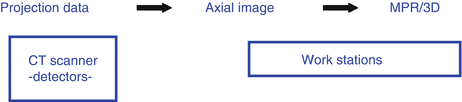
Projection data (PD) described by Dalrymple et al. [23] are the initial raw data collected by the CT detectors. Without any mathematical conversion in computerized media, these data from the body part cannot be viewed directly or used to create 3D images. Projection data are first used to generate axial images on the CT scanner. The spatial properties of PD are determined by the scan settings by the technologist or radiologist on the console, and once the data is obtained, it cannot be altered later. As long as the projection data are kept on the CT scanner and not deleted, (1) the thickness of the axial images (either thin or thicker slices), (2) the body part of interest (field of view, FOV), and (3) the reconstruction algorithms (i.e., bone or soft tissue) can be generated [18].
The flow of the information from the detectors can be picturized as shown below:
Isotropic Imaging
Thin sections are preferable for imaging musculoskeletal system in order to create multiplanar reformatted (MPR) and 3D images with superb spatial resolution [21, 24]. Isotropic imaging obtained with thin slices (i.e., 0.5 mm slice thickness) will yield MPR and 3D images with high spatial resolution and fine detail without possible artifacts such as “stairstep” and blurring effects [24]. Performing CT with thinner slices warrants creation of images in any plane with high spatial resolution. This issue particularly comes into account when imaging traumatized patient comfortably in any position she/he can bear, without giving a specific position in order to image the injured body part [24]. Complex joints such as the wrist and foot can be examined in any plane with multiplanar reformatted images without any sacrifice in the image detail, as a consequence of isotropic imaging with thinner slices [24]. Thinnest slice section issue has its own drawbacks, however, in terms of the noise of the image and the radiation dose for the patient, which will be discussed below.
Multiplanar Reformatting: MPR
Images at any plane can be reconstructed by the doctor himself on the workstations by interactive means, which by this way one can observe any point of interest in each plane. In order not to miss subtle fractures, the MPR images should be viewed as thin as possible to the point of apparent noise level which produces somewhat granulation on the image. When images become noisy, this problem may be reduced by fusing thin slices into much thicker images [18].
Three-Dimensional (3D) Imaging Methods (Surface Shading/Volume Rendering)
With the aid of improved software systems and powerful computer-based workstations, 3D imaging is now possible with isotropic volumetric data set. Three-dimensional images can be colored as well, and clinicians become more familiar with and easily perceive the anatomy like the one he expects on the operating table. Though 3D images may facilitate the orientation to the anatomy, axial and MPR images should not be neglected, and 3D imaging should stay as an adjunct [18]. Three-dimensional images readily show the extent of tendons and muscles and their relationship to bones and joints when injured (Fig. 10.2). Three-dimensional images are also subject to less metal artifact when imaging those with metal armamentarium in their postoperative stage [22, 25, 26].
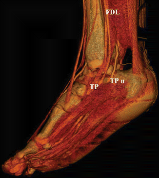
Fig. 10.2
It is possible to display the courses of the tendons, and their relationships with the bones as in the cadavers on 3D volume-rendered (VR) images by using advanced computer systems and softwares. TP tibialis posterior tendon, FDL flexor digitorum tendon, TPn tibialis posterior nerve
Radiation Dose
Multidetector CT being a high-dose imaging technique is well known [18]. The continuous recommendation for using the smallest slice thickness available to obtain isotropic data sets brings the concern for radiation dose to the patient. Especially when cervical vertebrae, facial bones and pelvis are examined with this technique, thyroid gland, lens and internal genitals which are sensitive to radiation, are subject to high doses during imaging. For this reason, the patient should be reevaluated for the indications of imaging the cervical vertebrae, skull, and pelvic region, and MDCT should not be used regularly for negligible trauma involving these regions [18].
The intensity of diagnostic X-rays from CT gantry can be reduced by decreasing the kilovolt and milliampere values when imaging the peripheral skeleton especially the forearm and leg since these structures are thin [18]. In the pediatric patient, radiation dose deserves much more importance; hence, it is wise to adhere the scanning parameters indicated in the literature for pediatric population [18]. Computerized tomography manufacturers also give hand to this problem by introducing new measures for reducing patient dose such as automatic tube current modulations which adjust the intensity of X-rays based on the thickness of the anatomy being scanned. Nevertheless, MDCT should be handled as a high-dose technique, and the benefit-to-risk ratio to the patient with this examination should be thoroughly evaluated.
General Approach to the Musculoskeletal Imaging
Oblique Image Acquisition Principle
When isotropic imaging is not possible due to the limitations of the scanner or thick slices (>0.5 mm) have been selected instead of thinner slices, the quality of the following MPR images can be sustained by “obliquity principle” [24]. In order to visualize much of the joint surface, it is recommended that the affected extremity be placed in the gantry according to the following rules: (1) position the extremity obliquely (45° is optimal) and (2) in the center of the gantry (resolution is maximized in the center) [24].
Extended Anatomic Coverage
In order to assess the extension of a lesion and the anatomic regions it affects, a sufficient portion in the neighborhood should be incorporated into the area of interest (Fig. 10.3).
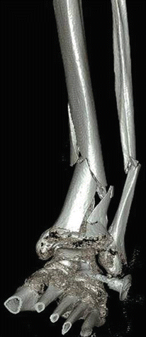


Fig. 10.3
Three-dimensional volume-rendered (VR) image with a large field of view that is acquired for demonstrating the fracture extension in a case with fragmented fracture at the tibia and fibula, with medial angulation of the long bones
< div class='tao-gold-member'>
Only gold members can continue reading. Log In or Register to continue
Related posts:

Stay updated, free articles. Join our Telegram channel
Full access? Get Clinical Tree
