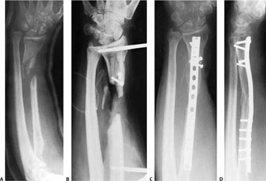
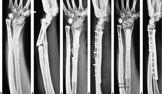
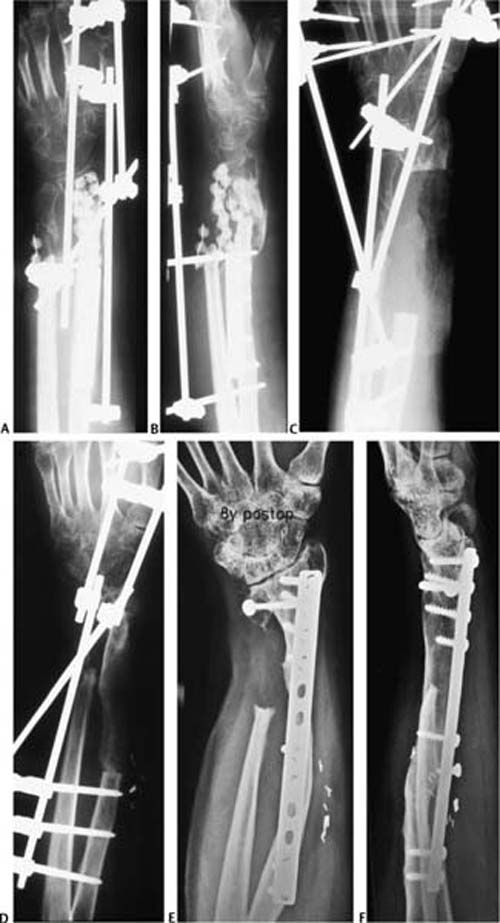
18 Nonunions, Malunions, and Synostosis of Forearm Fractures The forearm should be considered as a joint consisting of two long bones and three ligamentous restraints: the annular ligament complex, the interosseous membrane (IOM), and the triangular fibrocartilage complex (TFCC). There are two articular components as well: the proximal (PRUJs) and distal radioulnar joints (DRUJs), which permit rotation of the radius on a relatively fixed ulnar axis. If the forearm is now looked upon as a joint, diaphyseal fractures should be considered intraarticular and therefore deserve – as in any other fracture that disrupts an articular surface – accurate anatomic reduction to guarantee full restoration of function. This same principle should be taken into consideration for the surgical reconstruction of nonunited and malunited forearm fractures. Although open reduction and compression plate fixation of forearm fractures invariably restores anatomy and function with a relatively low rate of complications,1–5 surgical reconstruction of forearm nonunions and malunions represents a more difficult challenge in which despite achieving bony union, correcting deformity, and relieving pain, complete and symmetrical restoration of forearm rotation is difficult to obtain, but may be certainly improved to a reasonable functional arc of pronation and supination. This is due to the frequent concomitant derangement of the PRUJS and DRUJs as well as the IOM commonly associated with the bony deformity of the nonunited or malunited forearm bones. In both scenarios, symmetric shortening of both bones may not alter the congruity of the PRUJ or the DRUJ, provided there is no significant associated angular deformity. Conversely, shortening of a single forearm bone with or without angular deformity will automatically affect the articular anatomic relationships of either the PRUJ or DRUJ. Loss of the physiological bow of the radius is responsible for limited pronation, whereas reduction of the interosseous space associated with angular or ad latus (translation) deformity leads to secondary contracture of the IOM and decreases forearm rotation. Posttraumatic radioulnar synostosis is a less frequent complication, but its management, although currently well standardized, does not exclude recurrence in patients with special risk factors. In this chapter, we describe the current principles of management of both simple and complex diaphyseal forearm nonunions and malunions in adults, and present treatment recommendations for both primary and recurrent radioulnar synostosis. Because cast treatment of displaced forearm fractures in adults is practically “past history,” we are commonly confronted with nonunions following insufficient surgical stabilization with implants associated with extensive devascularization, infection, or extensive bone loss after high-energy injuries and open comminuted fractures. Occasionally, a well-vascularized nonunion may develop after a conservative trial of a minimally displaced forearm bone. These are treated with minimal decortication of the callus to provide a flat surface for the compression plate, and successful outcome is to be expected, provided that the construct is biomechanically sound. The types of presentation of a forearm nonunion include the following combinations: 1. Radius or ulna alone without radioulnar joint disruption 2. Radius or ulna alone with associated radioulnar joint disruption 3. Nonunion of one forearm bone with malunion of the other 4. Both bone nonunions Each of these may be further subgrouped according to Weber and Çech’s classification6 into 1. Vital or well-vascularized a. hypertrophic b. oligotrophic 2. Nonvital or avascular c. necrotic fragment d. comminuted necrotic area e. large defect f. atrophic The goals of treatment must extend beyond obtaining bony union to ensure anatomic restoration of skeletal alignment and both congruity and stability of the PRUJs and DRUJs. Preoperative assessment should include a careful clinical examination of the whole upper extremity to document the residual forearm function and that of the neighboring joints, the soft tissue condition, and the neurovascular status. Laboratory studies including an electromyogram for nerve lesions or aspiration and cultures in infected cases are routinely performed. For the radiographic assessment, comparative x-rays of both forearms including the elbow and wrist joints are considered essential for the preoperative planning and correction of deformity or shortening associated with the nonunion. Most of the simple, straightforward cases do not need additional imaging. Computed tomography (CT) may be helpful to rule out the presence of sequestrae in infected cases, and assess rotatory deformity and congruity of the DRUJ. Magnetic resonance imaging is reserved to evaluate additional soft tissue lesions such as the TFCC, the IOM, and muscle defects. Arteriograms are useful to document the patency of the vascular axes in nonunions following severe high-energy trauma, particularly when planning microvascular reconstruction of large bony defects. Disregarding the localization of the forearm nonunion, hypertrophic well-vascularized nonunions are managed with decortication of the callus and stable plate fixation, whereas atrophic nonunions need rigid fixation and autologous bone grafting. These principles of nonunion management advocated by Müller7 and Weber and Çech6 over 40 years ago have stood the test of time and are associated with a high success rate. Small diaphyseal defects may be bridged with a structural corticocancellous iliac graft, fixed to the plate. In larger diaphyseal bone defects up to 10 cm, in which a circumferential well-vascularized soft tissue bed is present, long bridging plates and morcellized iliac bone grafts are the procedure of choice because diaphyseal stability and skeletal continuity, length, and alignment are readily achieved along with rapid functional recovery.8 Vascularized bone grafts are reserved for those situations of large defects in which conventional bone grafting procedures have failed and in those cases with poor vascularized bed and massive scarring as in chronically infected cases or in situations with composite soft tissue and skeletal defects following mutilating injuries.9,10 Creation of a one-bone forearm for the treatment of large diaphyseal defects (most commonly in the ulna) is a valid alternative,11–13 but skeletal continuity is restored at the expense of loss of forearm rotation. For this reason this modality remains, in our view, the ultimate salvage procedure. Other valid alternatives of nonunion management such as electrical stimulation,14,15 the use of bone morphogenic protein,16,17 or the use of the Ilizarov technique18 will not be discussed because we do not routinely use these options. Our preferred surgical exposure for the radius is the extensile approach described by Henry,19 in which the whole bone from the radial head to the wrist joint can be exposed medially to the brachioradialis on the volar aspect of the forearm. In the proximal third the major advantage is that both the deep and superficial branches of the radial nerve can be protected as the supinator muscle is detached from the proximal shaft. Extension of the incision proximally to the elbow flexion crease further permits exposure of the anterior capsule of the elbow joint, particularly the lateral compartment and the PRUJ. In the midshaft area the insertion of the pronator teres is well visualized, and the insertion of the central part of the IOM can be exposed at this level. In the distal point the radius may be widely exposed partially elevating the insertion of the flexor pollicis longus muscle and further distally the pronator quadratus. The whole of the ulna is exposed with a longitudinal incision over its subcutaneous border between the extensor and flexor carpi ulnaris (FCU) muscles. If exposure of the DRUJ is needed, the incision may be extended distally by swinging it dorsally over the ulnar head at the neck level ending at the junction of the fourth and fifth carpometacarpal joints. In this manner the superficial branch of the ulnar nerve remains safely in the medial subcutaneous flap. The PRUJ is exposed through a posterolateral approach between the anconeus and extensor carpi ulnaris. (ECU) If scars of previous surgery are present, usually these are utilized to avoid additional soft tissue disruption and iatrogenic devascularization. Hypertrophic nonunions require minimal exposure of the nonunion site to preserve the well-vascularized a callus on both fragments. Limited decortication at the surface of plate application is all that is needed. Because these nonunions are generally elastic, the associated deformity is usually corrected by carefully molding the plate to the normal anatomy of the shaft segment. The plate is securely fixed to the proximal fragment and deformity correction is gradually obtained with strategic use of the compression device and bone clamps applied to the distal fragment. This works very nicely when the plate comes to lie on the convexity of the angulation, acting as a tension band implant. If the convexity of the angulation faces the opposite bone reducing the interosseous space, associated IOM release is mandatory. The prebent plate is then fixed in a bridging mode to which one screw is inserted in the most proximal and one in the most distal plate hole. Thereafter using Verbrugge bone clamps the ununited bone ends are approximated to the plate, thus recreating the interosseous space.6 Following reduction, axial compression can be achieved with eccentric introduction of screws of a dynamic compression (DC) plate. Molding of the plate with the appropriate curvature is particularly important in the midshaft area of the radius, whereas less plate bending is needed in the proximal and distal thirds. For the ulna, being practically a straight long bone, a slight bend at the nonunion site is helpful to obtain compression at the opposite cortex as in acute fracture fixation. Figure 18–1 (A,B) Large defect nonunion of the distal third of the radius. Notice distal radioulnar joint disruptions, severe shortening, and radial deviation of the distal fragment. Middle: Intraoperative roentgenograms with the distractor in place. Notice realignment of the distal radioulnar joint. (C,D) Radiographs at 12 months following volar bridge plating and autologous morcellized iliac crest cancellous bone grafting. Notice cortical remodeling of the interposed graft. Atrophic nonunions require a more careful preparation of the site including resection of necrotic areas to bleeding bone, as well as devitalized intermediate fragments. The obliterated medullary canal on both ends is routinely opened with an awl or a drill bit that accommodates to its diameter. Although relatively small defects created after débridement of a bone forearm nonunion may be stabilized with plates creating a symmetric skeletal shortening, whenever possible we prefer to maintain length using bridging plates and interpositional bone grafting. This is particularly important in the treatment of large defects of one bone, while the opposite bone is intact. If the radius presents with a pseudoarthrotic defect, there is severe radial deviation and shortening of the distal fragment, as well as disruption of the DRUJ with a positive ulnar variance, which equals the amount of radial shortening (Fig. 18–1). A pronatory rotational deformity is frequently present as well as palmar displacement of the distal fragment. Because these chronic nonunions are invariably associated with soft tissue contracture, restoration of length and realignment of the DRUJ can be obtained with a combination of soft tissue release and intraoperative temporary distraction. Soft tissue release includes resection of scarred nonviable tissue surrounding the nonunion area, detachment and partial resection of the contracted IOM, and subperiosteal detachment of the brachioradialis tendon from the distal radius. If severe contracture of the DRUJ is present, release of the pronator quadratus and re-section of the volar DRUJ capsule are recommended. Intraoperative progressive distraction of the nonunion is achieved with the use of the AO femoral distractor,20 placing one 4.5-mm Schanz pin into each radial fragment. Distraction should be performed slowly over a period of 30 minutes (Fig. 18–1). Visual control of the median nerve to avoid excessive sudden tension is important, although one stage restoration of posttraumatic skeletal shortening up to 2.5 to 3 cm is usually not associated with neurapraxia. However, this may occur if the peripheral nerve is tethered and adherent to scarred tissue. Therefore, inspection of the nerve and neurolysis should be performed accordingly. Realignment of the DRUJ is assessed with fluoroscopy, and passive forearm rotation is controlled at this point. If reduction of the DRUJ is not obtained despite distraction, a shortening osteotomy of the ulna may be performed simultaneously or delayed to a later date. If severe incongruity of the joint is present with secondary degenerative changes, prosthetic replacement is advocated. We no longer perform distal ulna resections because loss of the ulnar head invariably leads to radioulnar convergence and painful instability of the ulnar stump.21 Figure 18–2 (A,B) Oligotrophic nonunion of the radial shaft following insufficient internal fixation of an Galeazzi fracture and early removal of the plate. Notice severe palmar angulation, shortening, and complete disruption of the distal radioulnar joint. (C,D) Following débridement of the necrotic bone ends, the nonunion was realigned and stabilized with a 3.5 dynamic compression plate; the 3-cm defect was filled with a corticocancellous structural iliac graft fixed to the plate with one screw. (E,F) Radiographs 6 years after the operation. Notice full remodeling of the graft and restoration of the interosseous space. The patient regained full pronation and supination of her left forearm. While the distractor maintains alignment, the premolded plate is applied in a bridging fashion. Although most forearm nonunions are stabilized with 3.5 limited-contact dynamic compression (LCDC) plates with a minimum of 6 to 8 cortices for screw fixation on each fragment, the 4.5 narrow DC plate may be recommended as a stronger implant to bridge large defects. Locking plates with angular stability may be also used, especially in osteoporotic bone. Augmentation of screw holding power with bone cement is still a valid alternative to increase stability of plate fixation in such situations. The defect is then grafted with morcellized autologous bone grafts, taking care not to place them close to the IOM. If a soft tissue defect is present in this area, Gelfoam (Pfizer, New York, NY) is applied as a barrier to obliterate the dead space to reduce the danger of radioulnar synostosis. For smaller defects up to 3 cm, a corticocancellous strut graft may be interposed and fixed with screws to the plate (Fig. 18–2). The cortical border of the graft is placed opposite to the plate surface while the cancellous surface lies on the plate.22 Morcellized cancellous grafts are additionally placed in the proximal and distal junctions of the construct. Restoration of length in smaller defects is achieved by fixing the plate with screws to the distal fragment and applying the articulating tension device in distraction mode on the proximal end of the plate. This device permits a maximal excursion of 40 mm. Alternatively, for shorter distances and absence of soft tissue contractures a laminar spreader applied between the plate and a separate cortical screw can be used.23,24 A less common scenario is the nonunited ulna with a large defect and an intact radius. According to the shortening and angulation, a concomitant dislocation of the radial head may be present. Similar tactical steps as described for the radius are recommended. In longstanding cases, an open reduction of the radial head and annular ligament reconstruction may become necessary. If degenerative cartilage changes are present, prosthetic replacement of the radial head is preferred to resection, to obviate late proximal radius migration and secondary disruption of the DRUJ. If the nonunion presents with a draining sinus and active infection, an aggressive débridement; removal of implants, external fixation, temporary irrigation-suction; and a prolonged course of parenteral antibiotics are performed as the first stage of the treatment. Sequential débridements and repeat cultures may become necessary in cases where the inflammatory signs persist. Definitive reconstruction as described for the atrophic nonunion with large defects is indicated as soon as both the clinical and laboratory parameters of active infection have normalized. It must be kept in mind, that revascularization of free morcellized cancellous graft is directly dependent on the vascularity of the soft tissue envelope. If the soft tissues surrounding a chronic infected nonunion reveal massive scarring, are devitalized, or are associated with loss of muscle substance microvascular free tissue transfer is advisable (Fig. 18–3). The most commonly used graft is an osteocutaneous fibular graft pedicled on the peroneal vessels.25,26 The composite graft is placed into the defect having the peroneal vessels in an appropriate position for an optimal anastomosis to the radial or ulnar recipient vessels according to the particular scenario. To minimize instability and prevent delayed union, the graft junctions are stabilized to the recipient bone ends with plate or screw fixation or a combination of both. The cutaneous portion of the graft is sutured into the overlying soft tissue defect. Except in those cases where maximal implant stability cannot be achieved such as in patients with severe osteoporosis or in the free fibular grafts in which additional initial cast or removable splints are indicated, functional after-treatment with early range of motion (ROM) exercises of the forearm and neighboring joints are permitted. Forceful passive physiotherapy measures or dynamic splints to restore pronation and supination should be used with caution in the first 3 months following surgery. Thereafter, strengthening exercises and progressive loading is allowed as soon as radiographic signs of bony healing and incorporation of the grafts are present. The results reported in recent articles in which the above-mentioned techniques were used are highly satisfactory. Barbieri et al22 obtained union in 10 out of 12 cases treated with structural corticocancellous grafts in an average of 4 months. Ring and coworkers8 obtained complete union in 35 patients with an average defect of 2.2 cm (range: 1 to 6 cm) in an average time of 6 months. Jupiter et al27 reported the use of vascularized fibular grafts for segmental defects of the radius averaging 7.9 cm. Eight out of 9 patients treated (6 of whom had chronic osseous infection) presented radiographic union at both the proximal and distal junctions of the graft. The functional results were satisfactory and 6 out of 9 patients had returned to their preinjury occupation. Safoury28 recently reported 18 infected nonunions and segmental defects of the forearm treated with vascularized fibular grafts. In this series, all nonunions healed with resolution of infection in an average time of 4 months. Although the overall reported rate of complications in the articles reviewed is relatively low, the most common problems requiring subsequent surgery were the DRUJ (Darrach resections), failure to obtain union, persistence or reactivation of infection, and very seldom creation of an “iatrogenic” radioulnar synostosis. The functional outcome is undoubtedly multifactorial. Better results with a balanced arc of functional forearm rotation are usually observed in those patients with anatomic realignment of the forearm bones and radioulnar congruity. If the soft tissue and muscle envelope is contracted and scarred or suffered segmental loss as in sequelae of high-energy trauma or chronic infection, function will be limited despite restoration of skeletal stability. Affectation of the neighboring joints with residual stiffness will also influence the functional outcome. Revascularization of autologous cancellous or corticocancellous bone grafts require a well-vascularized soft tissue envelope along with environmental stability provided by rigid plate fixation. Although revascularization and incorporation of morcellized grafts is faster, remodeling into a “cortical-like” structure takes a longer time. However, successful long-term results have been recently reported using this technique for atrophic nonunions of the femur29,30 and humerus.31 The advantage of the use of “pressure resistant corticocancellous bone blocks,” as suggested by Weber and Çech,6 is the immediate reconstruction of the loading capacity of the cortical part of the graft opposite to the plate. If corticocancellous blocks are used, screw fixation to the plate is mandatory to reduce micromotion to a minimum and enhance undisturbed revascularization. His recommendation for this grafting modality was, however, for defects of not more than 3 cm. Figure 18–3 (A,B) Radiographs of an infected nonunion of the distal third of the radius following an open complex comminuted fracture. The radiographs demonstrate the presence of PMMA gentamicin beads and a diaphyseal radius plate after repeated débridements for a period of 10 months. Notice massive disuse atrophy of the carpus. (C,D) Following radical débridement and partial diaphysectomy, the defect was bridged with an iliac crest vascularized bone graft pedicled on the deep iliac circumflex artery and veins. Definite volar plate fixation through a Henry approach and a Sauvé–Kapandji procedure (undertaken because of the segmental loss of the distal third of the ulna) were performed 4 weeks after the microvascular reconstruction. At the same time the external fixator was removed. (E,F) Radiographs 8 years following reconstruction reveal a solid remodeling of the interposed vascularized graft. The patient regained a functional but limited arc of forearm rotation, adequate strength. There was no recurrence of the infection. Diaphyseal malunion in adults results after insufficient reduction following conservative or operative treatment, or may present as an iatrogenic deformity after attempted osteotomies. The deformity may include one or both bones of the forearm. Treatment goals in the care of forearm fractures are bone healing and restoration of function. Bone union of the forearm shaft in a nonanatomic position impairs motion of the wrist, especially the rotation of the forearm. This may present as restriction of motion, clicking, pain, and giving-way during forearm rotation (instability), or as a cosmetic problem. All of these complications can be avoided by correcting all components of the deformity: discrepancies in length, angulation, and rotation. Complex three-dimensional (3D) features of the bony architecture such as the bowing of the radius are equally important, thus its reconstruction must be considered as well. This is easily accomplished during initial fracture treatment, but once the bone has healed in malalignment, the chances for spontaneous correction are only realistic in early childhood. In children older than 10 years and adults, only a corrective osteotomy can reestablish normal bony architecture of the forearm bones. This together with the appropriate soft tissue release will reduce restriction of forearm rotation; with congruency of the radioulnar joints the stability will be restored as well. For the planning of the surgical correction the opposite healthy forearm is used, as there are no generally valid normative values. Additionally, 3D templates may be utilized. Our clinical experience with this preoperative assessment has been positive. The immature skeleton has a considerable potential to spontaneously correct residual deformities, that is, angulations after fractures.32–35 The amount of correction is defined by multiple factors as, for example, the distance between fracture and physis/growth plate, residual growth time until maturity, and, of course, the amount of angulation.32,36,37 Deformities in the vicinity of highly active growth plates have a better prognosis, as their growth arrest will arrive substantially later in comparison to less-active physical plates.38–42 Consequently, the lowest corrective potential is in the diaphysis. Several authors43–45 have observed residual deformity following diaphyseal fractures in children. After the age of 10, a complete correction of diaphyseal malunions during growth could not be observed either in boys or in girls.43 It was concluded that only an angulation of up to 10 degrees, ad latus deformity of 100%, and rotational malalignment up to 45 degrees should be accepted.44,45 A separate entity is the plastic deformation of the forearm.46
Diaphyseal Forearm Nonunion
Classification
Treatment
Surgical Treatment
General Principles
Tactics
Approaches
Preparation, Reduction, Fixation, and Bone Grafting
Infected Nonunions
Postoperative Rehabilitation
Results and Complications
Malunion of the Forearm Bones
Potential for Spontaneous Correction during Growth
Related posts:
 Open Reduction and Internal Fixation of Proximal Humeral Fractures Using Locking Plates
Treatment of Glenoid Fractures and Injuries to the Superior Shoulder Suspensory Complex
Open Reduction and Internal Fixation for Fractures about the Elbow in the Elderly
Acute Fracture-Dislocations about the Elbow
Nonunions of the Distal Humerus
Total Elbow Arthroplasty for Distal Humeral Fractures
Open Reduction and Internal Fixation of Proximal Humeral Fractures Using Locking Plates
Treatment of Glenoid Fractures and Injuries to the Superior Shoulder Suspensory Complex
Open Reduction and Internal Fixation for Fractures about the Elbow in the Elderly
Acute Fracture-Dislocations about the Elbow
Nonunions of the Distal Humerus
Total Elbow Arthroplasty for Distal Humeral Fractures
![]()
Stay updated, free articles. Join our Telegram channel

Full access? Get Clinical Tree


