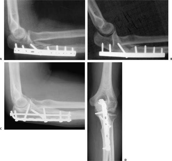
17 Nonunions and Malunions of Monteggia Fracture Dislocations Monteggia originally described a fracture of the ulna with dislocation of the proximal radioulnar joint.1 His name is now applied to a variety of injuries that fit this definition. Bado placed numbers on the variations that had long been recognized: apex anterior fracture of the ulna with anterior dislocation of the radial head (type I), apex posterior fracture of the ulna with posterior dislocation of the radial head (type II), apex lateral fracture of the ulna with lateral dislocation of the radial head (type III), and diaphyseal fractures of both the radius and the ulna with proximal radioulnar dislocation (type IV).1 Bado also introduced many “Monteggia variants,” which are not very useful because they are not truly forearm fracture dislocations as the Monteggia originally defined them.1 The posterior Monteggia lesion has been the focus of some dispute in this regard because the fractures that are close to the ulnohumeral joint are associated with relatively limited displacement of the proximal radioulnar joint.3 Jupiter and colleagues have emphasized that posterior Monteggia lesions occur on a spectrum from the diaphysis to intraarticular fractures.4 Although the more proximal fractures have less obvious proximal radioulnar dislocation, the injuries have so many factors in common that it is useful to consider them together. This holds true for the consideration of malunion and nonunion as well.5 Malunions and nonunions of Monteggia injuries in adults (often older women) occur mainly with posterior (Bado type II) Monteggia lesions that have inadequate plate fixation leading to collapse of the fracture and either malunion or nonunion.5 In many cases, a plate has been applied to the medial or lateral surface of the ulna and only two or three screws have been placed in the proximal metaphyseal fragment (Fig. 17–1A).5,6 Many of the patients are older women with osteoporosis. The proximal screws loosen resulting in recurrence of the apex posterior fracture deformity and posterior dislocation of the radial head from the radiocapitellar and proximal radioulnar joints (Fig. 17–1B). Patients often complain of pain and lack of supination. Associated factors that can challenge the treating physician are compromised soft tissues, osteopenia, failed hardware, and an already compromised ulnar nerve. The preoperative examination and documentation should include a detailed neurovascular examination, location of previous incision (s), and range of motion. The elbow should be scrutinized for signs of infection. Standard radiographs in two planes provide generally enough data for preoperative planning. In cases of associated instability and/or suspicion of a coronoid fracture, a preoperative computed tomography (CT) scan is helpful (despite often-present hardware). The type of nonunion should be noted, whether oligotrophic, atrophic, or hypertrophic. Other than routine preoperative laboratory studies, no specific blood tests are needed. In case of (suspected) infection, a complete blood count including differential, an erythrocyte sedimentation rate and C-reactive protein can be determined. Aspiration of the nonunion site to determine evidence of infection can be considered, but is seldom done in our practice. It is always reasonable to consider no treatment. Patients with anterior malunions often have good elbow mobility and limited pain, particularly children. Adults with anterior malunions often present after decades of good elbow function and it is sometimes unclear what finally brings them in. If they desire greater elbow flexion, this can often be accomplished with radial head resection. Surgeries for pain or deformity are less satisfying and not recommended. Figure 17–1 (A) A 51-year-old woman was treated with initial fixation of a posterior Monteggia with a laterally placed 3.5-mm dynamic compression plate. (B) Five weeks later, she was referred with failed fixation and recurrent posterior luxation of the radial head. (C,D)
Patient Assessment
Physical Examination
Radiographic Assessment
Laboratory Studies
Treatment
Nonsurgical Treatment
Anterior Malunions
Related posts:
 Open Reduction and Internal Fixation of Proximal Humeral Fractures Using Locking Plates
Open Reduction and Internal Fixation for Fractures about the Elbow in the Elderly
Acute Fracture-Dislocations about the Elbow
Percutaneous Pinning of Proximal Humeral Fractures
Nonunions, Malunions, and Synostosis of Forearm Fractures
Total Elbow Arthroplasty for Distal Humeral Fractures
Open Reduction and Internal Fixation of Proximal Humeral Fractures Using Locking Plates
Open Reduction and Internal Fixation for Fractures about the Elbow in the Elderly
Acute Fracture-Dislocations about the Elbow
Percutaneous Pinning of Proximal Humeral Fractures
Nonunions, Malunions, and Synostosis of Forearm Fractures
Total Elbow Arthroplasty for Distal Humeral Fractures
![]()
Stay updated, free articles. Join our Telegram channel

Full access? Get Clinical Tree


