Neurogenic Bladder and Bowel


Neurogenic Bladder and Bowel


Todd A. Linsenmeyer

James M. Stone

Steven A. Steins


Voiding dysfunctions are commonly encountered in patients who are referred for rehabilitation. These voiding problems may result from medications, cognitive changes, physical impairments, or neurologic etiologies. Timely identification of voiding dysfunctions, treatment, and follow-up are important. This is particularly true in the rehabilitation setting, where voiding dysfunctions may cause patient embarrassment, interruption of therapy, and increased morbidity and ultimately may make the difference between reintegration into the community and being confined to a home or nursing home.


ANATOMY AND PHYSIOLOGY OF THE UPPER AND LOWER URINARY TRACTS


Upper Urinary Tracts

The kidney is composed of two parts: the renal parenchyma, which secretes, concentrates, and excretes urine; and the collecting system, which drains urine from multiple renal calyces into a renal pelvis. The renal pelvis then narrows to become the ureter. The point at which the renal pelvis becomes the ureter is known as the ureteropelvic junction (1).

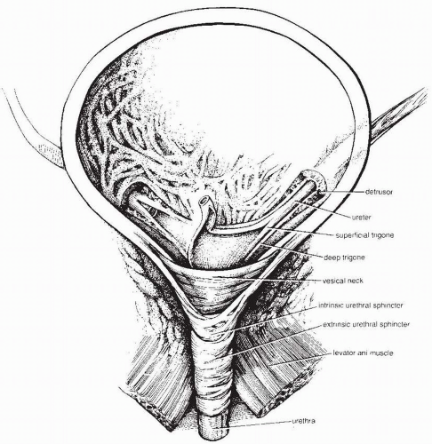
The ureter travels down to the bladder. It is approximately 30 cm in length in the adult. The ureter has three areas of physiologic narrowing that take on clinical significance with respect to possible obstruction from stones. These areas are the ureteropelvic junction, the crossing over of the iliac artery, and the ureterovesical junction (2,3). The ureterovesical junction is the place where the ureteral orifice opens up into the bladder. Its function is to allow urine to flow into the bladder but prevent reflux into the ureter. This can be accomplished because the ureters traverse obliquely between the muscular and submucosal layers of the bladder wall for a distance of 1 to 2 cm before opening into the bladder (Fig. 51-1). Any increase in intravesical pressure simultaneously compresses the submucosal ureter and effectively creates a one-way valve (4). Presence of ureteral muscle in the submucosal segment also has been shown to be important in preventing reflux (5). However, if there is high intravesical pressure the submucosal segment can be compressed and prevent urine from draining down into the bladder, causing a physiological obstruction.


Normal Urine Transport from the Kidneys to the Bladder

Urine transport is the result of both passive and active forces. Passive forces are created by the filtration pressure of the kidneys. The normal proximal tubular pressure is 14 mm Hg, and the renal pelvis pressure is 6.5 mm Hg, which slightly exceeds resting ureteral and bladder pressures. Active forces are the result of peristalsis of the calyces, renal pelvis, and ureter. Peristalsis begins with the electrical activity of pacemaker cells at the proximal portion of the urinary collecting tract (6).

For the ureter to propel the bolus of urine efficiently, the contraction wave must completely coapt the ureteral walls (7). Ureteral dilation for any reason results in inefficient propulsion of the urine bolus, and this can delay drainage proximal to that point. This can result in further dilation and, over time, lead to hydronephrosis.


Lower Urinary Tracts

Anatomically, the bladder is divided into the detrusor and the trigone. The detrusor is composed of smooth muscle bundles that freely crisscross and interlace with each other. Near the bladder neck, the muscle fibers assume three distinct layers. The circular arrangement of the smooth muscles at the bladder neck allows them to act as a functional sphincter. The trigone is located at the inferior base of the bladder. It extends from the ureteral orifices to the bladder neck. The deep trigone is continuous with the detrusor smooth muscle; the superficial trigone is an extension of the ureteral musculature (see Fig. 51-1) (4).

There is no clear demarcation of the musculature of the bladder neck and the beginning of the urethra in a man or woman. In a woman, the urethra contains an inner longitudinal and outer semicircular layer of smooth muscle. The circular muscle layer exerts a sphincteric effect along the entire length of the urethra, which is approximately 4 cm long.

In a man, the urethra runs the length of the penis. It begins at the meatus and is surrounded by the spongy tissue of the corpora cavernosus. In addition to the corpora cavernosus, which runs on the underside of the penis, the penis is made up of two corpora cavernosa that contain the spongy erectile tissue. The urethra is divided into the posterior or prostatic urethra, extending from the bladder neck to the urogenital diaphragm, and the anterior urethra, which extends to the meatus. The junction between the anterior and posterior urethra is known as the membranous urethra.


FIGURE 51-1. Anatomy of the bladder and related structures in a woman. Note how the ureter tunnels for a distance through the bladder wall, helping to prevent vesicoureteral reflux. Also note that there is not a clear demarcation between the bladder neck and sphincter mechanism. (From Hinman F Jr. Bladder repair. In: Hinman F Jr, ed. Urological Surgery. Philadelphia, PA: WB Saunders; 1989:433.)


Urinary Urethral Sphincters

Traditionally, the urethra has been thought to have two distinct sphincters, the internal and the external or rhabdosphincter. The internal sphincter is not a true anatomic sphincter. Instead, in both men and women, the term refers to the junction of the bladder neck and proximal urethra, formed from the circular arrangement of connective tissue and smooth muscle fibers that extend from the bladder. This area is considered a functional sphincter because there is a progressive increase in tone with bladder filling so that the urethral pressure is greater than the intravesical pressure. These smooth muscle fibers also extend submucosally down the urethra and lie above the external rhabdosphincter (8).

In a man, the external or urethral rhabdosphincter often is diagrammatically illustrated as a thin circular band of striated muscle forming a diaphragm just distal to the prostatic urethra (i.e., membranous urethra). In an anatomic study, however, Myers et al. (8) reconfirmed earlier studies showing that the urethral external striated sphincter does not form a circular band but has fibers that run up to the base of the bladder. The bulk of fibers are found at the membranous urethra (9). This sphincter
is under voluntary control. The striated muscular fibers in both man and woman are thought to have a significant proportion of slow-twitch fibers with the capacity for steady tonic compression of the urethra. In a woman, striated skeletal muscle fibers circle the upper two thirds of the urethra (9).


STRUCTURE AND FUNCTION OF THE MALE AND FEMALE CONTINENCE MECHANISM

In a man, the structures responsible for continence at the level of the membranous urethra include the mucosa, longitudinal smooth muscle of the urethra, striated sphincter, and levator ani musculature. Traditionally, the striated sphincter has been considered responsible for maintaining continence. However, experimental paralysis of the striated sphincter and levator ani following surgery for prostate outlet obstruction did not result in incontinence. This demonstrated the important role of the smooth muscle fibroelastic component of the membranous urethra. The increased tone at the bladder outlet (i.e., internal sphincter) also helps maintain continence (10). In a woman, there are three important factors in maintaining continence:

Adequate pelvic floor support from the endopelvic fascia and anterior vagina.

Good sphincter function.

Maintenance of the intra-abdominal position of the proximal urethra.

During an increase in intra-abdominal pressure, continence is maintained by the downward-moving pelvic viscera compressing the urethra against the layer of endopelvic fascia and distribution of the increase of intra-abdominal pressure to the proximal intra-abdominal urethra. The urethral epithelium, which is sensitive to estrogen, is believed to help maintain continence by forming a mucosal seal (9).


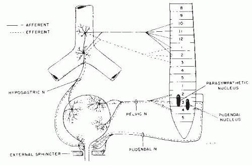
FIGURE 51-2. Peripheral innervation of the bladder and urethra. Sympathetic stimulation responsible for storage travels through the hypogastric plexus. Parasympathetic stimulation causing bladder contractions travels through the pelvic nerve. (From Blaivas JG. Management of bladder dysfunction in multiple sclerosis. Neurology. 1980;30:73.)


NEUROANATOMY OF THE LOWER URINARY TRACT

Urine storage and emptying is a function of interactions among the peripheral parasympathetic, sympathetic, and somatic innervation of the lower urinary tract. Additionally, there is modulation from the central nervous system (CNS).


Bladder Neuroanatomy


Efferent System

The parasympathetic efferent supply originates from a distinct detrusor nucleus located in the intermediolateral gray matter of the sacral cord at S2 to S4. Sacral efferents emerge as preganglionic fibers in the ventral roots and travel through the pelvic nerves to ganglia immediately adjacent to or within the detrusor muscle to provide excitatory input to the bladder. After impulses arrive at the parasympathetic ganglia, they travel through short postganglionics to the smooth muscle cholinergic receptors. These receptors, called cholinergic because the primary postganglionic neurotransmitter is acetylcholine, are distributed through the bladder. Stimulation causes a bladder contraction (11,12).

The sympathetic efferent nerve supply to the bladder and urethra begins in the intermediolateral gray column from T11 through L2 and provides inhibitory input to the bladder. Sympathetic impulses travel a relatively short distance to the lumbar sympathetic paravertebral ganglia. From here, the sympathetic impulses travel along long postganglionic nerves in the hypogastric nerves to synapse at α- and β-adrenergic receptors within the bladder and urethra. Variations in this anatomic arrangement do occur; sympathetic ganglia sometimes also are located near the bladder, and sympathetic efferent fibers may travel along the pelvic as well as the hypogastric nerves (Fig. 51-2) (11,12).

Sympathetic stimulation facilitates bladder storage because of the strategic location of the adrenergic receptors.
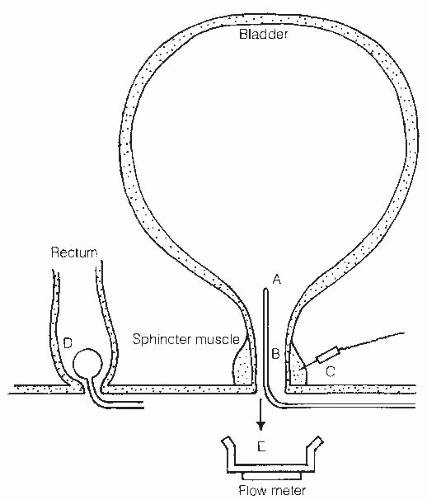
Beta-adrenergic receptors predominate in the superior portion (i.e., body) of the bladder. Stimulation of β-receptors causes smooth muscle relaxation. Alpha receptors have a higher density near the base of the bladder and prostatic urethra; stimulation of these receptors causes smooth muscle contractions and therefore increases the outlet resistance of the bladder and prostatic urethra (Fig. 51-3) (11, 12, 13).


FIGURE 51-3. Location of bladder receptors. Bladder storage is maintained by simultaneous sympathetic α-adrenergic receptor (contraction) (A) and β-adrenergic receptor (relaxation) stimulation (B). Bladder emptying occurs with parasympathetic cholinergic receptor stimulation (C).

After spinal cord injury (SCI), several changes occur to the bladder receptors that alter bladder function. There is evidence that when smooth muscle is denervated, its sensitivity to a given amount of neurotransmitter increases (i.e., denervation supersensitivity). Therefore, smaller doses of various pharmacologic agents would be expected to have a much more pronounced effect in those with SCI as compared with those with nonneurogenic bladders (14).

A change in receptor location and density may also occur. Norlen et al. (15) found that after complete denervation there was a change from a β-receptor predominance to an α-receptor predominance. Because α-receptors cause contraction of smooth muscle, a change in receptors may be one reason for some individuals to have poor compliance of the bladder after SCI.

Animal studies have revealed that, although the previously described long postganglionic neurons exist, there are ganglia close to the bladder and urethra in which there are both cholinergic and adrenergic fibers. This has been termed the urogenital short neuron system. These ganglia are composed of three cell types, adrenergic neurons, cholinergic neurons, and small intensely fluorescent cells, which are believed to be responsible for this interganglionic modulation of the adrenergic and cholinergic neurons. Further work is needed to define this system in humans (16).


Afferent System

The bladder afferent system transmits the mechanoreceptive input that is essential for voiding. The most important afferents that stimulate voiding are those that pass to the sacral cord via the pelvic nerves. These afferents include two types of afferents, small myelinated A-delta and unmyelinated (C) fibers.

The small myelinated A-delta fibers respond in a graded fashion to bladder distention and are needed for normal voiding. The unmyelinated (C) fibers have been termed silent C-fibers because they do not respond to bladder distention and therefore are not essential for normal voiding. However, these silent C-fibers do exhibit spontaneous firing when they are activated by chemical or cold temperature irritation at the bladder wall. Additionally, the unmyelinated (C) fibers (rather than A-delta afferents) have been found to “wake up” and respond to distention and play an important role in stimulating uninhibited bladder contractions in following suprasacral SCI.

This role of the unmyelinated (C) fibers has been demonstrated in studies using intravesical administration of capsaicin and resiniferatoxin. Both of these agents are potent C-fiber afferent neurotoxins. In non-SCI animals (with A-delta afferents), there was no blockage of bladder contractions with bladder distention. However, in SCI animals (with “awake” C-fiber afferents), capsaicin completely blocked rhythmic bladder contractions induced with bladder distention (16). While intravesical capsaicin and resiniferatoxin are considered investigational drugs, similar results have been found in human studies in those with suprasacral SCI and multiple sclerosis (17). These findings have important potential therapeutic implications. However, further work is needed to determine the optimal dosage and vehicle for intravesical instillation (see Section “Management of Voiding Dysfunctions” later in this chapter).


Bladder Neurotransmitters

It is known that there are more transmitters than acetylcholine and norepinephrine, including nitric oxide, vasoactive intestinal polypeptide, endogenous opioid peptides, and neuropeptide Y.
These transmitters may work independently or help modulate the classic neurotransmitters. Nitric oxide and vasoactive intestinal polypeptide have smooth muscle relaxant effects. This helps explain the concept of atropine resistance. It has been found that a single neurotransmitter-blocking agent such as atropine fails to suppress 100% of the bladder or urethral activity (14,18). This explains why a combination of agents may be more effective than a higher dose of a single agent.


Urethral Sphincter Innervation

The external urethral sphincter classically has been described as having somatic innervation, allowing the sphincter to be closed at will. Somatic efferents originate from a pudendal nucleus of sacral segments from S1 to S4. Somatic efferents then travel through the pudendal nerve to the neuromuscular junction of the striated muscle fibers in the external urethral sphincter.

The internal urethral sphincter has been described as being under control of the autonomic system. This area has a large number of sympathetic α-receptors, which cause closure when stimulated. Animal studies have revealed that nitric oxide is an important parasympathetic neurotransmitter mediating relaxation of the urethral smooth muscle (8,18).

The distinction between the internal and external sphincter is becoming less clear. Elbadawi and Schenk (19) reported histochemical evidence of a triple innervation pattern of the external sphincter (i.e., rhabdosphincter) in five mammalian species, with dual sympathetic and parasympathetic autonomic components superimposed on the somatic component. Sundin and Dahlstrom (20) demonstrated sprouting and increasing adrenergic terminals after parasympathetic denervation in cats. Crowe et al. (21) reported a substantial invasion of adrenergic nerve fibers in smooth and striated muscle in the urethra in SCI patients with lower motor neuron lesions.


Influences of the Central Nervous System on the Lower Urinary Tract

Facilitation and inhibition of the autonomic nervous system are under control of the CNS. There are several theories of how this occurs. Denny-Brown and Robertson (22) suggested that micturition was primarily mediated by a sacral micturition reflex. According to their theory, descending nervous system pathways modulate this micturition reflex (21). Barrington, Bradley, and de Groat thought that facilitative impulses to the bladder originated from a region of the anterior pons termed “Barring-ton’s center” (23).

De Groat and associates additionally stressed the importance of the sympathetic nervous system in facilitating urine storage (24). Carlsson provided evidence that this pontine mesencephalic area also plays a role in coordinating detrusor and sphincter activity. Stimulation of Barrington’s center significantly decreased electromyographic (EMG) activity in the periurethral-striated sphincter while causing a bladder contraction (25).

Transection experiments in cats suggest that the net effect of the cerebral cortex on micturition is inhibitory. This also is true for the basal ganglia and corresponds to clinical findings of detrusor hyperreflexia in those with basal ganglia dysfunction (e.g., Parkinson’s disease). The cerebellum is thought to maintain tone in the pelvis floor musculature and influence coordination between periurethral striated muscle relaxation and bladder emptying (15,25).


Normal Voiding Physiology

Micturition should be considered as having two phases: the filling (storage) phase and the emptying (voiding) phase. The filling phase occurs when a person is not trying to void. The emptying phase occurs when a person is attempting to void or told to void.

During filling (filling or storage phase), there should be very little rise in bladder pressure. As filling continues, low intravesical pressure is maintained by a progressive increase in sympathetic stimulation of the β-receptors located in the body of the bladder that cause relaxation, and stimulation of the α-receptors located at the base of the bladder and urethra that cause contraction. Sympathetic stimulation also inhibits excitatory parasympathetic ganglionic transmission, which helps suppress bladder contractions. During the filling phase, there is a progressive increase in urethral sphincter EMG activity (26). Increased urethral sphincter activity also reflexly inhibits bladder contractions. When a bladder is full and has normal compliance, intravesical pressures are between 0 and 6 cm H2O and should not rise above 15 cm H2O. Filling continued past the limit of the viscoelastic properties of the bladder results in a steady progressive rise in intravesical pressure (27). This part of the filling curve usually is not seen in a person with normal bladder function, because this much distension would cause significant discomfort and not be tolerated.

When a patient is told to void (voiding or emptying phase), there should be cessation of urethral sphincter EMG activity as well as a drop in urethral sphincter pressure and funneling of the bladder neck. There is no longer reflex inhibition to the sacral micturition center from the sphincter mechanism. This is followed by a detrusor contraction. The urethral sphincter should remain open throughout voiding, and there should be no rises in intra-abdominal pressure during voiding. In younger individuals, there should be no postvoid residual, although postvoid residuals may increase with aging.


Geriatric Voiding Physiology

The aging process often affects voiding physiology. The kidneys undergo an age-related decrease in glomerular blood flow and renal blood flow (28). The elderly also experience a loss in concentrating ability and often excrete most of their fluid intake at night, even in the absence of medical conditions such as prostate outlet obstruction, diabetes, remobilization of lower-extremity edema, and use of evening diuretics (29).

Detrusor overactivity has been reported as the most common type of voiding dysfunction in incontinent elderly men and women (29). More recently, this has been called an overactive bladder. This may be associated with a central nervous lesion (such as a stroke, head injury, cervical disk disease, etc.).
A replacement of normal muscle cell junctions by novel “protrusion junction cells” and ultraclose abutments, connecting cells into chains, which facilitates and increases spontaneous smooth muscle activity, has been found in incontinent elderly patients (30). There is often a combination of causes that result in an overactive bladder. Detrusor hyperactivity may coexist with impaired contractility (DHIC) of the bladder wall, resulting in both incontinence and retention. This combination was found to be one of the most common urodynamic findings in the elderly incontinent nursing home population (29). A combined pattern may also occur if there are cognitive/mobility issues and a person begins to void (incontinence) and then stops voiding.

Outlet obstruction by prostatic hypertrophy is the second most common cause of incontinence in men, although most men with outlet obstruction do not have incontinence (29). Outlet obstruction results in a significant increase in collagen, leading to bladder trabeculation. This in turn can cause a decrease in the viscoelastic properties for storage as well as the ability to contract and may be one reason for increasing postvoid residuals and decreasing bladder capacity with aging (31). It should be noted that outlet obstruction in women is rare; however, it may occur from various etiologies, such as urethral stenosis or kinking from a large cystocele or a previous bladder neck suspension. A trabeculated bladder appearance in older women without obstruction usually results from a thinning of the bladder wall with more prominent muscle bundles rather than deposition of collagen (31).

Stress incontinence is the second most common cause of incontinence in elderly women (29). In younger women, stress incontinence frequently results from pelvic laxity; however, in elderly women, it is also caused by a decrease in urethral closure pressure. This decrease in urethral pressure has been attributed to a decrease in estrogen, which causes a loss of muscle bulk and atrophic changes of the urethra and vagina. This, in turn, can cause inflammation and friability of these tissues, decreased periurethral blood flow, further laxity of pelvic structures, and possible urethral prolapse (31).

Detrusor underactivity may also occur with aging. At a cellular level, this has been characterized by widespread degenerative changes of both muscle cells and axons without accompanying regenerative changes (32). Ouslander et al. (33) reported that approximately 25% of elderly patients evaluated had postvoid residuals greater than 100 mL. Approximately 10% of geriatric incontinence has been attributed to overflow incontinence (29).


Pediatric Voiding Physiology

Voiding physiology also changes with age in children. In the newborn, the sacral micturition reflex is primarily responsible for voiding. Because the brain stem is intact, there is coordination of the bladder contraction with sphincter relaxation; however, there is little inhibition of the micturition reflex from the cerebral cortex. As the child grows, the voided volume increases and voiding frequency decreases. By 3 years of age, most children have some voluntary control of voiding. This control usually is complete by the age of 4 years. There are some neurologically intact children, however, for whom complete control of voiding may take 5 or 6 years (34).


CLASSIFICATION OF VOIDING DYSFUNCTION

There have been a wide variety of classifications to describe voiding dysfunctions. These classifications have been based on neurologic lesion (e.g., Bors-Comarr, Bradley), urodynamic findings (e.g., Lapides,), functional classification (e.g., Wein), and combination of bladder and urethral function based on urodynamics (e.g., International Continence Society) (35, 36, 37, 38, 39). The International Continence Society classification: The Standardisation of Terminology of Lower Urinary Tract Function: Report from the Standardisation Sub-committee of the International Continence Society has become widely accepted not only because it provides a standardization of terminology describing bladder and urethral function but also because a description of each of these terms is provided (39). Table 51-1 shows some highlights of
the urodynamic terminology from International Continence Society Classification. Table 51-2 shows Wein’s classification. This classification is helpful at directing treatment because it is based on a clinical problem (incontinence or retention), which can then be applied to specific urodynamic findings. A urodynamic basic SCI data set has recently been developed, which should be helpful, collecting information in a standardized way regarding urodynamic bladder and sphincter function in those with SCI (40).


TABLE 51.1 Urodynamic Terminology—Highlights From International Continence Society Classification


Bladder


Bladder—function


Normal (during filling)


Detrusor overactivity (during filling)


Neuropathic detrusor overactivity


Idiopathic detrusor overactivity


Normal (during emptying)


Underactive (during emptying)—inadequate magnitude or duration of a contraction to empty the bladder)


Acontractile (no contractions during urodynamics)


Areflexic (no contractions due to neurogenic cause)


Bladder—sensation


Normal


Increased


Reduced


Absent


Nonspecific


Bladder capacity


Small (350 mL)


Average (350-650 mL)


Large (650 mL)


Compliance


Low (hypocompliance)


Normal (350-650 mL with little pressure rise)


High (hypercompliance)


Urethra


Normal


Incompetent (during filling)


Obstructive (during voiding)


Mechanical (stricture, bladder outlet obstruction)


Overactive (detrusor/[external] sphincter dyssynergia)


TABLE 51.2 Urodynamic and Functional Classification


Incontinence


Caused by the bladder


Detrusor overactivity


Decreased capacity


Low bladder wall compliance


Normal (cognitive/mobility issue)


Caused by the outlet


Incompetent sphincter


Retention


Caused by the bladder


Detrusor areflexia


Large capacity/high compliance


Normal (cognitive/mobility issue)


Caused by the outlet


High voiding pressure with low flow rate


Internal sphincter dyssynergia


External sphincter dyssynergia


Overactive sphincter mechanism (i.e., sphincter or pseudosphincter dyssynergia)


Retention and incontinence


Caused by the bladder


Uninhibited contractions with underactive detrusor


No contractions


Normal (cognitive/mobility issue)


VOIDING DYSFUNCTIONS FOUND IN COMMON NEUROLOGIC DISORDERS


Suprapontine Lesions

Any suprapontine lesion may affect voiding. Lesions may result from cerebrovascular disease, hydrocephalus, intracranial neoplasms, traumatic head injury, Parkinsons disease, and multiple sclerosis. It should be noted that multiple sclerosis is unique among the suprapontine lesions because it also affects the white matter of the spinal cord and often has a relapsing and remitting nature. The expected urodynamic finding following a suprapontine lesion is detrusor hyperreflexia without detrusor-sphincter dyssynergia. Because of various factors such as medications, prostate obstruction, and possible normal bladder function but poor cognition, the voiding dysfunctions may be very different from expectations. Voiding dysfunction following cerebrovascular accident (CVA), Parkinson’s disease, and multiple sclerosis has been studied more extensively than those associated with other suprapontine lesions and is reviewed in the following discussion.


Cerebrovascular Accidents

After a CVA, some patients initially have acute urinary retention. The reason for this detrusor areflexia is unknown. Urinary incontinence, however, is the most common urologic problem following an acute CVA. Various series have reported that 40% to 60% of patients are incontinent 1 week post-CVA (41, 42, 43). In the inpatient rehabilitation setting, a 33% incidence of incontinence during the first 3 months post-CVA has been reported (44). It also has been well documented that this problem significantly improves or resolves in the majority of patients. At 1 month, the percentage of incontinent patients dropped to between 29% and 42%. By 6 months to 1 year post-CVA, 14% to 15% of patients still were incontinent, which is similar to the 15% to 30% incidence in the general geriatric population (41, 42, 43). Risks for incontinence poststroke include age greater than 75 years, dysphagia, motor weakness, and visual field defects. At 2 years poststroke, incontinent patients versus continent patients had higher case fatality rates (67% vs. 20%), higher institutional rates (39% vs. 16%), and grater disability (39% vs. 5%) (43). A recent study has further categorized poststroke urinary incontinence as urge incontinence or impaired awareness urge incontinence. Those with impaired awareness urge incontinence had poorer attention and outcomes. Impaired awareness incontinence was also found to be a strong predictor factor for mortality and nursing home residence at 1 year. It would be expected that those with impaired awareness incontinence would not do well (45).

Detrusor overactivity with uninhibited bladder contractions is the most common urodynamic finding following a stroke. It has been reported to occur 70% to 90% of the time (42,46). One hypothesis for this finding is the release of the spinal micturition reflexes from the inhibitory higher centers. Symptoms, however, often do not correlate with urodynamic findings. Linsenmeyer and Zorowitz evaluated 33 consecutive incontinent patients who were 1 to 3 months post-CVA. They found that whereas 82% of men had uninhibited contractions, 43% also had urodynamic evidence of outlet obstruction. Six percent of the incontinent group had no bladder contractions and twelve percent had normal urodynamic findings (46). Voluntary sphincter contractions (i.e., pseudodyssynergia) to keep from voiding should not be misinterpreted as true detrusor-sphincter dyssynergia. In a review of 550 patients, Blaivas (47) reported that patients with CVAs do not develop true detrusor-sphincter dyssynergia. EMG studies by Siroky and Krane (48) gave similar results.


Parkinson’s Disease

Symptoms of bladder dysfunction have been reported in 37% to 72% of patients with Parkinson’s disease. These symptoms may be frequency or urgency (57%), obstruction (23%), or a combination of the two (20%). Detrusor overactivity has
been the most common urodynamic finding (72% to 100%) (49, 50, 51). Detrusor overactivity is thought to occur because of loss of the inhibitory input from the basal ganglia on the micturition reflexes; however, detrusor instability also has been associated with benign prostatic obstruction. Detrusor areflexia may result from bladder decompensation through a combination of bladder outlet obstruction and chronic use of anticholinergic and α-adrenergic medications (51).

EMG studies of the external sphincter reveal that patients may have pseudodyssynergia or bradykinesia but not true detrusor-sphincter dyssynergia (49, 50, 51). The majority of patients (63% to 75%) have normal sphincter function (49, 50, 51).


Multiple Sclerosis

Only 6% of patients with multiple sclerosis first present with urologic symptoms (52). Bemelmans and associates, however, reported that 50% of asymptomatic patients with early multiple sclerosis had urodynamic abnormalities that needed further follow-up, and 50% of these required therapeutic intervention (53). As the disease progresses, urologic symptoms become common, eventually affecting at least 50% of men and 80% of women (54). The type of voiding dysfunction often is difficult to predict because of the diffuse involvement and changing nature of the disease.

Goldstein et al. reported that in a series of 86 symptomatic patients, 49% had incontinence, 32% had urgency and frequency, and 19% had obstructive hesitancy and retention. They also documented that patients with similar neurologic findings may have different voiding dysfunctions and that urologic signs and symptoms do not accurately reflect the voiding dysfunction (55). Wheeler et al. found that 55% of patients who were studied had changes in their urodynamic picture. The urodynamic pattern varied from detrusor areflexia to detrusor overactivity and vice versa (56).

Because suprapontine and suprasacral plaques occur most frequently, detrusor hyperreflexia is the most common urodynamic finding; however, as many as 50% of patients have poorly sustained uninhibited bladder contractions with inefficient bladder emptying. Detrusor areflexia is found in approximately 20% of patients with urologic symptoms. This is believed to be a result of sacral plaque involvement (57).

True detrusor-sphincter dyssynergia may occur in multiple sclerosis when there is involvement of the suprasacral spinal cord. Approximately 15% to 20% of patients develop detrusor-sphincter dyssynergia. Blaivas and Barbalias (58) reported that this was an ominous sign because of the potential for upper-tract damage and development of reflux as a result of the increased intravesical pressures needed to force urine past the dyssynergic sphincter. Upper-tract pathologic processes, including pyelonephritis, renal calculi, reflux, and hydronephrosis, have been reported to occur in 10% to 20% of patients with multiple sclerosis (58,59).


Suprasacral Spinal Cord Lesions

Traumatic SCI is the most common suprasacral lesion affecting voiding. Other suprasacral lesions include transverse myelitis, multiple sclerosis, and primary or metastatic spinal cord tumor.

Patients with suprasacral spinal cord lesions would be expected to have detrusor hyperreflexia with detrusor-sphincter dyssynergia. However, in cases of partial lesions, occult lesions of the sacral cord, or persistent spinal shock, this is not always the case (60).

Traumatic suprasacral SCI results in an initial period of spinal shock, in which there is hyporeflexia of the somatic system below the level of injury and detrusor areflexia. During this phase, the bladder has no contractions, even with various maneuvers such as water filling, bethanechol supersensitivity testing, or suprapubic tapping. The neurophysiology of spinal shock and its recovery are not known. Recovery of bladder function usually follows recovery of skeletal muscle reflexes. Uninhibited bladder contractions gradually return after 6 to 8 weeks (61).

Clinically, a person with a traumatic suprasacral SCI may begin having episodes of urinary incontinence and various visceral sensations, such as tingling, flushing, increased lower-extremity spasms, or autonomic dysreflexia with the onset of uninhibited contractions. As uninhibited bladder contractions become stronger, the postvoid residuals decrease. Rudy et al. (62) reported that voiding function appears optimal at 12 weeks postinjury. However, detrusor hyperreflexia has been reported to have a delayed onset of up to 22 months postinjury. Eventually, all of these patients did develop uninhibited contractions (63). Bors and Comarr (35) considered the bladder “balanced” when postvoid residuals were less than 20% of the total bladder capacity in those with detrusor hyperreflexia. Graham (64) reports that 50% to 70% of patients will develop balanced bladders without therapy. Unfortunately, high intravesical voiding pressures usually are required for the development of a balanced bladder. These high pressures may cause renal deterioration.

Traditionally, it has been thought that there is decreased activity of the external urethral sphincter during acute spinal shock. However, Downie and Awad (65) noted in dogs that with surgical transection between T2 and T8, there was no change in the activity of the periurethral striated musculature despite detrusor areflexia. In humans, Nanninga and Meyer (66) found that in 44 patients in spinal shock with suprasacral lesions, all had a positive bulbocavernosus reflex, and 30 of 32 had sphincter activity despite detrusor areflexia within 72 hours of injury. Koyanagi et al. noted that external sphincter electrical activity was not affected during acute spinal shock but was likely to increase after recovery from spinal shock. This increase was more marked in those with high suprasacral lesions than in those with low suprasacral lesions (67).

Detrusor-external sphincter dyssynergia often occurs following suprasacral lesions. Blaivas et al. (68) noted that it occurred in 96% of patients with suprasacral lesions. They found several different patterns of striated sphincter dyssynergia. Rudy et al. proposed that detrusor-sphincter dyssynergia is an exaggerated continence reflex. The continence reflex is the normal phenomenon of increasing urethral sphincter
activity with bladder filling. They believed that the patterns described by Blaivas et al. (62) represented variations of the single continence reflex.

In addition to the detrusor-external sphincter dyssynergia, internal sphincter dyssynergia also has been reported, often occurring at the same time as detrusor-external sphincter dyssynergia.


Sacral Lesions

There are a variety of lesions that may affect the sacral cord or roots. These include spinal trauma, herniated lumbar disk, primary or metastatic tumors, myelodysplasia, arteriovenous malformation, lumbar stenosis, and inflammatory process (e.g., arachnoiditis). In Pavlakis et al. series, trauma was responsible for conus and cauda equina lesions more than 50% of the time. The next most common cause was L4/L5 or L5/S1 intervertebral disc protrusion. The incidence of lumbar disc prolapse causing cauda equina syndrome is between 1% and 15% (69). Damage to the sacral cord or roots generally results in a highly compliant acontractile bladder; however, particularly in patients with partial injuries, the areflexia may be accompanied by decreased bladder compliance, resulting in progressive increases in intravesical pressure with filling (70). The exact mechanism by which sacral parasympathetic decentralization of the bladder causes decreased compliance is unknown (70,71).

It has been noted that the external sphincter is not affected to the same extent as the detrusor. This is because the pelvic nerve innervation to the bladder usually arises one segment higher than the pudendal nerve innervation to the sphincter (72). The nuclei also are located in different portions of the sacral cord, with the detrusor nuclei located in the intermediolateral cell column and the pudendal nuclei located in the ventral gray matter. This combination of detrusor areflexia and an intact sphincter helps contribute to bladder overdistention and decompensation.


Peripheral Lesions

There are multiple etiologies for peripheral lesions that could affect voiding. The most common lesion is a peripheral neuropathy secondary to diabetes mellitus. Other peripheral neuropathies that have been associated with voiding dysfunction include chronic alcoholism, herpes zoster, Guillain-Barré syndrome, and pelvic surgery (73,74). A sensory neuropathy is the most frequent finding in diabetes. Urodynamic findings, including decreased bladder sensation, chronic bladder overdistention, increased postvoid residuals, and possible bladder decompensation, may result from bladder overdistention secondary to decreased sensation of fullness. Andersen and Bradley (75) reported that in their series, mean bladder capacity was 635 mL, with a range of 200 to 1,150 mL. An autonomic neuropathy also may be responsible for decreased bladder contractility. Guillain-Barré syndrome and herpes zoster are predominantly motor neuropathies. Transient voiding symptoms, predominantly urinary retention, have been reported to occur in 0% to 40% of patients and are thought to represent involvement of the autonomic sacral parasympathetic nerves. Detrusor hyperreflexia occasionally has been found in those with Guillain-Barré syndrome (76). Voiding dysfunctions resulting from pelvic surgery or pelvic trauma usually involve both motor and sensory innervation of the bladder (75).


COMPREHENSIVE EVALUATION OF VOIDING DYSFUNCTION


Neurourologic History

The urological history should first focus on the patient’s voiding symptoms. The history should establish whether the onset of the current symptoms is new, has become worse, or has remained unchanged since the neurologic insult. This will allow for more meaningful discussions when patients and family ask about “returning to normal.” A preexisting problem such as urinary frequency may cause incontinence from decreased mobility.

Because symptoms often correlate poorly with the actual voiding problem, it is best not to initiate pharmacological or surgical treatment based solely on symptoms. Katz and Blaivas (77), in a prospective study of 425 consecutive patients, found that the clinical assessment based on symptoms did not correlate with the objective urodynamic findings in 45% of patients thought to have storage problems, in 25% believed to have emptying problems, and in 54% of those believed to have storage and emptying problems. It has also been well documented that urodynamics should be included in the evaluation of those with SCI since bladder and sphincter function cannot be predicted by history and physical examination. Ouslander et al. (78) found in the geriatric female population that presenting symptoms were predictive of the urodynamic diagnosis in only 55% of those with pure urge incontinence. It has also been well documented that urodynamics is essential in the evaluation of those with SCI since bladder and sphincter function cannot be predicted by history and physical examination (79).

Significant past history includes additional medical problems that may contribute to present problems, such as diabetes, previous CVAs, hypertension, and use of diuretics. The past history also needs to focus on surgery that may affect voiding, such as previous transurethral resection of the prostate, surgery for stress incontinence, or pelvic surgery. Questions about past and present bowel and erectile function should be asked. Potentially reversible causes of voiding disorders need to be investigated. A helpful mnemonic coined by Resnick and Yalla (80) to describe the reversible causes of incontinence in the elderly is DIAPPERS. These same factors also may be responsible for problems with retention. The mnemonic can be broken down as follows:

Delirium

Infection

Atrophic vaginitis, urethritis

Pharmaceuticals

Psychological


Endocrine

Reduced mobility

Stool impaction

The physiatric history that has particular significance for voiding dysfunction is hand function, dressing skills, sitting balance, ability to perform transfers, and ability to ambulate. These factors not only play a role in why a person may be incontinent but also are important considerations in developing management strategies.


Neurourologic Examination

The neurourologic physical will not give objective evidence about the bladder and sphincter function but may suggest potential contributory causes of a voiding dysfunction. The examination should focus on the abdomen, external genitalia, and perineal skin. The rectal examination is important to evaluate for cancer. Prostate obstruction cannot be determined by prostate size alone since it is not the overall size of the prostate but the amount of prostate growing inward that causes obstruction. Therefore, a urodynamic study, which measures the actual pressure within the bladder during a contraction and the resulting flow of urine, is important to objectively diagnose outflow obstruction.

In postmenopausal women, the urethra and vaginal introitus should be examined for atrophic changes suggestive of estrogen deficiency. In women, the examination also should focus on the degree of pelvic support. A determination of masses producing extrinsic compression on the bladder should be made during the vaginal examination.

The mental status portion of the neurourologic examination should, as a minimum, evaluate the patient’s level of consciousness, orientation, speech, long- and short-term memory, and comprehension. Voiding disorders may be secondary to or made worse by disorientation, inability to communicate the desire to void, or lack of understanding when the patient is told to void.

The sensory examination should focus on determining the level of injury in those with SCI. Especially important is establishing whether the level of injury is above T6, which would make the patient prone to autonomic dysreflexia. Sacral sensation evaluates the afferent limb (i.e., pudendal nerve) of the sacral micturition center. Loss of pinprick and light touch sensation in the hands and feet is suggestive of a peripheral neuropathy.

The motor examination helps establish the level of injury and degree of completeness in those with SCI. Hand function should be assessed to determine the ability to undress or possibly perform intermittent catheterization (IC). Upperand lower-extremity spasticity with sitting, standing, and ambulating also needs to be evaluated. Anal sphincter tone is also important. Decreased or absent tone suggests a sacral or peripheral nerve lesion, whereas increased tone suggests a suprasacral lesion. Voluntary contraction of the anal sphincter tests sacral innervation, suprasacral integrity, and the ability to understand commands.

Cutaneous reflexes that are helpful to the neurourologic examination are the cremasteric (L1 to L2), bulbocavernosus (S2 to S4), and anal reflex (S2 to S4). Absence of these cutaneous reflexes suggests pyramidal tract disease or a peripheral lesion. The bulbocavernosus reflex is a useful test to evaluate the sacral reflex arc. However, it may be unreliable. A false negative often results from a person being nervous and already having his or her anal sphincter clamped down at the time of the examination. Muscle stretch reflexes also should be evaluated. A sudden increase in spasticity may indicate a urinary tract infection (UTI). In addition, pathologic reflexes (e.g., Babinski reflex) may help localize the neurologic lesion.


UROLOGIC ASSESSMENT OF THE UPPER AND LOWER URINARY TRACT


Indications for Testing

A variety of tests can be performed to evaluate the upper and lower urinary tract. The exact types of tests and follow-up depend on the disease process, the patient’s clinical course, and any preexisting urologic problems needing further follow-up.

If the disease process is one that is not known generally to affect the upper tracts, such as a stroke, hip replacement with retention, or peripheral neuropathy, then the evaluation can be directed at the lower urinary tract. Evaluation of the upper tracts in these individuals should be undertaken if there is any suggestion of upper-tract involvement, such as pyelonephritis or hematuria.

Patients with disease processes that may affect the upper tracts, such as multiple sclerosis, should undergo baseline testing of the upper tracts and then periodic screening. Emphasis otherwise is directed primarily at the lower tract with the use of urine analysis, culture and sensitivity, postvoid residual, and urodynamics. Testing usually is done annually, but may be needed more or less frequently depending on the patient’s clinical course.

Spinal cord-injured patients, particularly those with potential high intravesical voiding pressures, need constant surveillance of the upper tracts as well as lower tracts. Although there is no agreement on exactly which tests and the frequency at which testing should be done, there is agreement that upperand lower-tract testing is necessary. The American Paraplegia Society has developed recommendations for the urologic evaluations of those with SCI (81).

Institutions often will have SCI patients undergo a yearly evaluation for the first 5 to 10 years, and if their upper tracts are stable, then evaluations are every other year. However, there is evidence that bladder function continues to change even after 20 years postinjury, suggesting that yearly evaluations should be considered (82). People with an indwelling suprapubic or Foley catheter often will get yearly cystoscopy to rule out stones and bladder tumors.


Specific Upper- and Lower-Tract Tests

Tests designed to evaluate the upper tracts include an intravenous pyelogram (IVP), renal ultrasound, 24-hour urine
creatinine clearance, and quantitative mercaptoacetyltriglycine (MAG) 3 renal scan and computerized tomography (CT). When ordering a test one must consider whether the information is needed about the function of the upper tracts or about the anatomy of the upper tracts. For example, a renal ultrasound is excellent to detect anatomical changes; however, it will not suggest any problems with renal function until hydronephrosis develops. Conversely, a renal scan detects stasis of the upper tracts before hydronephrosis develops, but is unlikely to detect a small kidney stone.

A 24-hour urine creatinine clearance and quantitative renal scan evaluate upper-tract function, whereas renal ultrasound and CT are used to evaluate upper-tract anatomical features. An IVP evaluates both function and anatomy. Despite evaluating both function and anatomy, there are a number of disadvantages to an IVP. These disadvantages include potential allergic reactions, radiation exposure, and patient inconvenience, specifically getting an IVP laxative preparation the night before the test. Therefore, IVP is used infrequently.

The primary purpose of quantitative MAG 3 radioisotope renal scan is to monitor renal function and drainage. It has been found to be a safe and effective modality in those with SCI (83,84). While serum creatinine and creatinine clearance are easy to obtain and inexpensive, there are a number of possible problems when using them to monitor kidney function in those with SCI (85). Since individuals with SCI frequently have less muscle mass than able-bodied individuals, their serum creatinine should be less than that of able-bodied individuals. Therefore, a normal serum creatinine lab value may actually represent a high value for those with SCI. In addition, serum creatinine will not rise until there is at least a 50% decline in renal function. When ordering a 24-hour urine collection be aware that overcollection of urine (>24 hours) will overestimate kidney function and undercollection will underestimate kidney function (85).

The renal ultrasound is helpful for detecting hydronephrosis and kidney stones. When evaluating renal anatomy, ultrasound has largely replaced IVP (86). The major advantages of ultrasound are that it is noninvasive and does not involve any contrast agents. The major disadvantages of ultrasound are that it is user-dependent and does not show renal function (87).

If further anatomic definition is needed to evaluate for stones or tumors, CT should be considered. It has largely replaced IVPs in a number of institutions. In a prospective study of nonenhanced helical CT scans versus IVP, CT correctly identified 36 of 37 ureteral stones with one false positive. CT had a sensitivity 97%, specificity of 96%, and accuracy of 97% at detecting ureteric stones. This was double that of IVP (88).

Tests to evaluate the lower tracts include cystogram, cystoscopy, and urodynamics. Because each of these involves instrumentation, it is best to obtain a urine culture and sensitivity test, and give antibiotics if positive before the testing. An untreated infection or bacterial colonization has the potential to cause bacteriemia and increased bladder overactivity.

Some indications for cystoscopy in those with voiding disorders include hematuria, recurrent symptomatic UTIs, recurrent asymptomatic bacteriuria with a stone-forming organism (i.e., Proteus mirabilis), an episode of genitourinary sepsis, urinary retention or incontinence, pieces of eggshell calculi obtained when irrigating a catheter, and long-term indwelling catheter. Cystoscopy also is indicated when one is removing an indwelling Foley catheter that has been in place for more than 2 to 4 weeks or changing to a different type of management, such as IC or a balanced bladder. Cystoscopy can reveal a pubic hair or eggshell calculus that may be missed on radiography and serve as a nidus for bladder stones.


Urodynamics

Urodynamics provides objective information on voiding function (Table 51-2).

Urodynamics in general terms is defined as the study of normal and abnormal factors in the storage, transport, and emptying of urine from the bladder and urethra by any appropriate method (40). When deciding on an appropriate urodynamic test, one needs to consider whether information is needed about the filling phase, emptying phase, or both phases of micturition.

The following are some of the more common indications for an urodynamics evaluation:

Recurrent UTIs in a patient with neurogenic bladder

Urinary incontinence

Urinary frequency

Large postvoid residuals (i.e., retention)

Deterioration of the upper tracts

Monitoring of voiding pressures

Evaluation and monitoring of pharmacotherapy

The physician’s presence is important to help direct the urodynamics study. Typical decisions include how much water to put in the bladder, whether to repeat the study, and whether to have the patient sit or stand to void. Observing the patient during urodynamics also will help in getting an idea of factors that might influence the test, such as patient anxiety or inability to understand when told to void.

Blood pressure monitoring is particularly important in SCI patients prone to autonomic dysreflexia. Urodynamics is particularly helpful for detecting autonomic dysreflexia in men with SCI at T6. Autonomic dysreflexia may occur with bladder distention or more commonly when bladder distention provokes an uninhibited contraction. This causes the sphincter to contract, which causes a significant rise in blood pressure and other symptoms of autonomic dysreflexia. However, 43% of the men with SCI at T6 and above may have “silent dysreflexia” (elevated BP without any symptoms) during voiding. This would not be detected without urodynamics and simultaneous blood pressure monitoring (89).

In order to have an accurate urodynamic evaluation, it is important that a person does not have a UTI. Bladder wall inflammation is likely to cause the bladder to lose some of its compliance, resulting in a smaller bladder capacity than normal.
The inflammation is also likely to trigger uninhibited contractions and cause the bladder to be more overactive than usual. A recent prospective study found that 9.7% of SCI individuals who had asymptomatic bacteriuria developed a symptomatic UTI post testing. Nearly 40% of SCI individuals with sterile urine developed asymptomatic bacteriuria post testing (90).

It would be expected that one or two doses of an antibiotic would clear the bacteria from the urinary tract and reduce the risk of an infection. However, it has been our practice to obtain a urine culture and sensitivity 1 to 2 weeks prior to the test. Those with pyuria or a symptomatic UTI are treated for 5 days prior to testing with the goal not only to eradicate the bacteria in the bladder but also to give adequate time to reduce inflammation of the bladder wall. Those with sterile urine or asymptomatic bacteriuria are given one or two doses of an antibiotic prior to testing. A person who presents for testing with cloudy urine or other symptoms of a UTI is rescheduled.


Evaluation of Bladder Filling (Storage Phase)

The simplest type of bladder test to evaluate bladder filling is known as a bedside cystometrogram. This test involves attaching a cylinder such as 50 mL filling syringe without the plunger to a Foley catheter. Water is then poured into the cylinder and allowed to drain by gravity into the bladder. The blood pressure and volume of fluid going into the bladder are recorded. The Foley is sometimes attached by means of a Y-connector to a manometer, which is used to measure the actual rise in water pressure. This test can be used to evaluate sensation (whether or not a person is aware of the bladder being filled), stability (whether or not there is a rise in the column of water signifying a bladder contraction), and capacity (the volume at which the bladder contraction occurs). It can also be used as a screening test to determine if an SCI patient has come out of spinal shock. There are several limitations to the bedside cystometrogram, however. It is difficult to determine if small rises in the water column result from intra-abdominal pressure (i.e., straining) or a bladder contraction. An iatrogenic bladder contraction can be elicited if the tip of the Foley catheter rubs against the trigone pressure sensors, which can then trigger bladder contractions. Most important, the voiding phase cannot be evaluated (91).

The carbon dioxide urodynamics has been largely replaced with water-fill urodynamics. Although the gas is cleaner and neater to use than water, the major disadvantage is that the voiding phase of micturition cannot be evaluated. Therefore, this test is of little use when trying to evaluate bladder and sphincter function during emptying.


Evaluation of Bladder Emptying

One of the easiest screening tests to evaluate bladder emptying is a postvoid residual; however, it should not be used to characterize the specific type of voiding dysfunction. The postvoid residual can be determined with catheterization or bladder ultrasound. A younger person should have no postvoid residual; however, an elderly person with no voiding symptoms may have a postvoid residual of 100 to 150 mL. A normal postvoid residual does not rule out a voiding problem. For example, a postvoid residual may be normal despite significant outflow obstruction (e.g., benign prostate hypertrophy, sphincter-detrusor dyssynergia) as a result of a compensatory increase in the strength of detrusor contractions or of absent bladder contractions in the presence of increasing intra- abdominal pressure (e.g., Valsalva maneuver, Crede maneuver). Caution also has to be taken in interpreting a large postvoid residual. It may be abnormal because it was not taken immediately after voiding, because of poor patient understanding, or because of an abnormal voiding situation (e.g., the patient was given a bedpan at 2:00 A.M.).

A multichannel water-fill urodynamic study is the gold standard to evaluate bladder function because it measures both the filling and the emptying phase of micturition. Multichannel refers to the fact that each of the various urodynamics parameters is measured as a separate channel such as detrusor pressure, abdominal pressure, and flow rate. Urodynamic studies also may incorporate urethral pressure recordings, urethral sphincter or anal sphincter EMG, videofluoroscopy, and the use of various pharmacologic agents, such as bethanechol (Fig. 51-4).


FIGURE 51-4. Waterfill urodynamics setup. Simultaneous monitoring of various urodynamics parameters is shown. Intravesical pressure minus intra-abdominal pressure will produce the detrusor pressure (Pdet). A: Intravesical pressure, Pves. B: Urethral sphincter pressure, Pur. C: Urethral sphincter electromyography. D: Intra-abdominal pressure, Pabd. E: Urine flow rate.


Multichannel Water-fill Urodynamic Study

A water-fill urodynamic study evaluates two distinct phases of bladder function. The first is the filling (storage) phase, during which water is being infused into the bladder. Urodynamic parameters that can be evaluated during this phase include bladder sensation, bladder capacity, bladder wall compliance, and bladder stability (whether or not there are uninhibited contractions). The second portion of the study is the voiding (emptying) phase. The voiding phase is considered to begin when a person is told to void. In those who have neurogenic bladders and reflexly void, the voiding phase is considered to begin when the person has an uninhibited contraction and voiding begins. Urodynamic parameters that can be evaluated during the voiding phase include opening or leak-point pressure (bladder pressure at which voiding begins), maximum voiding pressure, urethral sphincter activity (EMG or actual pressure), flow rate, voided volume, and postvoid residual. In those who have the potential for autonomic dysreflexia, changes in blood pressure before, during, and after voiding can also be evaluated.

With an empty bladder, there should be no sensation of fluid within the bladder. During the filling phase, the first sensation that a person has of having a full bladder (first sensation of fullness) usually occurs with 100 to 200 mL within the bladder. The sensation of fullness occurs around 300 to 400 mL, and the onset of urgency usually occurs between 400 and 500 mL. There is, however, variability in bladder capacity, which ranges between 400 and 750 mL in adults. There should be little to no rise in the intravesical pressure, which indicates normal bladder wall compliance. Additionally, there should be no involuntary bladder contractions during this part of the study.

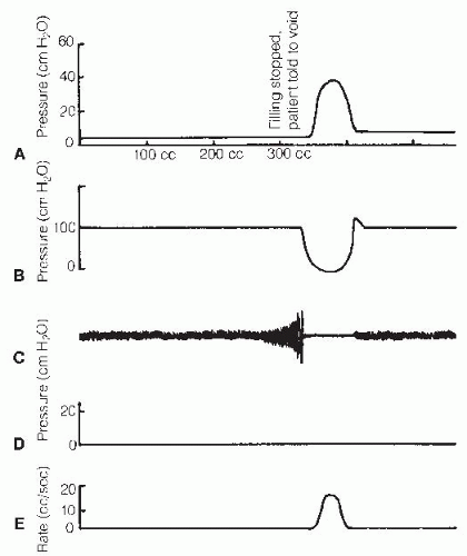
During the voiding phase, the detrusor pressures usually are less than 30 cm H2O in women and between 30 and 50 cm H2O in men. A normal maximum flow rate is 15 to 20 mL/s and should not be less than 10 mL/s in any age group. The patient should have at least 150 mL in the bladder because the flow rate depends on the voided volume (44). The flow usually has a bell-shaped curve, progressively increasing to its maximum rate and then decreasing. The urethral sphincter should remain open throughout voiding, and there should be no rises in intra-abdominal pressure during voiding. As previously discussed, there should be no postvoid residual, although postvoid residuals increase with age.

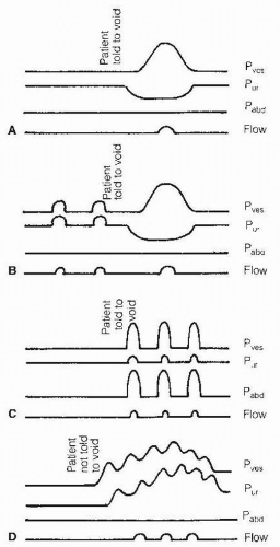
A single elevated postvoid residual during urodynamics should be interpreted with caution because the patient may be nervous and voluntarily stop the urine stream. Several catheterized or ultrasound postvoid residual tests should be done to confirm an increased urodynamic postvoid residual (Fig. 51-5). Urodynamics is able to characterize specific types of voiding patterns (Fig. 51-6).


Special Considerations in Children

At one time, urodynamic evaluation was delayed until a child was school-aged and definitive corrective surgery was to be performed. However, reflux and renal deterioration often occur during the first 3 years of life. McGuire and colleagues reported that there was a high incidence of renal deterioration in patients with urethral leak-point pressures greater than 40 cm H2O (92). Therefore, it is recommended that all myelodysplastic newborn children be evaluated as soon as possible (93).


FIGURE 51-5. Normal urodynamic findings. There is a minimal rise in intravesical pressure during the filling phase. The voiding phase is initiated with quieting of EMG activity, and relaxation of the external urethral sphincter is followed by a bladder contraction. A: Intravesical pressure (Pves). B: Urethral sphincter pressure (Pur). C: Urethral sphincter electromyography. D: Intra-abdominal pressure (Pabd). E: Urine flow rate.

It is difficult to obtain high-quality water-fill urodynamic studies on children younger than 4 or 5 years old. In younger children, it sometimes is necessary to use sedation or general anesthesia. It is important that children feel comfortable with the physician, nurses, and test. As a general principle, the amount of additional information gained from insertion of EMG needles usually is not enough to warrant the risk of obtaining poor urodynamics results from a crying, fearful child. This is especially true if it is anticipated that the child will come back for follow-up studies.


MANAGEMENT OF VOIDING DYSFUNCTIONS

A useful way to organize management of voiding dysfunctions is to base treatment options on a modification of the Wein classification: incontinence caused by (a) the bladder
or (b) the outlet (e.g., bladder neck, sphincter, or prostate) or retention caused by (a) the bladder or (b) the outlet. Management can be categorized as behavioral, pharmacological, surgical, or supportive. Table 51-3 shows various treatment options. Although each of these modalities is listed separately, it is important to note combinations within the same category (e.g., behavioral—timed voiding, fluid restriction, biofeedback) and separate categories (e.g., behavioral—timed voiding, pharmacologic—anticholinergics).


FIGURE 51-6. Schematic representation of various voiding patterns. A: Normal. B: Uninhibited contractions occur with filling. The sphincter is attempting to inhibit contractions. The patient has a normal voiding phase. C: No bladder contractions. Rises in bladder pressure result from rises in abdominal pressure (i.e., Valsalva voiding). D: Uninhibited contractions occur with simultaneous sphincter contractions (i.e., detrusor sphincter dyssynergia). Pabd, intra-abdominal pressure; Pur, urethral sphincter pressure; Pves, intravesical pressure.

It is important to characterize the type of voiding dysfunction that a person has with an urodynamics evaluation, particularly when considering pharmacological and surgical options (see Table 51-3). In addition to the type of voiding dysfunction, the physician needs to consider the type of disease process (i.e., progressive, stable, or remitting), cognition, mobility, family support, and medical conditions when recommending a bladder-management program. Empirical pharmacotherapy should be discouraged because there is a risk of potential side effects of drugs that may have no benefit or make the problem worse.


TABLE 51.3 Treatment Options for Voiding Disorders


Incontinence Caused by the Bladder


Behavioral: Scheduled (timed) voiding, limited fluid intake, biofeedback


Pharmacologic: Oral anticholinergics, antispasmodics, tricyclic antidepressants, DDAVP (Vasopressin), intravesical oxybutynin, intravesical C-fiber afferent neurotoxins (capsacin, resiniferatoxin),a calcium antagonists,a prostaglandin inhibitorsa


Surgical: Augmentation cystoplasty, urinary diversion, interruption of innervation, neurostimulation, botulinum toxin injections into the bladder walla


Supportive: Diapers, external condom catheter, intermittent catheterization, indwelling catheter


Incontinence Caused by the Sphincter


Behavioral: Scheduled voiding, pelvic floor exercises, biofeedback


Pharmacologic: Alpha-adrenergic agonists, estrogen, injectable periurethral bulking agent


Surgical: Artificial sphincter, urethral suspension, neurostimulationa


Supportive: Same as with bladder


Retention Caused by the Bladder


Behavioral: Scheduled voiding (↓ cognition/mobility), suprapubic tapping (bladder hypocontractility), Valsalva, Credé


Pharmacologic: Cholinergic agonists, intravesical prostaglandin,a narcotic antagonistsa


Surgical: Sphincterotomy, neurostimulation (if bladder contraction present)


Supportive: Intermittent catheterization, indwelling catheter


Retention Caused by the Sphincter/Outlet


Behavioral: Biofeedback, suprapubic tapping, anal stretch/scissoring


Pharmacologic: Alpha-adrenergic blockers, baclofen, diazepam, dantrolene


Surgical: Sphincterotomy, botulinum toxin injections, pudendal neurectomy, bladder outlet surgery, urethral stents, balloon dilationa


Supportive: Same as with bladder


a Investigational use.


The following are goals of management in patients with voiding dysfunctions:

Prevent upper-tract complications (e.g., deterioration of renal function, hydronephrosis, renal calculi, pyelonephritis).


Prevent lower-tract complications (e.g., cystitis, bladder stones, vesicoureteral reflux).

Develop a bladder management program that will allow patient to reintegrate most easily back into the community.


Therapy for Incontinence Caused by the Bladder


Behavioral Treatment Options

Many patients with incontinence caused by the bladder benefit from a scheduled (timed) voiding regimen. Patients with incontinence resulting from poor cognition, aphasia, or poor mobility but normal bladder function often are also helped by being placed on a commode or offered a urinal at set intervals. This is known as timed voiding. Patients who have an overactive detrusor may decrease incontinent episodes by voiding by the clock rather than waiting for a sense of fullness. They are taught to void before reaching their full bladder capacity because uninhibited contractions often become more forceful and frequent as the bladder is reaching its full capacity. Once the contraction begins it is very difficult to get undressed and go to the bathroom in time.

Another type of behavioral intervention is bladder training. This is done by progressively increasing the time between voiding by 10 to 15 minutes every 2 to 5 days until a reasonable interval between voiding is obtained (94). Bladder training often is effective for a person who has recovered or is recovering from a neurologic lesion (e.g., head injury, stroke) with improved bladder function but is voiding frequently out of habit or from fear of incontinence based on past experience.


Pharmacological Treatment Options

Pharmacologic treatment often is needed in addition to timed voiding in patients with incontinence caused by detrusor overactivity. There are currently a wide variety of medications available, which have anticholinergic effects. If a person does not tolerate one type of anticholinergic, he or she may tolerate another type of anticholinergic medication. Anticholinergic agent’s primary action is to block acetylcholine receptors competitively at the postganglionic autonomic receptor sites. Some agents, such as oxybutynin, also have a localized smoothmuscle antispasmodic effect distal to the cholinergic receptor site and a local anesthetic effect on the bladder wall (95). Some of the more common potential side effects of anticholinergic medications include dry mouth, pupillary dilatation and blurred vision, tachycardia, drowsiness, and constipation from decreased gastrointestinal motility. Newer anticholinergic agents have been developed to be more selective to the bladder receptors, or have a slow sustained release or topical patch developed in an attempt to lessen anticholinergic side effects, particularly dry mouth and constipation. It has not been shown whether these agents maintain their effectiveness for an entire 24-hour period. Twenty-four-hour effectiveness is important in those with SCI at or above T6 who have the potential to develop autonomic dysreflexia if their medications “wear off” and they begin to develop uninhibited contractions. In those with neurogenic bladders the object is frequently to completely shut down the bladder and cause retention so IC can be performed. Therefore, those with neurogenic detrusor overactivity often require more than the “standard” doses used for able-bodied individuals.

Tricyclic antidepressants sometimes are used alone or in combination with anticholinergic agents. These medications are thought to have a peripheral anticholinergic effect and a central effect. They have been found to suppress uninhibited bladder contractions, increase bladder capacity, and increase urethral resistance (96). There have been several reports of severe autonomic dysreflexia in SCI patients secondary to overdistention of the bladder with urine. Therefore, caution should be taken in giving these medications that depend on uninhibited contractions to void (reflex voiders).

Intravesical instillation of medications is sometimes used because oral anticholinergic medications have a number of side effects. The major advantage of intravesical medications is that there are minimal to no systemic side effects due to less systemic absorption from the bladder wall. This is particularly helpful for those with a neurogenic bowel, because anticholinergic medications frequently cause constipation that could lead to fecal impaction. Anticholinergic medications may also cause a dry mouth, which can be particularly difficult to tolerate for those trying to limit their fluids because of being on IC.

Intravesical lidocaine has been shown to be effective at suppressing uninhibited bladder contractions in those with overactive bladders (97). Although it has a rapid onset of action, it does not have a long duration of action. Therefore, this medication is best reserved for use in acute problems. For more long-term suppression of uninhibited bladder contractions, oxybutynin can be used for intravesical instillation (98). It not only has an anticholinergic effect but also a topical anesthetic effect. This medication is effective at suppressing uninhibited bladder contractions, but it still has the disadvantage of only being effective for 4 to 6 hours. One study reported using 5 to 10 mg dissolved in 15 to 30 mL of normal saline instilled into the bladder three to four times a day; this dose noted in the seven men resulted in an improvement in body image and enhanced sexuality because of the significant improvement in incontinence (99). Another investigator examined 32 patients, comparing standard dosages of intravesical oxybutynin (0.3 mg/kg body weight per day) with increasing dosages in steps of 0.2 mg/kg body weight up to 0.9 mg/kg body weight per day. Twenty-one of thirty-two (66%) patients became continent with the standard dose. Seven of the eleven failures at the lower dose became continent with a median dose of 0.7 mg/kg body weight for an overall success rate of 28 of 32 (87%). Four of the eleven (12.5%) had no improvement, and two of the eleven patients had side effects with a dosage of 0.9 mg/kg body weight per day (100). Despite its effectiveness, a number of individuals abandon using intravesical instillations because it is so labor intensive.

Intravesical instillations may, however, assume a more important role of helping to control uninhibited contractions with the development of longer-acting agents. Of particular interest is afferent C-fiber neurotoxins. The prototype
medication is capsaicin, which is effective at suppressing uninhibited contractions for several months at a time (101). Unfortunately, capsaicin frequently causes discomfort or suprapubic pain, urgency, hematuria, and autonomic dysreflexia, which can last to up to 2 weeks postinstillation.

A newer afferent C-fiber neurotoxin, resiniferatoxin, is being investigated. It is 1,000 times stronger than capsaicin and is long-acting. It has an extremely rapid onset of action at desensitizing the C-fiber afferent neurons, which causes minimal discomfort when it is instilled. In a study, 14 patients with detrusor hyperreflexia were instilled with 100 mL (or the bladder capacity if lower than that volume) of 50 to 100 nm resiniferatoxin instillation in 10% alcohol in saline. Treatment improved or abolished incontinence in 9 of 12 (75%) patients. Mean cystometric capacity increased from 182 to 330 mL. Maximal detrusor pressure was not modified by treatment. The effects were long-lasting, up to 12 months in seven patients (102). It is our understanding that Food and Drug Administration (FDA) multicenter trials evaluating resiniferatoxin in the United States have been stopped due to funding issues.

Desmopressin acetate has been found to decrease the number of episodes of nocturia in patients with multiple sclerosis. However, further studies are needed to determine its usefulness in the elderly because of the high prevalence of contraindications such as renal insufficiency, heart failure, and risks of inducing hyponatremia and fluid retention (29).

Another modality that has been gaining increasing popularity to help suppress uninhibited contractions in those with an overactive bladder is botulinum-A toxin injected into the bladder wall. While there have been encouraging data from early open-labeled and randomized, controlled trials with regards to efficacy and tolerability in both nonneurogenic and neurogenic bladder overactivity, the most experience has been with neurogenic bladder overactivity (103, 104, 105). Botulinum toxin inhibits acetylcholine release at the neuromuscular junction, which in turn blocks neuromuscular contraction and relaxes muscles that are either spastic or overactive. Doses ranging between 100 to 300 units have been confirmed by cystometry to suppress an overactive detrusor (103, 104, 105). Because it may take 1 to 4 weeks to completely deplete the acetylcholine at the neuromuscular junction, maximal effects at quieting the bladder do not occur immediately. Since there is reinnervation and sprouting at the neuromuscular junctions, the effects usually wear off after 3 to 6 months, so the injections usually need to be repeated. It appears that injections into the bladder wall may last closer to 6 months. Recent systematic reviews confirmed that botulinum toxin injections into the detrusor provide a clinically significant improvement in adults with neurogenic detrusor overactivity and incontinence refractory to other pharmacological therapy. It was well tolerated. The reviews point out that more studies are needed to evaluate issues such as the optimal dose, number and location of injections, and duration of effect (103, 104, 105). The panel for the SCI Consortium Guidelines on Management For Adults with SCI listed the use of botulinum injections into the bladder (detrusor) muscle as a bladder management option for those with SCI on IC with detrusor overactivity (106). Botulinum toxin to treat detrusor-sphincter dyssynergia is discussed in the pharmacologic agent section “Therapy for Retention Caused by the Outlet or Sphincter.”

In summary, there is a wide variety of pharmacologic agents that may be used by themselves or in conjunction with treatment modalities. A number of pharmacological agents discussed in this section as well as in the following sections are still undergoing investigation or “off-label” use. One reason is that individuals with “neurogenic bladders” are frequently excluded from initial FDA trials or do not make up a large enough population to have drug companies initiate trials in those with SCI. When using any pharmacologic agents for treatment, potential side effects and contraindications must be weighed against potential benefits.


Surgical Treatment Options


Bladder Augmentation

Bladder augmentation is a surgical technique that is frequently used to create a large bladder capacity with low intravesical pressures. Because this is a surgical procedure, other alternatives, such as pharmacologic treatment, should first be tried.

Surgical treatment is sometimes needed to improve bladder capacity in adult patients who are incontinent and want to perform IC. The SCI Consortium Bladder Management Panel recommended that bladder augmentation be considered for individuals with SCI who have (a) intractable involuntary bladder contractions causing incontinence, (b) the ability and motivation to perform IC, (c) the desire to convert from reflex voiding to an IC program, (d) a high risk for upper-tract deterioration secondary to hydronephrosis and/or ureterovesical reflux as a result of high-pressure detrusor-sphincter dyssynergia (106). They also recommended to consider avoiding bladder augmentation in individuals with (a) inflammatory bowel disease, (b) pelvic irradiation, (c) severe abdominal adhesions from previous surgery, and (d) compromised renal function (106).

An extensive preoperative evaluation is important. The history should include questions about any gastrointestinal problems. Urodynamics should be done to evaluate bladder and sphincter function. Various treatments may be needed to treat the sphincter if there is a low leak point pressure. Screening laboratory work should include liver and renal function. To help reduce the risk of significant acidosis and metabolic abnormalities, bladder augmentation is best reserved for those with serum creatinine less than 2.0 mg/dL. A cystogram should be done to evaluate for vesicoureteral reflux. Ureteral reimplantation may be considered if there is significant reflux. Upper-tract evaluation is important both to rule out any problems and to serve as a baseline for follow-up post augmentation (107).

There are a number of different techniques of bladder augmentation in which different segments of bowel can be used. The most common type of bladder augmentation is the clam cystoplasty. This procedure involves isolating a piece of
intestine, being careful to keep it attached to its mesentery, detubularizing it, and sewing it onto the bladder, which is first partially bivalved. Various bowel segments can be used and depend on the surgeon’s preference.

There are predictable metabolic abnormalities depending on the segment being used. The stomach mucosa has secretory epithelium with little resorptive function. Gastric mucosa secretes hydrochloric acid in conjunction with systemic bicarbonate release. Therefore, hypochloremic metabolic alkalosis can result if the stomach is being used, particularly if there is poor renal function. However, the stomach has the least absorptive properties and is best if one is concerned about metabolic acidosis from reabsorption of urinary solutes through the bowel wall. However, this is technically more difficult than an intestinal segment closer to the bladder.

The jejunal mucosa is different from the ileum and large intestine in that it secretes sodium and chloride and may result in hyponatremia, hypochloremia, and hyperkalemia. This segment is most likely to result in metabolic abnormalities and rarely used in diversions. The ileum and colon have similar transport mechanisms. Ammonia and chloride are reabsorbed. This can lead to hyperchloremic metabolic acidosis.

The most frequent changes that occur after this are an increase in mucus noted in the urine, possible metabolic changes, abnormal drug absorption (especially those that are absorbed by the gastrointestinal tract and excreted unchanged by the kidneys, such as Dilantin and certain antibiotics), osteomalacia from chronic acidosis, and stones, particularly in those with urea-splitting organisms and hyperchloremic metabolic acidosis. Long-term consequences of bowel attached to bladder are unknown. There have also been case reports of cancer in those with bladder augmentations, ileal conduits, and colon conduits. These have been adenocarcinomas, undifferentiated carcinomas, sarcomas, and transitional cell carcinomas (108, 109, 110, 111, 112, 113, 114). There have been a few reports of patients who had an augmentation cystoplasty more than 10 years previously developing adenocarcinoma in the bladder (110).

Only gold members can continue reading. Log In or Register to continue

Stay updated, free articles. Join our Telegram channel

May 25, 2016 | Posted by in PHYSICAL MEDICINE & REHABILITATION | Comments Off on Neurogenic Bladder and Bowel

Full access? Get Clinical Tree
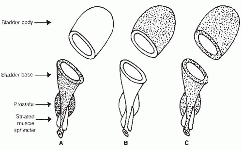
Get Clinical Tree app for offline access