Introduction
The first four lumbar nerves form the lumbar plexus, which lies embedded in the psoas muscle in the posterior abdominal wall. A second sacral plexus is formed from the fifth lumbar to the fourth sacral spinal nerves. These spinal nerves of the sacral plexus are part of the cauda equina (see Chapter 4, Figure 4.2) and enter the pelvis through the anterior foramina of the sacrum.
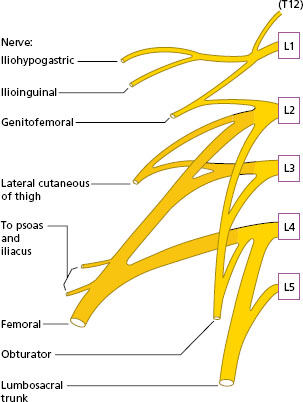
Figure 9.1 shows the position of the lumbar and sacral plexuses in relation to the lumbar spine, the pelvis and the hip. The course of the muscular branches of the lumbar and sacral plexus will be described; the cutaneous branches will be mentioned in outline only.
Lumbar plexus: position and formation
The upper four lumbar nerves (L1–L4) form the lumbar plexus, which lies in the psoas muscle alongside the lumbar vertebrae in the posterior abdominal wall. Branches direct from the plexus supply the hip flexors (psoas and iliacus) and quadratus lumborum (see 10). The branches of the plexus emerge from the psoas. Most of the fibres of L4 join the lumbar plexus, but the remainder join L5 to form the lumbosacral trunk, which is part of the sacral plexus. Figure 9.2 shows the roots of the lumbar plexus and the formation of the main terminal branches.
Terminal branches of the lumbar plexus
Three important nerves are formed from the lumbar plexus:
- the femoral nerve supplying the anterior muscles of the thigh;
- the lateral cutaneous nerve supplying the skin on the lateral side of the thigh;
- the obturator nerve supplying the medial muscles of the thigh.
Figure 9.1 shows the three nerves in relation to the pelvis and the hip.
Figure 9.2 Lumbar plexus: roots and main terminal branches.
Femoral nerve
This is the largest nerve of the lumbar plexus. It lies in the psoas muscle in the pelvis, and then emerges from the lateral border of the muscle to lie between the psoas and iliacus, leaving the pelvis anteriorly under the inguinal ligament. In the thigh, the femoral nerve branches to supply the quadriceps group of muscles. The saphenous nerve, a branch of the femoral nerve in the thigh, becomes cutaneous at the medial side of the knee and continues on to the medial side of the ankle (Figure 9.3).
Lateral cutaneous nerve
This nerve emerges from the lateral side of the psoas muscle and crosses the iliacus obliquely to the anterior superior spine of the ilium (Figure 9.4). The nerve passes under the lateral end of the inguinal ligament and becomes cutaneous in the lateral thigh. Pressure on the nerve in the area of the iliac spine causes loss of sensation on the lateral side of the thigh.
Figure 9.3 Right femoral and saphenous nerve.
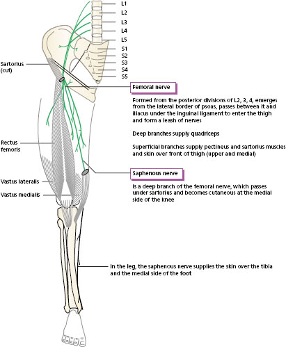
Figure 9.4 Right obturator nerve and lateral cutaneous nerve.
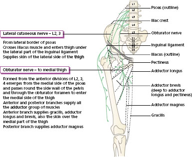
Obturator nerve
This leaves the medial side of the psoas muscle at the brim of the pelvis and passes through the obturator foramen of the hip bone to reach the medial side of the thigh (Figure 9.4). In the medial compartment of the thigh, the obturator nerve supplies all of the adductor group of muscles.
Functional importance of the femoral and obturator nerves
The femoral and obturator nerves are both important in walking. The quadriceps, supplied by the femoral nerve, stabilises the knee during support. It propels the body upwards in standing up from sitting. In climbing stairs the quadriceps acts concentrically to lift the body on to the next step, and eccentrically in coming down. The hip adductors, supplied by the obturator nerve, are active to shift the body weight over the supporting foot when the other leg is off the ground. If the adductors are weak owing to damage of the obturator nerve, the leg swings outwards instead of forwards in the swing phase in walking.
Other nerves of the lumbar plexus
Three other cutaneous nerves are formed from the first two nerves of the lumbar plexus. The first lumbar nerve divides into two, the iliohypogastric and the ilioinguinal nerves, which supply the skin of the buttock and groin, respectively. A third cutaneous nerve, the genitofemoral, is formed from L1 and L2, and supplies a small area of skin on the upper front part of the thigh.
Sacral plexus: position and formation
The sacral plexus is formed in the pelvis from the joining of the lumbosacral trunk (L4, L5), the first three and part of the fourth sacral nerves. The landmark to find the position of the sacral plexus in the pelvis is the piriformis muscle, lying across the posterior aspect of the hip joint. The main part of the plexus passes backwards with the piriformis to enter the posterior compartment of the thigh. Figure 9.1 shows the emerging sacral nerves lying on the anterior surface of the sacrum. Figure 9.5 represents the roots of the sacral plexus and the main terminal branches.
Related posts:


Stay updated, free articles. Join our Telegram channel

Full access? Get Clinical Tree


