Injury to peripheral nerves can be broken down into two pathologic changes: damage to the myelin or damage to the axon (i.e., Wallerian degeneration). In demyelination, destruction of the myelin sheath occurs without axonal damage. Demyelinating injuries can slow electrical conduction over the entire length of the nerve, multiple segments of the nerve, a focal area of the nerve, or produce a conduction block (when focal demyelination is so severe that nerve action potential propagation across that segment does not occur) [3]. In Wallerian degeneration, the axon degenerates distally following transection or severe injury to the nerve. The time required for degeneration varies between sensory and motor segments and is related to the size and myelination of the fiber [4].
Mononeuropathy is characterized by injury to a single peripheral nerve. Nerve damage may be caused by vascular, muscular, or tumor compression; bony entrapment of the nerve; trauma (sharp, blunt, repetitive stress); toxins; metabolic syndromes (diabetes); vascular ischemia; and iatrogenic injury. The aim of this chapter is to provide a comprehensive overview of mononeuropathies around the hip with discussion of the evaluation of nerve injury, relevant anatomy, etiology, clinical presentation, and management. The mononeuropathies of the hip that will be discussed in this chapter include:
1.
Iliohypogastric neuropathy
2.
Ilioinguinal neuropathy
3.
Genitofemoral neuropathy
4.
Lateral femoral cutaneous neuropathy
5.
Obturator neuropathy
6.
Femoral neuropathy
7.
Sciatic neuropathy
8.
Superior gluteal neuropathy
9.
Inferior gluteal neuropathy
10.
Pudendal neuropathy
Evaluation of a Nerve Injury
The most important element in evaluating a peripheral nerve injury is precise knowledge of the course of the nerve, the terminal muscle innervations of the nerve, the spinal nerves that contribute to its motor branches, and the other muscles that these spinal nerves innervate. Knowledge of dermatomes, although important, often does not provide as clear of a distinguishing picture between peripheral nerve injury and radiculopathy, unless it is a purely sensory nerve. The knowledge of nerve anatomy and associated motor function will enhance the physical examination of the patient as well as the interpretation of the electrodiagnostic studies (Table 2). Most mononeuropathies can be preliminarily diagnosed by history and clinical exam; however, the gold standard for diagnosis of a mononeuropathy is electrodiagnostic (EDX) studies involving electromyography (EMG) and nerve conduction studies (NCS).
Table 2
Motor and sensory innervation of each peripheral nerve around the hip
Lumbosacral plexus | |||
|---|---|---|---|
Peripheral nerve | Root level | Muscle innervation | Cutaneous innervation |
Iliohypogastric | T12–L1 | • Transversus abdominis • Abdominal internal oblique | Anterior cutaneous branch: • Small area of the skin above the pubis Lateral cutaneous branch: • Upper buttock as far as the greater trochanter |
Ilioinguinal | L1 | • Transversus abdominis • Abdominal internal oblique | • Groin • Proximal medial thigh • Base of the penis and the upper part of the scrotum in men • Mons pubis and labium majorum in women |
Genitofemoral | L1–L2 | • Cremaster muscle in males • No motor innervations in women | Genital branch: • Pubis • Scrotum in men or mons pubis and labium majorum in women Femoral branch: • Upper part of the femoral triangle • Small patch of the skin on the proximal anterior thigh |
Lateral femoral cutaneous | L2–L3 | • No motor innervations | • Anterolateral aspect of the thigh |
Obturator | L2–L4 | Anterior branch: • Adductor longus • Adductor brevis • Gracilis • Pectineus Posterior branch: • Obturator externus • Adductor brevis • Proximal portions of adductor magnus | Anterior branch: • Distal 2/3rds of the medial thigh Posterior branch: • Articular capsule, cruciate ligaments, and synovial membrane of the knee joint |
Femoral | L2–L4 | • Iliopsoas • Pectineus Anterior branch • Sartorius Posterior branch • Quadriceps: rectus femoris, vastus lateralis, vastus medialis, vastus intermedius | Anterior branch (intermediate and medial cutaneous nerves) • Anteromedial aspect of the thigh Posterior branch (saphenous nerve) • Anteromedial aspect of the leg, medial malleolus, and arch of the foot |
Sciatic | L4–S3 | Tibial division: • Semitendinosus • Semimembranosus • Long head of biceps femoris • Ischiocondylar part of adductor magnus • Gastrocnemius/soleus muscles • Posterior tibialis • Long toe flexor Peroneal division: • Short head of biceps femoris • Anterior tibialis • Long toe extensor • Peroneus longus/brevis | • Posterior gluteal region • Posterior thigh • Entire lower leg, ankle, and foot (except for the medial lower leg) |
Superior gluteal | L4–S1 | • Gluteus medius • Gluteus minimus • Tensor fasciae latae | • No sensory innervations |
Inferior gluteal | L5–S2 | • Gluteus maximus | • No sensory innervation |
Pudendal | S2–S4 | Inferior rectal nerve • External anal sphincter Perineal nerve • Muscles of the perineum • Erectile tissue of penis • External urethral sphincter | Inferior rectal nerve • Lower anal canal and perianal skin Perineal nerve • Perineum • Scrotum/labia Dorsal nerve of penis or clitoris • Skin of penis/clitoris |
Physical Examination
The evaluation of a mononeuropathy is anchored on a through neurologic exam that includes manual muscle testing, sensation testing to light touch, and reflex testing. Recommended manual muscle testing for a mononeuropathy of the hip includes hip flexor, hip abductor, hip adductor, knee extensor, knee flexor, ankle dorsiflexor, ankle plantar flexor, and great toe extensor muscle testing. Sensation testing to light touch should include L2–S1 dermatomes. Reflex testing should include bilateral patellar, medial hamstring, and Achilles reflexes. A Tinel’s sign over the specific nerve in question is onsidered special testing for a mononeuropathy. A Tinel’s sign is elicited by gentle percussion by a finger or hammer along the course of an injured nerve. A positive Tinel’s sign is acquired by the presence of transient tingling or pain in the distribution of the injured nerve that recreates typical pain. Tingling or pain isolated to the focal area of percussion does not constitute a positive Tinel’s sign. When suspicious for a nerve injury, the physical examination should be used to help distinguish between mononeuropathy, mononeuropathy multiplex, peripheral neuropathy, and radiculopathy.
Electrodiagnosis
EDX studies are the gold standard for diagnosis of most mononeuropathies. An EDX examination is not necessary for every patient with suspected nerve injury, but can be extremely helpful in localizing the lesion, assessing severity of the injury, and estimating prognosis for recovery. EDX studies involve two types of examination: EMG and NCS. In many of the smaller and deep nerves around the hip, it is very difficult to obtain reliable NCS; therefore, EMG is needed to confirm the diagnosis and rule out other potential pathologies (e.g., radiculopathy, plexopathy, or myopathy). There are many nuances to the performance and analysis of EDX testing that make it complicated and beyond the scope of this chapter. There is some variability in the interpretation of EDX studies. It is important to refer the EDX studies to physiatrists or neurologists that are comfortable in performing advanced EDX for the hip. A basic understanding of electrophysiology can help one assess the quality of the EDX study.
Nerve Conduction Studies
There are two different types of NCS: motor and sensory. Motor NCS involves the electrical stimulation of a peripheral nerve at two points (proximal and distal). The stimulation of the nerve generates action potentials that propagate down to evoke a response in the muscle distally. The amplitudes, onset latencies, and velocities of the compound muscle action potential (CMAP) for each motor nerve tested are recorded and compared to normative data or to the asymptomatic contralateral side. The CMAP represents the summation of electrical signals discharged by the muscle fibers’ depolarizing membranes. Sensory NCS records the sensory nerve action potential (SNAP) on the skin after proximal stimulation of the nerve. The SNAP is the summation of electrical signals discharged by the sensory neurons’ depolarizing membranes. The peak latency and conduction velocity of the SNAP is also compared to normative data. The amplitudes of SNAP vary widely and, therefore, are not useful in the analysis for neuropathy.
Prolonged onset latency (motor NCS) or peak latency (sensory NCS) implies that the conduction of the electrical impulse down the nerve was delayed. This can mean that there is some injury to the myelin; however, the preferred way to assess the integrity of the myelin sheath is by conduction velocity. A prolonged motor NCS conduction velocity is more specific for a demyelinating injury and can even localize the specific segment of nerve that is injured.
The amplitudes of the CMAP are used to determine the presence of axon loss, conduction block, or both. Axonal loss is characterized by decreased CMAP amplitudes at both the distal and proximal sites of stimulation to the motor nerve. There is no axonal loss associated with conduction block, but the injury to the myelin is so severe that it essentially blocks most, if not all, conduction down the nerve giving the false appearance that the axons are no longer intact. It is characterized by diminished proximal and preserved distal CMAP amplitudes. A mixed picture of conduction block with axonal loss is associated with a pattern of decreased distal CMAP amplitude and an even greater decrease in proximal CMAP amplitude.
Electromyography
EMG involves insertion of a needle electrode directly into the muscle. A comprehensive EMG involves testing numerous muscles specifically selected based on the suspected pathology in question. There are two portions of an EMG conducted for each muscle tested: insertional activity (irritation of the sarcolemma during a resting state of the muscle) and muscle activation (voluntary activation of the muscle at varying degrees of effort). During the insertional activity phase of testing, abnormal spontaneous discharges implies acute denervation of that muscles. During the muscle activation phase of testing, the patient is asked to engage the muscle at minimum and maximum strength. This will lead to active firing of motor units. The motor unit action potential (MUAP) morphology and recruitment patterns can distinguish muscles with chronic denervation and potential reinnervation.
The role of EMG in the diagnosis of mononeuropathy is straightforward. For a purely demyelinating lesion of the nerve, you would expect a completely normal EMG. Severe axonal pathology accompanying a mononeuropathy will result in abnormal EMG findings in all the muscles innervated by that nerve. For a comprehensive EMG exam, one would also test muscles of different peripheral nerves, but common spinal nerve roots to rule out a lumbosacral radiculopathy, mononeuropathy multiplex, or a lumbosacral plexopathy .
Ordering Electrodiagnostic Studies
The timing of the EDX study is critical to the interpretation of the study. False conclusions can occur if the EDX study is not ordered at the appropriate time. The ideal time to order an EDX study is approximately four weeks after the onset of symptoms. One can order EDX studies earlier as long as they understand the limitations of the study at each time point. Immediately after injury to a nerve, EDX findings are subtle for both demyelinating and axonal lesions and can be missed. Seven days after a complete nerve lesion Wallerian degeneration will have progressed to the point where distal stimulation of motor axons elicits no motor response. Ten days after onset of a complete lesion, SNAPs will be absent as well. Demyelinating injuries can be distinguished from axonal injuries at 7–10 days after onset of injury. The needle EMG portion will start to show active signs of denervation 2–3 weeks after onset of injury [5].
Interpreting the Electrodiagnostic Study
Understanding the varying degrees of demyelinating injuries due to a mononeuropathy and how to interpret them on EDX studies can help with prognosis. For instance, some subtle neurapraxias can demonstrate completely normal EDX results; therefore, they carry the most favorable prognosis for recovery. A demyelinating injury to the nerve with the presence of prolonged latencies and conduction velocities on NCS is the least severe type of EDX-confirmed mononeuropathies. The quantitative difference between the observed abnormal latencies and conduction velocities compared to the normative standards or contralateral asymptomatic side can also provide information on the severity of the injury. Evidence of a conduction block is considered a more severe injury to the myelin. A mixed conduction block with axonal loss is considered a progressively worse injury to the nerve. Finally, EMG evidence of denervation in a muscle supplied by the peripheral nerve in question connotes the most severe type of mononeuropathy, with the worst prognosis for recovery. In the setting of severe axonal injury, MUAP morphology can reveal acute denervation, chronic denervation, and evidence of reinnervation. These features can be used to follow a nerve injury over time and add to the prognosis for recovery.
Clinical Presentations of Nerve Injury Around the Hip
Nerve injury can result in considerable morbidity. Due to the diverse features of every nerve, the distinct clinical presentation of each mononeuropathy of the hip varies widely. All nerves around the hip originate from the lumbosacral plexus which is formed within the psoas major muscle by the merging of the lumbar plexus (anterior primary rami of L1–L4) and the sacral plexus (anterior primary rami of L5–S3) (Fig. 1).
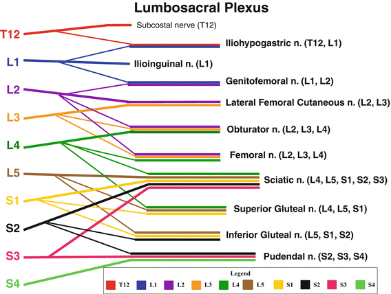
Fig. 1
Lumbosacral Plexus Anatomy. Pictoral representation of the lumobosacral root contribution to each peripheral nerve
Iliohypogastric Neuropathy
Anatomy
The iliohypogastric nerve (IHN) is a branch of the lumbar plexus arising from the primary ventral rami of L1 and a communicating branch of T12. The IHN passes through the psoas muscle and then descends. Approximately halfway between the anterior superior iliac spine (ASIS) and the highest point of the iliac crest, the nerve pierces the muscles of the abdominal wall [6]. The nerve supplies the lower fibers of the transversus abdominis and internal oblique muscles. It divides into lateral and anterior cutaneous branches. The lateral cutaneous branch crosses the iliac crest to innervate a patch of skin in the upper buttock, and the anterior cutaneous branch courses just above the inguinal ligament to supply a small area of skin above the pubis.
Etiology
Disorders of the IHN are rare. The most common causes of injury are surgical procedures involving transverse lower abdominal incisions such as hysterectomy, inguinal herniorrhaphy, and appendectomies. The main trunk of this nerve can be damaged by retroperitoneal tumors or large surgical incisions, producing sensory abnormalities in the distribution of the nerve and bulging of the lower abdominal muscles [6].
Clinical Presentation
Injury to the IHN is characterized by anesthesia, pain, and paresthesias to the lower abdomen and groin. Patients with injury to the anterior branch of the IHN report a suprapubic sensory disturbance, whereas patients with injury to the lateral branch report an isolated sensory disturbance over the upper buttock. There is some dermatomal overlap with the ilioinguinal nerve, which makes it clinically difficult to differentiate them. Weakness of the lateral abdominal wall musculature can be present; however, these muscles receive other innervation from the lower intercostals nerves. Furthermore, lateral abdominal muscle testing is difficult to reliably assess.
Diagnosis
The gold standard of diagnosis is a local anesthetic block of the nerve. Pain relief after the block confirms the diagnosis. These blocks are currently being performed under ultrasound guidance. There is no reliable EDX that can be performed to validate the diagnosis. Needle EMG of the lower abdominal musculature may serve as an adjunct in the diagnosis, although it is not commonly performed. Differential diagnosis includes upper lumbar or lower thoracic radiculopathy.
Ilioinguinal Neuropathy
Anatomy
The ilioinguinal nerve (IIN) arises as a branch of the lumbar plexus and is derived from the L1 ventral rami. The nerve pierces through the psoas muscle and follows a parallel course to the IHN as it runs distally and anteriorly, staying deep to the abdominal musculature. Adjacent to the ASIS, the nerve sends motor contributions to the inferior portions of the transversus abdominis and internal oblique muscles and cutaneous contributions to innervate a strip of skin over the iliac crest. The remainder of the nerve enters the inguinal canal and divides to provide cutaneous innervation for the groin, proximal medial thigh, base of the penis, and upper part of the scrotum, mons pubis, and labium majorum.
Related posts:
 Neuromuscular Hip Disorders: Focus on Cerebral Palsy
 Surgical Technique: Bone Graft for Avascular Necrosis of the Hip
 Rehabilitation of Non-Operative Hip Conditions
 Surgical Technique: Open Proximal Hamstring Repair
 Subspine Impingement and Surgical Technique
 Atraumatic Instability and Surgical Technique
Stay updated, free articles. Join our Telegram channel

Full access? Get Clinical Tree


