Myopathy


Myopathy


Jay J. Han

David D. Kilmer


Representing a diverse group of disorders primarily affecting skeletal muscle, myopathies are an important cause of disability affecting patient mobility, self-care, and independence. In addition to weakness, many myopathies have associated dysfunction in other organ systems, such as the cardiac and pulmonary systems. The disability associated with muscle disease depends on the specific type, extent of clinical involvement, and rate of progression.

The number and type of different muscle disorders under the umbrella term myopathy are vast and expanding. With our increasing knowledge about genetic and molecular basis for these disorders, classification and nomenclature regarding myopathies are constantly being reevaluated and modified. A detailed discussion of all the different myopathies is beyond the scope of this chapter; however, this chapter is intended to provide the physical medicine and rehabilitation specialist with an overview of diagnostic approach, clinical characteristics, and care and management of patients with myopathies, with emphasis on few of the most common diagnoses that a physiatrist may encounter.

Although most myopathies remain largely incurable, as is the case for most neuromuscular diseases at this time, they are not untreatable. Rehabilitation specialists have an important role in the care of patients with myopathies to maximize their functional capacities, prolong or maintain independent locomotion and function, prevent physical deformity and medical complications, and provide access for integration into the community with quality of life in mind. The comprehensive management of all the varied clinical problems associated with myopathies and other neuromuscular diseases often requires specialists from neurology, cardiology, pulmonology, and orthopedic surgery as well as clinical specialists in physical therapy, occupational therapy, speech therapy, and orthotics. Coordination of this difficult and demanding task may be best handled by a neuromuscular and rehabilitation medicine specialist knowledgeable in various myopathies. It is important for the rehabilitation physician to understand these diseases in order to appropriately treat the functional problems caused by muscle weakness as well as provide comprehensive interdisciplinary rehabilitation through awareness of other manifestations of the disease.


DEFINITION AND CATEGORIES OF MYOPATHY

In the peripheral nervous system, a primary defect may occur at the level of the anterior horn cell, peripheral nerve, neuromuscular junction, or muscle. A disease process in which the primary abnormality is at the level of the muscle itself is termed myopathy. A brief overview of the various myopathies is presented in this section to help the reader classify numerous myopathies in an orderly fashion. A more detailed discussion of the individual myopathies pertinent to rehabilitation specialists is found in the subsequent sections.

There are three basic categories of myopathy: hereditary, acquired, and myopathies associated with systemic disease (Table 30-1). In the hereditary myopathies, all of the myopathies have their inheritance pattern characterized or gene mutations identified. This category of myopathy is further subdivided into muscular dystrophies, congenital, distal, metabolic, and mitochondrial myopathies. In general, the muscular dystrophies present with significant structural defect of the muscle cell due to mutations in genes crucial for its normal function. Typically, the muscular dystrophies are accompanied by progressive muscle fiber degeneration, atrophy, and inflammation as noted by histopathological evaluation. The specific muscular dystrophies that are more commonly encountered, with its characteristic inheritance pattern, affected genes or mutations are listed (Table 30-2). Congenital myopathies are a group of relatively nonprogressive muscle diseases that present during infancy or early childhood and are classified largely based on clinical features and muscle biopsy morphological findings. Distal myopathies, as the name implies, have more pronounced weakness involving distal limb muscles rather than the typical proximal weakness seen with majority of other myopathies. Metabolic myopathies are caused by gene mutations that result in either abnormal glycogen or lipid metabolism. Usually, deficiency of an enzyme results from a gene mutation and causes an abnormal accumulation of substrate or a deficiency of the product of the enzymatic pathway. Mitochondrial myopathies are a group of muscle disorders with maternal inheritance pattern associated with abnormal structure and function of mitochondria. Since mitochondria are important in energy production throughout the body, other organ system involvements are also common including the nervous, cardiac, gastrointestinal, pulmonary, and endocrine systems. Diagnosis is often based on a combination of clinical findings and associated biochemical defects, along with histological abnormality noted as “ragged red fibers” on the modified Gomori trichrome stain. Another group of myopathies called channelopathies include primary muscle disorders caused by inherited abnormalities of various ion channels found on cell membranes. These include myotonia congenita, paramyotonia congenita, and primary hyperkalemic and hypokalemic periodic paralysis.


TABLE 30.1 Myopathies


Hereditary Myopathies


Acquired Myopathies


Muscular dystrophies


Inflammatory myopathies


Duchenne muscular dystrophy (DMD)


Polymyositis (PM)


Becker muscular dystrophy (BMD)


Dermatomyositis (DM)


Myotonic muscular dystrophy (DM1 and DM2)


Inclusion body myositis (IBM)


Facioscapulohumeral muscular dystrophy (FSHD)


Toxic myopathies


Limb-girdle muscular dystrophy (LGMD)


Corticosteroid myopathies


Congenital muscular dystrophy (CMD)


Lipid-lowering agent myopathies


Oculopharyngeal muscular dystrophy (OPMD)


Alcohol-related myopathies


Emery-Dreifuss muscular dystrophy (EDMD)


Myopathies related to other medications


Congenital myopathies


Endocrine myopathies


Central core, nemaline, centronuclear, multicore


Myopathies with glucocorticoid abnormalities


Fiber type disproportion, reducing body


Myopathies with thyroid disease


Fingerprint, cytoplasmic body, myofibrillar


Myopathies with parathyroid disease


Metabolic myopathies


Myopathies associated with pituitary dysfunction


Disorders of glycogenoses


Myopathies related to electrolyte disturbance


Disorders of lipid metabolism


Infectious and granulomatous myopathies


Respiratory chain defects


Viral, bacterial, fungal, tuberculous, parasitic


Distal myopathies


Sarcoid myopathy


Welander, Markesbery-Griggs-Udd, Nonaka, Miyoshi, Laing


Mitochondrial myopathies


Myopathies associated with systemic disease


Kearns-Sayre’s syndrome, Progressive External Ophthalmoplegia (PEO)


Critical illness myopathy


Mitochondrial Encephalomyopathy Lactic Acidosis Stroke (MELAS)


Myoclonic Epilepsy Ragged Red Fibers (MERRF)


Neuropathy Ataxia Retinitis Pigmentosa (NARP), Myopathy and external ophthalmoplegia Neuropathy Gastro-Intestinal Encephalopathy (MNGIE)


Electrolyte disturbances


Leber’s Hereditary Optic Neuropathy (LHON), Leigh’s syndrome


Channelopathies


Paraneoplastic


Myotonia congenita


Paramyotonia congenita


Primary hyperkalemic and hypokalemic periodic paralysis


TABLE 30.2 Inheritance Pattern and Gene Mutations of Muscular Dystrophies


Muscular Dystrophies


Inheritance Pattern


Gene Loci


Gene (Mutations)


DMD


X-linked


Xp21


Dystrophin


BMD


X-linked


Xp21


Dystrophin


DM1


AD


19q13.3


DMPK (expansion of CTG repeat)


DM2


AD


3q21


ZNF9 (expansion of CCTG repeat)


FSHD


AD


4q35


DUX4 (deletions of D4Z4 repeats)


LGMD (type 1s)


AD


Multiple loci


Myotilin, laminin A/C, caveolin-3


LGMD (type 2s)


AR


Multiple loci


Calpain-3, dysferlin, sarcoglycans, Fukutin-related protein (FKRP)


OPMD


AD or AR


14q11.2-q13


PABN1 and PABP2 (Polyadenylate-binding protein, Nuclear) (expansion of GCG repeat)


EDMD 1


X-linked


Xq28


Emerin


EDMD 2


AD


1q21.2


Laminin A/C


CMD


AR


6q22


Merosin


AD, autosomal dominant; AR, autosomal recessive.


The second category, acquired myopathies, consists of inflammatory, endocrine, toxic, granulomatous, and infectious myopathies. Under inflammatory myopathies are polymyositis (PM), dermatomyositis (DM), and inclusion body myositis (IBM). Muscle disorders associated with various endocrinopathies are now well recognized. These include myopathies associated with thyroid dysfunction (hyperthyroidism or hypothyroidism), adrenal disease, pituitary dysfunction, and parathyroid dysfunction (hyperparathyroidism or hypoparathyroidism). Myopathies can also result from electrolyte disturbances, including abnormalities of serum potassium, sodium, calcium, magnesium, and phosphorus. Under the toxic myopathy category, the most common agents associated with myopathy include HMG-CoA reductase inhibitors (cholesterol-lowering agents), corticosteroids, fibric acid derivatives (lipid- and cholesterol-lowering agents), chloroquine and amiodarone (amphiphilic drugs), colchicine and vincristine (antimicrotubular agents), zidovudine (HIV medication), and alcohol. Of the toxic myopathies, alcohol-related myopathy is thought to be the most common with both acute and chronic manifestations, often associated with heavy and prolonged alcohol use. Although typically asymptomatic in terms of muscle manifestation, sarcoidosis can present in the form of a granulomatous myopathy. Lastly, infectious myopathies are associated with essentially all types of infectious agents including viral (coxsackievirus, HIV, HTLV-1), bacterial, fungal, tuberculous, as well as parasites.

The last category is myopathies associated with systemic disease. Under this category are myopathies that have significant systemic processes that result in derangement of muscle function and health. The most common etiologies are severe multiorgan failure with sepsis, electrolyte disturbances associated with systemic disease, and underlying neoplasms.

Clinical features and progression vary within and between these categories as pathophysiology of each muscle disorder is different. Some of the myopathies, partly due to their time course of progression, involvement of other organ systems, prevalence in the population, and the availability of rehabilitative treatment options, may be more or less pertinent to rehabilitation specialists. However, in order to devise an appropriate rehabilitation plan, the rehabilitation physician should understand the expected disabilities and prognosis associated with the specific cause of myopathy.


EVALUATION OF THE PATIENT WITH SUSPECTED MYOPATHY


History

The primary symptom of a patient with suspected myopathy is weakness, defined as a reduction in maximal force generated by a muscle or muscle group (Table 30-3). This weakness may be fairly acute or insidious. Because the weakness is typically in the proximal musculature, certain functional problems should alert the clinician to the possibility of myopathy: difficulty getting up from a chair or toilet seat, trouble descending and climbing stairs, or difficulty with overhead activities, such as dressing, grooming, or reaching cabinets (Table 30-4). Symptoms suggesting distal weakness, such as problems opening jars, may be prominent with certain myopathies. The symptom of muscle fatigue, defined as the inability to sustain a given level of force for a certain period, is often difficult to assess. Although it may be associated with myopathy, when fatigue is the predominant symptom, other pathologic processes, such as neuromuscular junction disease and upper motor neuron disease, are more likely.


TABLE 30.3 Clinical Features and Laboratory Findings Suggestive of Myopathies


Proximal symmetric weakness


Normal sensation


Normal or mildly diminished tendon reflexes


Elevation of serum CK


EMG demonstrating brief, low-voltage, polyphasic potentials


Normal nerve conduction studies


Muscle biopsy with muscle fiber necrosis and regeneration, with central nuclei


Muscle pain, or myalgia, is a common presenting symptom, particularly in the inflammatory myopathies. However, the absence of pain should not distract the clinician from strongly considering the diagnosis of myopathy. When myalgias are the predominant symptom without demonstrated weakness, other disorders are more likely. History of myoglobinuria associated with weakness or fatigue symptoms should be sought and can help in the workup of muscle disorders, especially the metabolic myopathies. The presence of paresthesias or dysesthesias on history is certainly helpful, because they make the presence of myopathy very unlikely. A rare patient might interpret myalgias with descriptors sounding like sensory symptoms, distracting the clinician.

One of the most critical pieces of information is the family history. Whenever a myopathy is suspected that may have a genetic cause, a detailed family history and pedigree chart are essential. In an X-linked recessive disorder such as Duchenne
muscular dystrophy (DMD), men on the maternal side of the family are affected about 50% of the time and women are carriers in an equal percentage. Autosomal recessive disorders, such as many limb-girdle syndromes, frequently have no family members involved, making the diagnosis of a familial disorder more difficult. In an autosomal dominant disorder such as myotonic muscular dystrophy or facioscapulohumeral dystrophy (FSHD), typically 50% of offspring within a pedigree are affected. Sporadic cases resulting from new genetic defects occur with most autosomal dominant and sex-linked dystrophies, making a dystrophy possible even in the absence of a suspicious family history.


TABLE 30.4 Key Historical Questions for Suspected Myopathy


Does the patient relate difficulty with climbing or descending stairs, squatting, rising from a chair, or managing overhead activities?


Is there a family history of weakness or unexplained use of a wheelchair?


What is the developmental history in terms of birth, motor milestones, keeping up with peers as an adolescent, or difficulty with school physical education?


Is myalgia or fatigue the primary symptom or secondary to the motor weakness?


In a child presenting with weakness, a developmental history should include milestones of age for head control, independent sitting, standing, and walking. Additional factors related to ambulation include toe walking, excessive lordosis, falls, and running ability.


Physical Examination

Examination of the patient with suspected myopathy begins with observation (Table 30-5). In myopathies, muscle atrophy may not be obvious until late in the disease because of a wide normal range of variation in the population and the typical symmetry of muscular involvement. Calf enlargement may be noted in dystrophic myopathies, particularly in DMD and Becker muscular dystrophy (BMD). This “pseudohypertrophy” is caused by increased fat and connective tissue volume, rather than muscle fiber hypertrophy (1) (Fig. 30-1). Observation of facial features, such as a long thin face with temporal and masseter wasting with frontal balding, is typical for myotonic muscular dystrophy.

Other physical examination findings that may be particularly helpful in the evaluation of myopathies are the presence and distribution of rash, contractures, and ligamentous laxity. These may be useful when considering diagnoses such as DM, Emery-Dreifuss muscular dystrophy (EDMD), and muscle diseases with associated collagen dysfunctions. Cardiac examination is also important as some myopathies have an associated conduction abnormality or a cardiomyopathy. Examination of the pulmonary system can provide clues to an accompanying restrictive lung disease process or an aspiration pneumonia secondary to swallowing difficulties.

Because weakness is the predominant symptom, determination of muscle strength is critical. Unfortunately, the manual muscle test typically used by clinicians is only a very rough measure of strength. It is well known that up to 50% strength loss may occur before a muscle is graded as 4/5 using the Medical Research Council (MRC) scale (2). The more powerful pelvic proximal muscles are particularly difficult to measure, because the patient should be able to overcome the examiner’s resistance. The handheld dynamometer is a quantitative device to measure strength, but it shares the same limitation when strong muscles are being tested. It has been shown to provide reliable data in persons with neuropathic weakness (3). Because of a wide range of normality, the handheld dynamometer is probably better suited to measure serial strength than to quantify a specific muscle group as “normal” or “abnormal.”


TABLE 30.5 Key Physical Examination Points for Suspected Myopathy


Proximal > distal weakness, including neck and facial muscles


Observation of facial features


Sensation—should be normal


Muscle tendon reflexes preserved or mildly decreased


Presence of clinical myotonia


Waddling gait with Gowers’ sign on standing


Positioning of the shoulder girdle


FIGURE 30-1. Calf pseudohypertrophy in an 8-year-old boy with DMD.

Probably the most reasonable method to test strength in the clinic is to observe repetitive maneuvers, such as rising from a squat, repeatedly standing on the toes, or raising the arms overhead with resistance. The clinician should observe for Gowers’ sign: The patient rises from a low surface by pushing against the knees and moving the hands up the thighs to substitute for knee and hip extensor weakness.

Facial and neck muscle weakness predominates in several myopathies such as FSHD. The ability to “bury” the eyelashes or the ability of the examiner to easily overcome forced eye closure (because of orbicularis oculi weakness) and difficulty whistling (because of orbicularis oris weakness) are reasonable screening tests. The presence of ptosis or ophthalmoplegia
should also be noted. The neck flexor muscles are usually much more affected than the neck extensors and are the earliest muscle group to show abnormality in DMD (4).

Myotonia, a state of delayed relaxation or sustained contraction of muscle, is common to the myotonic muscular disorders. Action myotonia may be demonstrated by asking the patient to grip the examiner’s fingers tightly and then quickly attempt to relax. Extension of the fingers will be difficult. Alternatively, percussion myotonia may be elicited by tapping the thenar eminence with a reflex hammer, causing a local involuntary contraction of the thenar muscles. Muscle tendon reflexes are generally preserved in myopathies until there is profound loss of strength, an important differentiating factor from neuropathic disorders.

Careful observation of gait is very helpful in evaluation of the patient with myopathy, and a classic pattern of gait progression may be noted in progressive dystrophic myopathies. One of the earliest features in patients with myopathy is hyperlordosis of the lower back, a compensation for hip extensor weakness by maintaining the weight line behind the hip joints. Waddling is typical during gait because of weakness of the hip abductor musculature, resulting in the necessity to bring the trunk over the weight-bearing limb during stance phase, the so-called “gluteus medius lurch.” When knee extensor weakness becomes significant enough to cause knee buckling, the ankle is postured into progressive plantar flexion, producing a knee extension moment at heel strike and positioning the weight line anterior to the knee during later stance, which stabilizes the knee. This pattern predominates in DMD and BMD. In other myopathies, “back knee” or genu recurvatum during stance phase provides stability by bringing the weight line in front of the knee joint. In the unusual myopathy in which distal weakness predominates, such as myotonic muscular dystrophy and an occasional FSHD, weakness of the ankle dorsiflexors and evertors occurs early. These patients may ambulate with steppage gait and footslap at floor contact, very similar to the neuropathic disorders.

Positioning of the shoulder musculature and scapulae may be helpful in discerning myopathy. In FSHD and limb-girdle muscular dystrophy (LGMD), involvement of the latissimus dorsi, lower trapezius, rhomboids, and serratus anterior results in superior and lateral displacement of the scapula, giving the shoulders a forward-sloped appearance. There is associated scapular winging of the medial border, and the upward positioning of the scapula into the trapezius can mimic hypertrophy of this muscle.


Laboratory Evaluation

The most important blood study for suspected myopathy is measurement of serum creatine kinase (CK). With muscle fiber injury, this enzyme leaks into the serum. Particularly high elevations of CK (50 to 100 times normal) may be found in acute inflammatory myopathies and the early stages of DMD and BMD. The more slowly progressive dystrophies may have mild to moderate elevations in CK. However, CK is not the ideal screening test for all myopathies because the congenital myopathies, slowly progressive dystrophies, chronic inflammatory myopathies, and myopathies of systemic disease may have normal values. The clinician should be cautious not to overinterpret one mildly elevated CK level, because it may be elevated in healthy persons for several days after vigorous exercise. Conversely, once there is significant muscular atrophy, CK values may be low or normal based on the paucity of remaining muscle tissue to release the enzyme. Other serum transaminases, aldolase, and lactate dehydrogenase are often elevated in myopathy but are much less specific because they are found in liver in equally high amounts. In the metabolic myopathies, measurement of blood lactate and pyruvate may be helpful, particularly arterial lactate levels during ischemic or exercise stress. With abnormalities of glycogen metabolism, there will be no rise in lactate because patients cannot catabolize glycogen.


Electrodiagnosis

Electrodiagnostic studies (electromyography [EMG]) can be extremely important in the evaluation of the patient with myopathy to localize the pathology to the muscle rather than nerve or anterior horn cell. The pattern of EMG findings may indicate the best muscle for biopsy, and certain abnormalities on the EMG occasionally suggest a specific myopathic disease. However, electrodiagnostic studies in myopathy may be normal as well, so a myopathic disorder is not ruled out by normal EMG studies.

Nerve conduction studies should be normal in myopathic disorders, with the exception of a low compound motor action potential obtained when recording over muscles with severe atrophy. With needle EMG, the presence of abnormal spontaneous activity (positive sharp wave/fibrillation potentials) is dependent on whether the myopathy is causing active muscle fiber degeneration. For example, the inflammatory myopathies and rapidly progressive dystrophies frequently demonstrate abnormal spontaneous activity, whereas it is not often encountered in the slowly progressive dystrophies or myopathies associated with systemic disease.

The hallmark needle EMG finding suggesting myopathy is the presence of low-amplitude, often polyphasic, brief-duration potentials with voluntary contraction. Because recruitment of each additional motor unit only slightly augments strength, the electromyographer often notes an excessive number of motor units for a given strength of contraction. These findings may be subtle or absent, particularly in slowly progressive disorders. Particularly important muscles to evaluate with possible myopathy include the paraspinal, supraspinatus and infraspinatus, glutei, and iliopsoas muscles.


Muscle Biopsy

The ideal muscle for biopsy is weak, but not profoundly atrophic. Electrodiagnostic abnormalities increase the likelihood that the muscle will demonstrate useful findings, although one should not biopsy a muscle that has recently been evaluated with a needle electrode because of possible needle-induced fiber damage. The most accessible muscles include the vastus lateralis in the lower limb and the deltoid or biceps brachii in the upper limb. Histologic findings suggestive of myopathy include fiber necrosis, central nuclei indicative of regeneration, atrophied fibers, inflammatory infiltrates, and proliferation of connective
tissue and fibrosis. Certain congenital myopathies, including centronuclear or myotubular, central core, and nemaline rod, have distinctive histologic and electron microscopy findings. In addition to histologic studies, immunohistochemical techniques can provide information about the amounts of dystrophin and other structural membrane proteins.


Molecular Genetic Studies

Recent advances in molecular genetic techniques have resulted in remarkable increases in the knowledge of various myopathies. The chromosomal location, causal gene, and mutations have been identified in many neuromuscular disorders and are frequently helpful in diagnostic evaluation. An example of the impact of molecular genetic studies is the evaluation for possible DMD or BMD. Both disorders are caused by mutations in an extremely large gene located on the X chromosome. The protein product of the gene, known as dystrophin, was determined to be an important component of the muscle membrane cytoskeleton, contributing to the stability of the muscle fiber (5). For diagnosis, a clinically available gene deletion study from a blood sample is diagnostic of a dystrophinopathy, but it is able to detect mutations present in only about 65% of DMD patients and 80% of BMD patients. Additional DNA analysis to detect smaller mutations in the dystrophin gene increases the detection rate to approximately 90% of patients with DMD (6). However, a positive test does not clearly distinguish between DMD and BMD. A muscle biopsy for immunohistochemical analysis of the dystrophin protein is necessary in patients testing negative for the mutation or to differentiate between a patient with a particularly severe form of BMD versus a patient with a milder form of DMD. Absent dystrophin or levels less than 3% is consistent with DMD whereas in BMD, the dystrophin may have an abnormal molecular weight or decreased in quantity.

The number of commercially available genetic tests has grown tremendously over the past several years and continues to expand. In addition, there are numerous research laboratories that specialize in specific myopathies and can even offer genetic testing for research purposes, when commercial tests are not available. A list of clinical and research laboratories offering genetic tests for various myopathies or neuromuscular disorders can be found at: www.genetests.org. Although genetic tests occupy an important place among diagnostic tools now available to a clinician, it should not replace a careful history, thorough physical examination, and clinical common sense in the evaluation of a patient with myopathy.


CLINICAL FEATURES OF SPECIFIC MYOPATHIC DISORDERS


Dystrophic Hereditary Myopathies


Duchenne Muscular Dystrophy

DMD is an X-linked disorder with the chromosomal abnormality at the Xp21 gene locus (7). As noted above, the gene codes for the protein dystrophin, an important cytoskeletal component of the muscle cell membrane. It appears that absence of dystrophin makes the muscle cell highly susceptible to mechanical stress, with eventual muscle fiber loss and replacement with fibrotic tissue (5, 8).

DMD is the most common form of childhood muscular dystrophy, with an incidence of approximately 1:3,500 male births (9). Although a male inheritance pattern is typical, as many as one third of cases may be due to new mutations, without any previous family history. Typical initial symptoms include abnormal gait, frequent falls, and difficulty climbing steps. Hypotonia and delayed motor milestones occur in earlier onset cases, but in 75% to 80% of cases, onset is noted before age 4 (
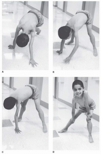
4). The abnormal gait is often noted by toe walking, which is a compensatory adaptation to knee extensor weakness, or increased lumbar lordosis as a compensation for hip extensor weakness. Another indication of pelvic girdle weakness is Gowers’ sign, demonstrated as the child rises from the floor. The patient generally begins by assuming a four-point stance, then brings the knees into extension while leaning the upper limbs forward, and sequentially moves the hands up the thighs until upright stance is achieved (Fig. 30-2A-D).

On examination, the earliest weakness is seen in the neck flexors, typically during the preschool years. Weakness of the proximal musculature of the shoulder and pelvic girdle is next, with steady progression, although the patient and family may feel that functional loss does not occur gradually but rather quite suddenly. This may relate to a critical point in weakness or range of motion when compensatory actions can no longer suffice to perform a task. Quantitative strength testing shows greater than 40% to 50% loss of strength by age 6 years (4), with fairly linear progression from ages 5 to 13 measured by manual muscle testing. Weakness appears to plateau after age 14 to 15, but this is probably a function of a floor effect and lack of sensitivity of the manual muscle testing scale (10, 11).

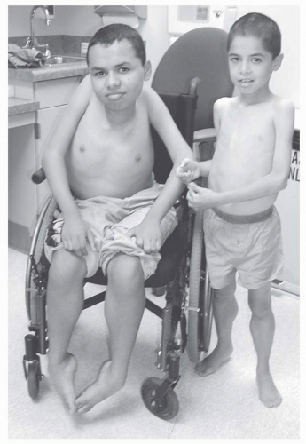
Rehabilitation concerns are summarized in Table 30-6. In patients not aggressively treated, the average age to wheelchair use is 10, with a range of 7 to 13 years of age. Prediction of transition to wheelchair use may be helped by using timed motor performance tests. In one natural history study, all DMD subjects who took more than 12 seconds to ambulate 30 ft lost the ability to ambulate within 1 year (4). Immobilization, even for an acute illness, may lead to permanent loss of ambulatory ability during this phase of the disease.


TABLE 30.6 Rehabilitation Concerns in DMD


Maintaining mobility, range of motion, and strength during childhood


Progressive scoliosis


Progressive restrictive lung disease


Cardiac dysrhythmias and cardiomyopathy


Obesity (early adolescence) and cachexia (late adolescence)


Psychosocial adjustment and social interaction


FIGURE 30-2. A-D: Gowers’ sign in an 8-year-old boy with DMD that is due to pelvic girdle weakness.


Unlike many myopathic disorders, joint contractures are a major concern in DMD. Nearly all affected boys older than 13 years have contractures (4, 12, 13), and these contractures most commonly occur first in the ankle plantar flexors, iliotibial bands, and hip flexors, with subsequent involvement of the knee flexors and elbow and wrist flexors. There does not appear to be a strong correlation between less than antigravity strength for a muscle group and the severity of joint contracture, nor for strength imbalance between antagonists across a joint (
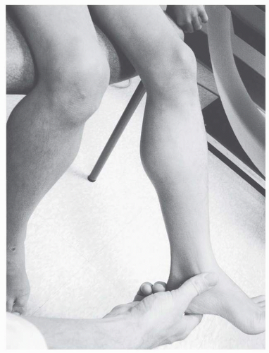
4). Clearly, lower extremity contractures become a problem after transition to a wheelchair for a significant part of the day. Natural history data suggest that progressive weakness, rather than heel-cord contractures, is associated with loss of ambulation as plantar-flexion contractures greater than 15 degrees are uncommon until after wheelchair reliance (4) (Fig. 30-3).

Scoliosis is a major clinical concern in DMD, and its prevalence is strongly related to age. Although significant curves often coincide with transition into wheelchair mobility, there does not appear to be a cause-and-effect relationship between scoliosis and wheelchair use (4, 14). Rather, factors such as the adolescent growth spurt and progressive involvement of the trunk musculature may be responsible for progression of scoliosis during the adolescent years. There is some evidence that severity of scoliosis may be predicted by the type of curve and early pulmonary function measurements (15). When the curves do not involve significant kyphosis or hyperlordosis and peak forced vital capacity (FVC) is greater than 2 L, severe progressive scoliosis appears less likely.


FIGURE 30-3. Brothers, ages 8 and 15, with DMD. In the older brother (left), note the presence of profound muscular wasting, scoliosis, and multiple joint contractures. The younger brother (right) demonstrates scapular retraction, increased lumbar lordosis, and stance phase plantar flexion (toe walking) to maintain a weight line posterior to the hip and anterior to the extended knee.

It is now clear that bracing does not slow the progression of spinal deformity (12, 16, 17). Decision making for surgical management of scoliosis is closely related to pulmonary function. Although FVC volumes increase during the first decade of life close to 100% predicted with DMD, maximal static airway pressure (both maximal inspiratory and expiratory pressures) are impaired by 5 to 10 years of age. After a plateau in the early part of the second decade, there is progressive, fairly linear decline of FVC during adolescence (4). A higher peak FVC obtained at age 10 to 12 may predict less severe restrictive lung disease and spinal deformity developing over the next few years (4). An FVC below 40% predicted may contraindicate spinal instrumentation for scoliosis because of increased perioperative mortality; however, with current improved pulmonary care this is not an absolute contraindication (18). Symptomatic respiratory failure in DMD typically manifests in later adolescence. Management of this complication is covered more in detail at a later section.

It is not surprising that cardiac function is affected in DMD, because the dystrophin protein has been shown to be present in both the myocardium and Purkinje fibers (19). Most DMD patients older than age 13 demonstrate electrocardiogram (ECG) abnormalities (4). The first abnormalities noted are Q-waves in the lateral leads, followed by elevated ST segments, poor R-wave progression, increased R/S ratio, and resting tachycardia and conduction defects (4). ECG findings have been used to predict death from cardiomyopathy and include R wave in lead V, less than 0.6 mV; R wave in lead V5 less than 1.1 mV; R wave in lead V6 less than 1.0 mV; abnormal T waves in leads II, III, aVF, V5, and V6; cardiac conduction defects; premature ventricular contractions; and sinus tachycardia (20). Sudden death from ventricular ectopy, a complication of the cardiomyopathy and left ventricular dysfunction, is well described in DMD (21, 22). However, progressive congestive heart failure is a more frequent sequela, and some investigators estimate that 40% to 50% of DMD patients die from this complication (23, 24). Cardiomyopathy is usually noted after 10 years of age and occurs in nearly all patients by age 18 (25). Cardiomyopathy is typically followed clinically with echocardiography, and the onset of systolic dysfunction is associated with a poor short-term prognosis (26). Once patients with DMD reach adolescence, regular screening with ECG, echocardiography, and Holter monitoring is prudent.


Considering the presence of a dystrophin isoform in brain tissue (27), it is not surprising that DMD patients show mildly decreased IQ scores compared with their peers and normative data (4). There may be a specific deficit with tasks requiring attention to complex verbal information, regardless of IQ (28). Mild impairments are noted on neuropsychological testing as well (4), without a specific area of strength or weakness.

Obesity from reduced physical activity is a major concern in DMD, particularly at the onset of wheelchair dependence (29, 30). Since many patients are now placed on corticosteroid treatment, weight gain is the most frequently reported side effect. At later stages of the disease (ages 17 to 21), significant weight loss becomes the predominant nutritional concern (30, 31). This probably results from nutritional compromise along with increased protein and calorie requirements during the later stages of DMD (32, 33), partially as a result of the increased work of breathing from restrictive lung disease.

At this time, there is no curative treatment available for DMD. Oral corticosteroids have been shown to increase muscle mass, increase strength, and slow muscle deterioration. However, the mechanism of its action is still unclear. Recent studies demonstrate additional potential benefits of corticosteroids including amelioration of cardiac, pulmonary, and scoliosis complications in DMD (34, 35, 36). Research involving other pharmacoagents that can increase muscle bulk and strength as well as research into the stem cell and gene therapy are ongoing.


Becker Muscular Dystrophy

BMD is similar to DMD as an X-linked recessive disorder. It has a similar pattern of muscle weakness, but generally presents with a later onset and a slower rate of progression (Table 30-7). Like DMD, the disorder has an abnormality in the gene location (Xp21) coding for the protein dystrophin. However, in this case, dystrophin levels are usually 20% to 80% of normal, or have the presence of the protein with an abnormal molecular weight. Mutation analysis of BMD has shown that majority are “in-frame” deletions, while DMD results from “frameshift” mutations. BMD is less common than DMD, with an overall prevalence recently estimated as 24 per million (37).

Without dystrophin analysis, it may be difficult to clinically discriminate between DMD and BMD. Although age of onset typically occurs later in BMD, there is significant overlap with DMD (38). The degree of CK elevation does not discriminate between the two diseases. The most useful clinical diagnostic discriminator is the ability to ambulate into adolescence. It is unusual for a patient with BMD to be wheelchair dependent before late adolescence, whereas even DMD outliers are dependent on the wheelchair for mobility by age 16. In fact, some BMD patients may be ambulatory well into middle age and beyond. There may be two distinct patterns of progression in BMD. In the first type, age of onset averages 7.7, and most patients have difficulty climbing stairs by age 20. In the more common milder form, age of onset averages 12, and there is no problem climbing stairs at age 20. The former group also seems to have a much higher rate of ECG abnormalities (39). The percentage of normal dystrophin cannot be used to predict clinical course with any certainty in BMD (40).


TABLE 30.7 Comparison of Clinical Features Between DMD and BMD


Duchenne Muscular
Dystrophy


Becker Muscular
Dystrophy


Age of onset


Childhood < age 5


Childhood/early adolescence


Pattern of weakness


Proximal


Proximal


Wheelchair dependence


Late childhood/early adolescence


Late adolescence or later


Scoliosis


Severe and progressive


Usually mild


Cardiac involvement


Significant


Significant


Pulmonary dysfunction


Severe and progressive


Usually mild


Cognitive involvement


Frequent


Unusual


FIGURE 30-4. A 36-year-old man with BMD with pseudohypertrophy of the posterior deltoid and infraspinatus resulting in a posterior axillary depression sign.

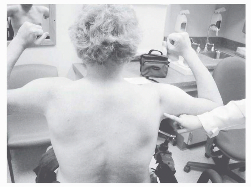
Findings on examination of the BMD patient mirror DMD, although milder. The neck flexors and proximal lower limb muscles are affected early, particularly the hip and knee extensors (38). Subsequently, there is gradual involvement of the proximal upper-limb muscles (Fig. 30-4). Extensors are
generally weaker than flexors (38). Calf enlargement occurs, and presence of Gowers’ sign is indicative of the proximal muscle weakness. On standing, there is increased lumbar lordosis, and hip abductor weakness results in a waddling gait with trunk lean over the weight-bearing limb.

Contractures are not a significant early functional problem in BMD (38, 39), becoming problematic only after wheelchair dependence. The joint locations for contractures are typical for one assuming the sitting posture, occurring in the hip flexors, knee flexors, and ankle plantar flexors. Significant scoliosis is much less common than DMD, and BMD patients rarely require spinal instrumentation (38, 39).

One particular clinical concern in BMD is the potential for significant cardiac disease out of proportion to other manifestations of the myopathy (39, 41, 42, 43, 44, 45). ECG abnormalities can be detected in about 75% of BMD patients (38, 46). Most common abnormalities include abnormal Q-waves, right or left ventricular hypertrophy, right bundle branch block, and nonspecific T-wave changes. Echocardiography demonstrates left ventricular dilatation in 37% of BMD patients, and 63% have subnormal systolic function that is due to global cardiac hypokinesia (46). Cardiac transplantation may even be necessary in some patients (47, 48). The degree of cardiac compromise may not be reflected by clinical symptoms, and these patients should be screened at regular intervals with ECG and echocardiographic studies.

Unlike DMD, significant pulmonary dysfunction is not a hallmark of BMD. FVC does not fall below the predicted level until the third to fifth decade of life. Because of relatively greater involvement of the intercostals and abdominal musculature compared with the diaphragm, maximum expiratory pressure is compromised at an earlier age than maximal inspiratory pressure (MIP), similar to DMD (38).

There are no consistent abnormalities on cognitive and neuropsychological testing in BMD other than a mild reduction in some patients (38).


Myotonic Muscular Dystrophy (DM1 and DM2)

There are two subtypes of myotonic muscular dystrophy, DM1 and DM2 (dystrophia myotonica type 1 and type 2). Both are muscular dystrophies that share similar clinical features of myotonia and distinctive effects on other organ systems. However, DM1 and DM2 are genetically separate entities with different genes involved. DM1 is caused by abnormal expansion of the CTG trinucleotide repeats in the dystrophia myotonica protein kinase (DMPK) gene on chromosome 19q13.3, while DM2 is caused by an abnormal expansion of the CCTG repeats in the zinc finger protein 9 (ZNF9) gene on chromosome 3q21 (49, 50, 51).

Myotonic muscular dystrophy type 1 (DM1) is the most common slowly progressive dystrophy in adults, with an incidence of 1/8,000 (9), while DM2 is much less common and thought to account for only about 2% of myotonic muscular dystrophy patients. Both are multisystem disorders affecting skeletal muscle, smooth muscle, myocardium, brain, and ocular structures (Table 30-8). This may manifest clinically with cataracts, cardiac conduction defects, endocrine abnormalities, swallowing dysfunction, and skeletal muscle weakness and myotonia.


Only gold members can continue reading. Log In or Register to continue

Stay updated, free articles. Join our Telegram channel

May 25, 2016 | Posted by in PHYSICAL MEDICINE & REHABILITATION | Comments Off on Myopathy

Full access? Get Clinical Tree

Get Clinical Tree app for offline access

TABLE 30.8 Rehabilitation Concerns in Myotonic Muscular Dystrophies