Introduction
The motor system moves the arms in skilful activity and the legs in walking, and at the same time controls the background posture of the whole body. The same system is involved in movements of the tongue, lips and larynx needed for speech. Movements are controlled by motor centres in the brain and activity passes down from these motor centres in descending pathways to the motor neurones of the cranial nerves in the movements of speaking, eating and facial expressions, and to the motor neurones of spinal nerves in the movements of the limbs and trunk.
Movement is executed in response to commands from the motor centres of the brain. The commands have been called motor programmes, which specify not only which muscles are activated but also the force, direction and timing of the activity. Motor programmes, developed with practice and stored in the brain, can be activated by internal decision making and/or input from the environment.
In simple ballistic movements, known as open loop, the action is planned and executed over a very short period. Examples of open-loop movements are pressing a key on a keyboard, throwing a ball and chopping vegetables. However, most actions take longer. In these closed-loop movements, the motor commands can be modified during the progress of the movement in response to feedback from the sensory system.
The cortical motor areas, together with the basal ganglia and the cerebellum, form the higher centres for the production and regulation of the motor commands to the muscles. Motor nuclei in the brain stem control the posture and the balance of the body as the movement proceeds. Motor commands reaching the spinal level are fine tuned as a result of a variety of influences from the descending pathways from the brain and from local spinal reflexes.
In this chapter, activity at the spinal level will be considered first, followed by the influence of descending pathways from the brain stem and the higher centres.
Spinal mechanisms
In the spinal cord, the lower motor neurones form the final common pathway for activation of the muscles in all movement, both voluntary and reflex. Chapter 1 included a description of how the cell bodies of the lower motor neurones lie in the anterior horn of the spinal cord and in the nuclei of the cranial nerves. The axons of the lower motor neurones lie in the peripheral nerves supplying the muscles (see Chapter 1, Figure 1.15).
The spinal cord also contains interneurones, in larger numbers than the motor neurones. An interneurone is a nerve cell found in the central nervous system with no branches in a peripheral nerve. The abundance of interneurones reflects the complex information processing performed by the spinal cord. Interneurones provide an inhibitory influence for the regulation of activity in the lower motor neurones and for the reciprocal inhibition of antagonist muscles. Interneurones are important in all bilateral movements when there is activity on both sides of the spinal cord, and in movement patterns when several adjacent spinal segments are involved. The spread of activity across the spinal cord by interneurones is the basis of associated reactions.
The associated reaction movements may become exaggerated when the interruption of descending pathways releases spinal reflexes from the control by higher centres.
Lower motor neurone activity controls the changes in the length and tension of all the active muscles as the body moves from one position to another. Two spinal reflexes provide the basis for the regulation of these changes to achieve co-ordinated movement. The two reflexes are the muscle stretch reflex and the Golgi tendon reflex.
Muscle stretch reflex
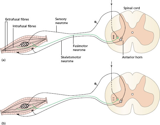
When the body holds a position, muscles are maintained at a constant length by activity in the muscle stretch reflex (see Chapter 1, Muscle tone). During movement, muscles change in length, and the level of stretch reflex activity is then modified by the influence of descending pathways in the spinal cord which change the setting of the spindles in the following way. Fusimotor (gamma) neurones supply the intrafusal muscle fibres of the spindles themselves. When these neurones are excited, the intrafusal fibres of the spindle contract. The spindle then becomes taut and more sensitive to length changes in the muscle (Figure 12.1a). When the influence from the descending tracts inhibits fusimotor neurones, the spindle becomes slack and only responds to marked changes in length of the muscle (Figure 12.1b). In this way, spinal stretch reflex activity is regulated during movement by the higher levels of the central nervous system.
Consider the hand performing fine manipulative movements, such as doing up buttons and tieing shoe laces. Stretch reflex activity must be dampened in the muscles of the hand to allow rapid length changes to occur. At the same time, muscles of the shoulder and arm perform background activity to hold the postion of the limb and allow the fingers to move accurately. The spindles in the supporting muscles are set at a high level, so that any change in length is resisted.
Static and dynamic intrafusal fibres
Looking in more detail at the structure of the muscle spindle, two different types of intrafusal fibre can be identified. Both types have a primary sensory ending wound round the central area, the annulospiral ending. In addition, some of the intrafusal fibres have secondary sensory endings towards the periphery of the fibre, known as flower-spray endings, which respond to the rate of change in length of the muscle during movement. The two types of intrafusal fibre are: (i) nuclear bag fibres with a bulge in the middle where the nuclei are found, and secondary sensory endings are present; and (ii) nuclear chain fibres, which are thinner and their nuclei are lined up in a row.
The nuclear bag (dynamic) fibres respond to rapid changes in length of the muscle, while the nuclear chain (static) fibres respond to prolonged slow stretch. The muscles spindles, therefore, relay detailed information to the spinal cord about both the length, and the rate of change in length, of a muscle during movement.
The muscle stretch reflex provides a feedback mechanism so that muscle groups producing movement change length appropriately and supporting muscles are held at the desired length.
Golgi tendon reflex
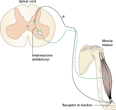
Golgi tendon organs are proprioceptors found at the junction between the muscle fibres and the tendon of a muscle. Each consists of a nerve ending embedded in collagen fibrils surrounded by a capsule. Since the Golgi tendon organs lie in series with the muscle fibres, they respond to an increase in tension in the whole muscle. (Remember that muscle spindles lie in parallel and respond to a change in length of a muscle.) When the tension in a muscle rises, the muscle pulls on the tendon and the Golgi tendon organs are stimulated. This activity stimulates sensory neurones, which synapse with interneurones in the spinal cord. The interneurones are inhibitory to the lower motor neurones of the same muscle (Figure 12.2) and also exert an influence on motor neurones supplying other muscles around the same joint. In this way, the Golgi tendon reflex provides a feedback mechanism to regulate muscle tension and to keep it within the limits required for the performance of any task.
Figure 12.1 Muscle spindle sensitivity: (a) high-descending pathways stimulate fusimotor neurones, the intrafusal fibres contract and the muscle spindle is very sensitive to distortion; (b) low-descending pathways inhibit fusimotor neurones, intrafusal fibres are slack and the muscle spindle is less sensitive to distortion.
It has been suggested that the Golgi tendon reflex can act as a protective mechanism to prevent damage to tendons when a sudden high level of tension develops in a muscle. In these conditions, stimulation of the tendon organs inhibits the lower motor neurones and a loss of muscle tension occurs. An example is a weight-lifter who experiences a sudden loss of power when attempting to lift a load that he cannot move.
Figure 12.2 Golgi tendon reflex.
The two spinal reflexes described, which are mediated by muscle spindles and Golgi tendon organs, co-operate to control the length and tension in the muscles at the correct levels during normal movement.
Spinal integration and synergy
The motor neurones in the spinal cord are organised systematically. Motor neurones of flexor muscles lie laterally and extensor motor neurones lie medially in each anterior horn of grey matter. Motor neurones of the proximal muscles of a limb lie towards the centre and those of the distal muscles lie towards the periphery in the grey matter. A large number of interneurones link all of these groups of neurones, forming a neural network.
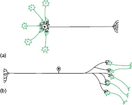
Within a network of neurones, the axon of each neurone branches to synapse with many other neurones (divergence) and each cell body receives branches from many other neurones (convergence) (Figure 12.3). Some of the neurones in a network will exert excitatory influences, while others will inhibit activity.
Inhibition is the term for the changes in the cell membrane of a neurone that make it more difficult for it to respond to activity from another source. The balance of excitatory and inhibitory influences within a network of neurones determines the final output. There are two main types of inhibition in the spinal cord: presynaptic inhibition and recurrent inhibition (Renshaw cells).
Figure 12.3 Neurone circuits: (a) convergence; (b) divergence.
Presynaptic inhibition occurs when the neurotransmitter substance from a terminal bouton of an inhibitory interneurone prevents the release of excitatory neurotransmitter by another neurone at a synapse (Figure 12.4a). An example of presynaptic inhibition is found in the pain gate mechanism that regulates the activity of the pain transmission cells (see Chapter 11). Another example is the suppression of stretch reflex activity by descending pathways from the brain via spinal interneurones.
Recurrent inhibition occurs in the skeletomotor neurones when collaterals or branches of the axons synapse with small inhibitory interneurones called Renshaw cells in the spinal grey matter. The inhibitory neurones, in turn, synapse with the cell bodies of the skeletomotor neurones supplying the same muscle and synergistic muscles (Figure 12.4b). It has been proposed that the Renshaw cells form a closed-loop feedback control system for alpha-motor neurone activity at the spinal level.
Functional movements involve the repetition of particular patterns of muscle activity to perform specific tasks. Reach and grasp movements require simultaneous activity in the flexors of the shoulder, the extensors of the elbow and wrist, and the flexors of the fingers. Walking involves the repetition of alternating flexion and extension movements of the hip, knee and ankle in a particular order. These patterns of movement are called synergies 1 Movement synergies are executed by spinal neural networks, containing motor neurones and interneurones, which generate repeated activity in the same groups of neurones.
Related posts:

Stay updated, free articles. Join our Telegram channel

Full access? Get Clinical Tree


