42
Metacarpal Shaft Fractures
Robert J. Goitz, Sokratis Varitimidis, and Dean G. Sotereanos
History and Clinical Presentation
A 37-year-old right hand dominant woman was involved in a motor vehicle accident and presented complaining of pain in her left hand. She denied any previous injuries to this hand and her past medical history was unremarkable.
Physical Examination
The patient’s left ring finger appeared to be rotationally malaligned (Fig. 42–1A), compared with the right hand (Fig. 42–1B). She was tender to palpation over the middle ring finger metacarpals as well as the index proximal phalanx. The hand was neurovascularly intact with normal two-point discrimination to less than 6 mm. Her skin appeared intact, without any lacerations, and there was good capillary refill.
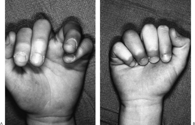
Figure 42–1. (A) Malalignment of the left ring finger overlapping the small finger. (B) Comparison view of normal contralateral right hand. All digits are aligned and point to the scaphoid tubercle.
Diagnostic Studies
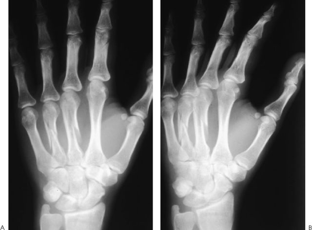
Three views of the left hand were obtained including anteroposterior, oblique, and lateral radiographs (Fig. 42–2). They confirmed fractures of the middle and ring finger metacarpals and index proximal phalanx. The middle finger metacarpal had a long spiral fracture that extended approximately three cortical diameter lengths along the metacarpal shaft. The ring finger metacarpal shaft was comminuted. The metacarpals were shortened relative to the normal cascade between the index and small fingers as evidenced by the relative height of the metacarpophalangeal (MP) joints. The index proximal phalanx fracture was minimal to nondisplaced.
PEARLS
- Three radiographic views of the hand are necessary to understand the fracture configuration.
- Fracture geometry implies stability and will dictate optional treatment.
- Open or multiple metacarpal fractures or those associated with malrotation are indications for open reduction and internal fixation (ORIF).
- If ORIF is chosen, adequate stability for immediate motion should be the goal to minimize adhesions and limitation of digital motion.
PITFALLS
- Malalignment associated with a metacarpal fracture is difficult to assess following a closed reduction in a splint.
- Prior to open fracture reduction, an accurate understanding of the fracture configuration is necessary to avoid poorly positioned screws, which could propagate or further comminute the fracture.
- Technical errors such as overtightening the screws or placing them less than two screw diameters from the fracture edge may complicate ultimate stability of the fixation and delay the initiation of therapy.
Diagnosis
Long spiral fracture of the long finger metacarpal shaft
Comminuted ring finger extraarticular shaft fracture with rotatory malalignment
Minimal to nondisplaced index finger proximal phalanx shaft fracture
Nonsurgical Management
Closed reduction of the ring finger metacarpal may be attempted by axial traction and splinting in an intrinsic-plus position. However, assessment of alignment is difficult once in the splint. Digits healed in a malaligned position resulting in digital overlap are not well tolerated. An osteotomy would be required to regain neutral alignment. If closed reduction is chosen to treat a metacarpal fracture for malalignment, assessment of the stability of the reduction is paramount. This can be accomplished by evaluating the fracture under fluoroscopy during active movement if a hematoma or wrist block was used for anesthesia or by using the tenodesis effect of digital motion during wrist flexion and extension.
Closed treatment of metacarpal fractures is indicated for nondisplaced or central isolated shaft fractures. The alignment and length are supported by the interosseous muscles, which originate from the adjacent metacarpal shafts, and the intermetacarpal ligament, which effectively suspends the metacarpal heads through their attachment to the volar plates. These supporting structures usually minimize the shortening of isolated oblique or spiral metacarpal shaft fractures to less than 5 mm. Displaced isolated transverse metacarpal shaft fractures can often be closed reduced under a hematoma or wrist block. A cast is used for 3 weeks, maintaining the MP joint in 70 degrees of flexion with the proximal and distal phalangeal joints exposed for active and passive mobilization. MP joint motion is indicated at 3 weeks, with a removable splint for an additional 3 weeks.
Surgical Management
The indications for surgical intervention for metacarpal shaft fractures include multiple metacarpal fractures, especially if they are comminuted, oblique, or spiral. These configurations would lack any bony or soft tissue support from adjacent digits. Open metacarpal fractures associated with soft tissue loss or extensor tendon injuries would also be an indication for operative intervention because early motion is often critical to minimize tendon adhesions and maximize digital motion.
Any rotational deformity of the digits is an absolute indication for surgical intervention. Excessive angulation or shortening is also an indication for operative intervention, but the amount tolerated is dependent on the digit involved. The ring and small finger metacarpals can accommodate greater angulation at the fracture site because motion at the carpometacarpal (CMC) joints is ∼30 degrees compared with the index and middle finger CMC joints, which are nonmobile. Therefore, ∼20 to 30 degrees of angulatory deformity may be accepted in the ring and small finger, but only 10 degrees in the index and middle fingers.
Metacarpal shortening is difficult to assess without comparative radiographs. Shortening of up to 1 cm is often reasonably well tolerated functionally and results in loss only in the contour of the metacarpal heads during gripping.
After operative intervention is determined to be necessary, optional internal fixation must be chosen. Isolated transverse or short oblique fractures can be treated satisfactorily with percutaneous pinning. They can be passed retrograde through the metacarpal head as an intramedullary pin or transversely to an adjacent metacarpal to add rotational stability. Comminuted and multiple transverse or short oblique fractures may be optionally treated with plate and screw fixation. Depending on the size of the metacarpal, 2.0-, 2.4-, or 2.7-mm plating systems may be chosen. A minimum of four cortices on either side of the fracture should be engaged. Spiral fractures greater than two cortical diameters in length may be treated with interfragmentary lag screw fixation.
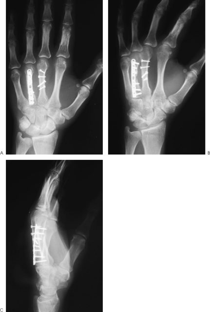
In the clinical example (Fig. 42–2), the middle finger metacarpal is a spiral fracture that can be optimally treated with lag screw fixation. The ring finger metacarpal is comminuted and may best be treated with a plate and screw system.
Surgical exposure of the two adjacent metacarpals was through a longitudinal incision made between the two shafts. Long dorsal sensory nerves are protected and transverse comminuting veins are ligated. The extensor tendons are retracted to either side. Each metacarpal is exposed separately by making a periosteal incision and subperiosteally removing the interosseous muscles from the dorsal metacarpal shaft. It is critical to fully examine the extent and direction of the fracture fragments prior to their reduction; if a screw is drilled too closely to the edge of the fracture, it may result in fracture propagation or an incompetent screw. Precision is an absolute necessity during fracture fixation.
The spiral fracture was addressed first with multiple lag screw fixation because assessment of the reduction is easier to determine. If the comminuted fracture is addressed first, the overall alignment may be difficult to assess because the adjacent digit is fractured, which may affect its rotatory alignment. In addition, anatomic alignment of a comminuted fracture may not be possible. The spiral fracture is prepared by clearing all soft tissue interposed between the fractured edges. They are then reduced and held provisionally with a reduction clamp placed strategically away from the planned screw position. As previously mentioned, screw placement should be determined prior to fracture reduction. In the clinical example presented, the spiral fracture was quite narrow and long, which resulted in the use of one 2.0-mm and three 1.5-mm screws. Standard lag screw techniques were used, which will be outlined for the 2.0-mm screws. First, a 1.5-mm drill is placed through both cortices openings perpendicular to the fracture line to maximize compression across the fracture site. The hole is countersunk to minimize prominence of the screw head and maximize contact of the head to the hole. The screw length is then determined with a depth gauge. Because most screws are self-tapping, the chosen screw should be at least 1 to 2 mm longer than the measured length to maximize screw purchase in the second cortex. The near cortex is then overdrilled with a 2.0-mm drill to ensure compression at the fracture site. Experience will determine the maximum torque provided to the screw, but more than “two-finger” tightness is never necessary. Less torque is often required in osteopenic bone.