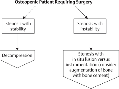
52 Osteoporosis is a skeletal disorder characterized by compromised bone strength that predisposes to fractures. Bone mineral density (BMD) accounts for ~ 70% of the strength of a bone and is the strongest predictor of fragility fracture. BMD can be directly measured in the spine or hip using dual-energy X-ray absorptiometry (DEXA). The magnitude of BMD is quantified by a T score, which is defined as the number of standard deviations (SDs) above or below the mean BMD for a healthy, young white woman. Osteoporosis has been defined as a T score of less than −2.5, whereas osteopenia is defined as a T score of between –1.5 and –2.5. The consequence of spinal osteoporosis is vertebral compression fractures (VCFs). Only one third of VCFs are acutely symptomatic. Long-term consequences of VCFs include kyphosis, mechanical back pain, gastrointestinal symptoms, and restrictive pulmonary disease. A 23% increase in age-adjusted mortality is seen in older women with a VCF. Spinal osteoporosis should be suspected in patients with nontraumatic vertebral body fractures. Multiple risk factors may contribute to osteoporosis (Table 52.1), but in up to one-third of women and 50% of men with osteoporosis, a secondary cause can be identified (Table 52.2). A detailed history, including risk factor assessment, and screening laboratory studies are appropriate in all patients (Table 52.3). Table 52.1 Risk Factors for Osteoporosis
Medical Management of Spinal Osteoporosis
![]() Workup
Workup
History
Female sex |
Increased age |
Estrogen deficiency White race |
Low weight and body mass index |
Family history of osteoporosis |
Cigarette smoking |
History of prior fracture |
Table 52.2 Secondary Causes of Osteoporosis
Endocrine: hyperthyroidism, hyperparathyroidism, hypogonadism, Cushing disease |
Medication: glucocorticoids, heparin, excess thyroid hormone replacement, anticonvulsants, omeprazole |
Nutritional: malabsorption, alcoholism, liver disease |
Malignancy: multiple myeloma |
Table 52.3 Laboratory Evaluation of Osteoporosis
Complete blood count |
Serum calcium, phosphorus, alkaline phosphatase, creatinine |
Serum albumin and total protein |
24-hour urine calcium |
Thyroid-stimulating hormone (if indicated by history) |
Parathyroid hormone (if indicated by screening laboratory) |
Serum free testosterone (men) |
25-hydroxyvitamin D |
Special Diagnostic Tests
In asymptomatic populations, BMD measurements have been recommended for all women over 65 and for women over 60 with risk factors for osteoporosis. Measurement of BMD in the spine most accurately reflects spinal bone mass. However, persons with significant lumbar spondylosis may have artificially elevated spinal BMD values with DEXA scanning, making hip BMD values more predictive of true bone mass in this patient population.
 Treatment and Outcome
All persons meeting BMD criteria for osteoporosis, with or without fractures, should be treated. In addition, treatment is recommended for individuals with a T score less than –2.0 or less than –1.5 with risk factors. While bone mineral density is an excellent predictor of fracture risk, density combined with clinical risk factors for fracture is a better predictor than density or clinical risk factors alone. The World Health Organization Fracture Risk Assessment Tool, FRAX, is a clinical software tool developed to calculate fracture risk on the basis of bone mineral density of the femoral neck as well as multiple clinical risk factors, including patient’s age, sex, height, weight, personal history of previous fracture, history of parental hip fracture, current smoking, history of long-term glucocorticoid use, rheumatoid arthritis, and daily alcohol consumption. The tool also incorporates the presence or absence of other secondary causes of osteoporosis. It estimates the 10 year probability of a major osteoporotic fracture. FRAX is available to clinicians online (www.shef.ac.uk/FRAX).
Ensuring adequate calcium and vitamin D intake is fundamental to osteoporosis therapy. Postmenopausal women not receiving estrogen should ingest 1500 mg of calcium daily from all sources. The optimal dose of vitamin D should be based on titration to normal serum 25-hydroxyvitamin D levels. Weight-bearing exercise such as walking should be encouraged. Back extensor strengthening exercises have been shown to decrease the risk of vertebral fracture as well as falls.
The antiresorptive bisphosphonates (diphosphonates) alendronate and risedronate are the first-choice agents for spinal osteoporosis, reducing vertebral fracture risk by ~ 50% in postmenopausal women with osteoporosis. Both drugs reduce risk of nonvertebral fractures to a similar extent. Another bisphosphonate, ibandronate, approved for the treatment of osteoporosis has also been shown to reduce the incidence of vertebral fracture by ~ 50%, but reduction in hip fracture risk remains unproven. An intravenous bisphosphonate, zolendronic acid, is administered once yearly and may be more effective than oral agents in vertebral fracture reduction (70% versus 50%). There is currently no consensus on how long to continue bisphosphonate therapy. However, stopping therapy after 5 years for some women may be reasonable, as there appears to be residual benefit on BMD and fractures for at least 5 years.
A newly available, novel antiresorptive agent, denosumab, is a human monoclonal antibody administered subcutaneously twice yearly for 36 months. Denosumab has been shown to decrease risk of vertebral, nonvertebral, and hip fractures in women with osteoporosis comparably to zolendronic acid. Unlike bisphosphonates, which interfere with osteoclast function, denosumab inhibits development of osteoclasts by inhibition of receptor activator of nuclear factor kappa-B ligand (RANKL).
The alternative antiresorptive agents raloxifene and calcitonin are probably less effective in bone preservation and fracture reduction. Raloxifene, acting as an estrogen receptor agonist in bone, reduces vertebral fracture risk by 30% to 50% but has not been shown to reduce nonvertebral fractures. Hormone replacement therapy with estrogen is no longer recommended as primary treatment for osteoporosis since the Women’s Health Initiative Study, which found that the combination of estrogen plus progesterone reduces fracture risk but increases the risk of breast cancer and cardiovascular events. Limited data suggest that salmon calcitonin nasal spray may reduce vertebral fracture risk by one-third in women with osteoporosis, but reduction of nonvertebral fractures has not been demonstrated.
Teriparatide (human parathyroid hormone 1–34) is the only currently available anabolic agent for treatment of osteoporosis, increasing bone density by ~ 10% and reducing vertebral fracture risk by 70% and nonvertebral fracture risk by at least 50%. Teriparatide is administered daily by subcutaneous injection and is considerably more expensive than oral bisphosphonates. Indications for teriparatide are evolving, but the drug should be considered in patients who sustain fractures or continue to lose bone mass on antiresorptive therapy. Antiresorptive therapy should be discontinued when teriparatide therapy is initiated.
 Complications
Complications include intolerance to medications or rarely osteonecrosis of the jaw, mainly in cancer patients receiving high-dose intravenous bisphosphonates. Recent reports have suggested increased risk of atypical subtrochanteric femoral shaft fractures in women treated with bisphophonates, but retrospective secondary analyses of large randomized bisphophonate trials failed to substantiate an increased risk.
Suggested Readings
Black DM, Kelly MP, Genant HK, et al; Fracture Intervention Trial Steering Committee; HORIZON Pivotal Fracture Trial Steering Committee. Bisphosphonates and fractures of the subtrochanteric or diaphyseal femur. N Engl J Med 2010;362(19):1761–1771 PubMed
A secondary analysis completed on three large randomized trials demonstrated no significant risk associated with bisphosphonate use and atypical femur fractures.
Cummings SR, San Martin J, McClung MR, et al; FREEDOM Trial. Denosumab for prevention of fractures in postmenopausal women with osteoporosis. N Engl J Med 2009;361(8):756–765 PubMed
A randomized controlled trial demonstrated reduction in the risk of vertebral, nonvertebral, and hip fractures in women with osteoporosis given denosumab subcutaneously twice yearly for 36 months.
Kanis JA, Johnell O, Oden A, Johansson H, McCloskey E. FRAX and the assessment of fracture probability in men and women from the UK. Osteoporos Int 2008;19(4):385–397 PubMed
This paper reviews the FRAX model and process of development.
Kennel KA, Drake MT, Hurley DL. Vitamin D deficiency in adults: when to test and how to treat. Mayo Clin Proc 2010;85(8):752–757, quiz 757–758 PubMed
This general review article provides an overview on vitamin D deficiency and supplementation.
This is an overview on osteoporosis treatment.
Nelson HD, Haney EM, Dana T, Bougatsos C, Chou R. Screening for osteoporosis: an update for the U.S. Preventive Services Task Force. Ann Intern Med 2010;153(2):99–111 PubMed
This article gives current recommendations for osteoporosis screening.

Stay updated, free articles. Join our Telegram channel

Full access? Get Clinical Tree


