Chapter 6 Measuring electrical skin resistance on acupuncture points
INTRODUCTION
For many decades, practitioners have used electrical devices to locate specific points for acupuncture, both in association with, or to replace, manual methods such as the pain pressure test (PPT). The main assumption underlying such electrical devices is that an acupuncture point is characterized by its different electrical resistance (or its reciprocal, conductance) compared to that of the skin surrounding it. This statement derives mainly from the work of some researchers in the 1950s, for example Voll1 and Nakatani and Yamashita.2 They found that a lower skin resistance value often coincided with spots that traditional Chinese medicine indicated as effective for acupuncture treatment. Although Niboyet3 meticulously re-examined the method, it was only in 1975 that experiments by Reichmanis4 and colleagues truly awakened interest in the topic. This author employed a rod electrode for roughly locating higher electrical conduction points on the skin, and then a roller-type electrode for tracing conductance curves along lines passing through those points.
More than 30 years of episodic research has produced no consensus on the effectiveness of measuring electrical resistance for identifying acupuncture points. The work up to the late 1970s has been reviewed by Mannheimer and Lampe5 and, more recently, a comprehensive review was presented by Ahn and Martinsen.6
Over the last decades many devices for locating acupuncture points based on resistance measurement have become commercially available. Their very simple electronics and low cost have made them widely accessible. However, acupuncturists rarely know more about them than that when an LED lights up or a beep is heard the machine has detected a variation in resistance. This is because very few commercial makers describe their devices or the principles behind them in depth.
ELECTRICAL SKIN RESISTANCE (ESR)
Normal commercial devices for locating low skin resistance spots have a large common ‘reference’ or ‘indifferent’ electrode held by the subject in one hand, generally a cylinder of 2–3 cm diameter or a similar contrivance (Fig. 6.1).
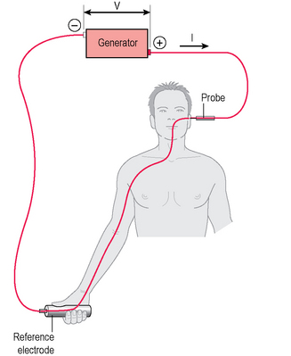
A ‘probe’ consisting of a narrow electrode is put into contact with the skin at the location to be investigated, usually with the help of a small spring to keep the contact pressure within a known range. The main prerequisite is for the area of skin contact at the reference electrode to be much larger than that at the measuring probe, so that the resistance at the reference electrode compared with that at the measuring probe can be disregarded. The current flows from the large to the small electrode, passing through a number of biological tissues and two skin/electrode interfaces. The usual approximation is to consider the electrical skin resistance (ESR) as V/I where V is known because it is pre-set by the generator, while I is measured.
The measured value of ESR includes contributions from several elements, but the most determining arises from the stratum corneum of the skin. On the derma lies a complex of tissues which constitute the epidermis, whose thickness is on average about 50–150 μm but can be as high as 1500 μm on the plantar aspect of the foot.7 It is made up of several superimposed specialized cell strata, of which the external is the corneum, consisting of regular rows of dead flat cells filled with keratin, a fibrous protein.
This stratum corneum continuously loses dead cells at its surface, which are replaced by new cells originating from the lower stratum. Its thickness (both absolute and as a fraction of the epidermis) depends on location. For instance, Sandby-Møller et al8 found a thickness from 11 μm on the shoulder to 18.3 μm at the dorsal aspect of the forearm, while Jacobi and Kaiser9 give 17 μm to 28 μm for the stratum corneum for porcine ear skin which they judge to give similar results in humans. The stratum corneum is characterized by very low water permeability, low ion mobility and high electrical resistance. Its interest here lies in the fact that, from experiments where the stratum corneum is removed and the decrease of resistance monitored, it is evident that skin resistance is produced mainly in the stratum corneum.
There are several well documented methods of skin ablation. The commonest, probably because it does not require any special equipment, consists of stripping the dead cells by successive applications of adhesive tape. Examples of this technique are described in Yamamoto and Yamamoto,10 Kalia and Guy,11 Kalia et al12 and Bashir et al.13 Another method consists of laser ablation,14 while the most recent method uses a flow of abrasive particles, as described in Gill et al.15
Tape stripping seems to increase ion mobility by two orders of magnitude (that is a hundredfold increase).12 This correlates very well with the findings on microdermabrasion, which decreases resistance from thousands of kΩ to tens of kΩ.15 Gill and colleagues15 give a value of about 20–80 kΩ for skin resistance after complete stratum removal.
To give an order of magnitude for skin resistance, in an earlier work Inada et al16 had given 12–120 kΩ/cm2, while Kalia and Guy11 found a value of 187 kΩ for the real part of the complex skin impedance in hydrated skin with intact stratum corneum at 1 Hz, with electrodes of 3.14 cm2. Oleson et al17 give, instead of resistance, the amount of current flowing through the skin under a voltage of 9 V. They found spots of low resistance allowing the passage of 300 μA, but unfortunately they do not specify the internal electrical resistance of the device used.
In fact, the very strong influence of the stratum corneum had been noted extremely early in articles seeking to locate acupuncture points by electrical methods. McCarrol and Rowley18 deliberately damaged the stratum corneum by applying a very high pressure (2 kg/mm2) at the tip of the searching electrode, observing a dramatic drop in skin impedance, which, in one case, amounted to 94% of the impedance measured previously using a tip loaded with 0.1 kg/mm2. Their experiment was conducted at 1000 Hz, but no details of the tip’s dimension are given. Their conclusion was that when trying to locate an acupuncture point by measuring skin conductance with a small electrode, the chances are that we are simply measuring the integrity of the stratum corneum. Therefore, we can suspect that any macroscopic variations are probably due to accidental stratum corneum abrasions.
There is almost nothing in the literature on this subject. A valuable contribution is the experiment by Terral and Rabischong.19 They made histological sections of the skin of rabbits on the spots where they had located acupoints and found that at locations with lower electrical resistance the connective tissue was more lax, and contained a number of nerve fibres and other structures. It is, however, to be noted that rabbits had been carefully shaved, therefore it is not possible to know how the stratum corneum had been altered.


