Fig. 6.1
Application of Coban wrapping on the fifth digit (University Medical Center Groningen, UMCG 2008)
6.1.1.3 Splinting
Splinting of athletes is a common technique for initial treatment of suspected fracture or ligament/tissue disruption [4]. Splinting requirements and purposes for the upper extremity in athletes depend on whether it takes place during primary care or during aftercare. During the early phases of rehabilitation, the goal of splinting is rest and protection of disrupted tissues. Depending on the type of injury and the amount of injured tissues, a choice should be made as to the type of splint, the joints to be immobilised and duration of wearing and weaning of the splint. Splinting techniques can range from very simple to rather complex.
6.1.1.4 Taping
Like splinting, taping plays an essential role in the care of sport injuries to the hand or fingers. Specific techniques are available and proven to be effective. It is important to note the general principles. Taping techniques should consider the angle of pull, the direction of pull and the tension needed to be effective.
During the acute phase, taping can be used to help stabilise and compress the healing tissues. It is considered that both conventional and medical taping over the skin might stimulate mechanoreceptors by sensing alteration of length and tension of the muscle fibres to deliver more signals to the central nervous system. The medical taping concept differs from conventional taping techniques, as it stresses ‘mobility’ instead of ‘immobilisation’ [5]. Several applications of medical taping are conceivable such as correcting posture, reducing inflammation and accumulation of fluid (oedema and haematoma) (Fig. 6.2), stimulating proprioception, correcting direction of movement and increasing stability (Fig. 6.3). At a later stage taping can be used in prevention (prophylactic taping) of stress to the newly built collagen.
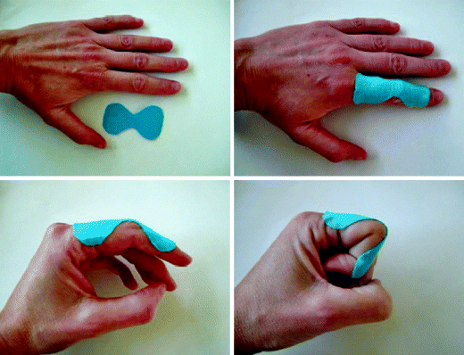
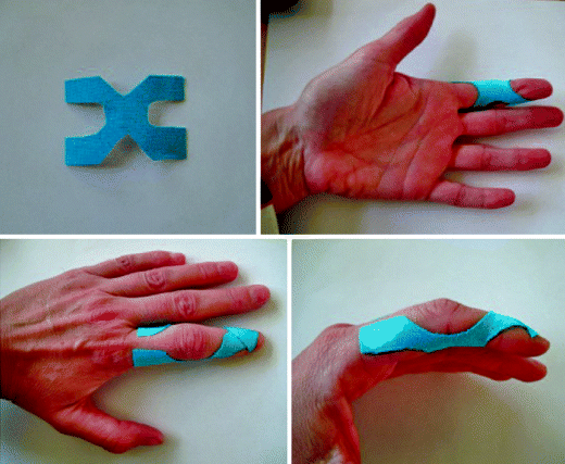

Fig. 6.2
Application of a dorsal ‘butterfly’ tape to stimulate drainage of oedema (University Medical Center Groningen, UMCG 2011)

Fig. 6.3
Application of a ‘H’ tape to prevent (prophylactic) hyperextension of the proximal interphalangeal joint (University Medical Center Groningen, UMCG 2011)
6.1.1.5 Thermotherapy
Thermal agents include superficial and deep heating agents and are also known to reduce pain, improve joint motion and enhance healing [3]. The purpose of heating is to increase circulation to the injured area, as plasticity of collagen fibres is temperature dependent. When temperature increases, flexibility of the collagen increases and vice versa [6]. Heating agents, e.g. paraffin, are usually applied in subacute or chronic injuries and increase circulation (vasodilatation), cellular metabolic rate and capillary permeability, thus reducing pain.
6.1.2 Aftercare
After the acute phase, the inflammatory tissue responses decrease. Therapeutic modalities can now aim at remodelling and realigning collagen fibres along lines of tensile strength [6, 7]. Therapeutic exercises can be reintegrated such as strength training, increase of range of motion, dexterity training and sensory re-education. The injured structures can be exposed to progressively increasing loads. However, therapists should remain alert for exacerbation of pain, swelling or appearance of other clinical symptoms, which might indicate overload of the tissue in repair. Most athletes are used to exercising and in this phase of wound healing the athletes can be provided with a home-exercise programme. In contrary, athletes are more likely to overload the tissue due to aggressive exercising. Therapists must therefore be aware of the timelines required, anticipate on each individual athlete and prevent aggressive training, which interferes with the process.
6.1.2.1 Range of Motion, Strength and Dexterity
Hand function problems after injury are often related to motor control, sensibility and sensory processing, decrease in strength and limitation in range of motion. In order to return to sports, the athlete must have enough strength and mobility to prevent the recurrence of injury. As healing progresses, controlled activity is required directed at normal mobility and strength. Loss of motion may be attributed to a number of factors, such as contracture of ligaments and joint capsule; resistance to stress from muscles, tendons or fascia and sometimes a combination of these two. It is critical for a therapist to determine the specific cause of mobility loss. A great number of mobilisation techniques exist but in general, mobility can be increased by using passive, place-hold and active exercises. The advantage of these techniques is that the athletes can perform them themselves, and they are easily combined with training of strength and dexterity. Many gadgets are available to help improve these functions such as Theraputty (Fig. 6.4), dumbbells to increase stability of the wrist (Fig. 6.5) or marbles, dice or small pins to progress dexterity (Fig. 6.6).
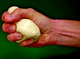

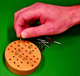

Fig. 6.4
Strength training by the use of Theraputty (University Medical Center Groningen, UMCG 2011)

Fig. 6.5
Wrist stabilisation programme using dumbbells (University Medical Center Groningen, UMCG 2011)

Fig. 6.6
Training of fine motor skills/manipulative skills (University Medical Center Groningen, UMCG 2011)
6.1.2.2 Sensory Re-education
Sensory re-education is a therapeutic programme using sensory stimulation to help patients recover functional sensibility in the damaged area and learn adaptive functioning. Sensory re-education aims at relearning and modulating the changed sensory code from the hand after an injury [8]. Among the goals of sensory re-education is the retraining of neural pathways and responses to stimuli in order to restore the patient’s sensory perception. Increased sensory input and activity may help to stimulate nerve regeneration and growth. In addition, previously unused neural connections may be trained to take over damaged pathways. This neural plasticity can be used to the advantage of the patient with nerve damage or impairment.
Recovery of sensation stems from the patient learning to make sense of unfamiliar somatic sensations through repeatedly matching them to vision and at the same time developing ‘tactics of perception’ in the form of purposeful exploratory movements of the hand. This dual process is not spontaneous but requires intensive and directed practice. Without this active retraining, the patient with a sensory-deficient hand quickly learns to use it as little as possible. Many techniques of sensory stimulation are used to provide input to sensory receptors and pathways. Some forms of stimulation used include electrical stimulation; stroking the skin with textured, friction-producing items such as Velcro and the use of specially modified tools and instruments (Fig. 6.7). This programme should be applied as a home-exercise programme since it has to be performed at least a few times a day to be effective.
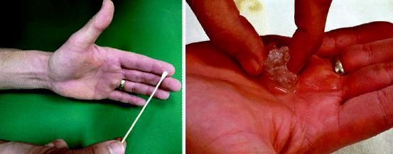

Fig. 6.7
Stimulation of sensory receptors of the skin (University Medical Center Groningen, UMCG 2011)
Key Points
Application of treatment modalities in acute stage after injury or during aftercare depends on and differs during all stages of the wound healing process. Inflammation demands modalities that limit the amount of swelling and pain, while therapy in the proliferation phase aims at increase of blood flow and lymphatic flow. Therapists supervise the athletes when performing a home-exercise programme to reach their goals while preventing overload by exercising. In the last phase of wound healing, therapy aims at realignment of collagen fibres and increase of tensile strength. During that last stage, athletes are supported in their return to sport. Each specific injury requires a different time set and depends on the tissues that were damaged or ruptured (Table 6.1).
Table 6.1
Overview of keywords related to therapeutic modalities, their applicability and relationship to the wound healing process and its different stages
Inflammation
Proliferation
Maturation-remodelling
Cryotherapy
Cold packs, ice bags, ice massage
Ice massage, contrast baths
–
Compression
Wrapping (Coban)
Wrapping (Coban), oedema massage
–
Splinting
Immobilisation/rest
Immobilisation/rest
Mobilisation/range of motion, protective playing splints
Mobilisation/range of motion
Taping
Stabilisation, compression, reducing inflammation and accumulations of fluid stimulating proprioception
Correction of posture, mobility stimulating proprioception
Prophylactic
Thermotherapy
–
Superficial heating: paraffin, hot packs
Deep heating: ultrasound, shortwave
Sensory re–education
Sensory re–education
Movement versus constant touch, sharp versus dull, location
Difference between different textures and objects.
Range of motion, strength and dexterity
Range of motion, strength and dexterity
Theraputty, dumbbells, manipulation
Theraputty, dumbbells, manipulation
6.2 The Role of the Sports Physician
Abstract
Common in sports traumatology and affecting mostly ‘small joints’, hand lesions are often underestimated or trivialised by the athlete, coach and teammates but also by uninitiated therapists. However, everything must be implemented very quickly and according to strict rules after an injury to preserve the three core functions of the hand as much as possible: strength, precision and agility. In turn, these functions depend on three basic parameters: stability, mobility and indolence. In case of recent hand trauma, the frequency or apparent insignificance of some lesions should not lead a sports physician to overlook rare, and often more serious, diagnoses. Only a thorough knowledge of the pathology and a complete clinical examination can help avoid sometimes inappropriate stereotypical attitudes, which often evolve towards functionally troublesome (oedema, stiffness) or even disabling (laxity, arthritis, etc.) sequelae. This is particularly important for top athletes, because the treatment of these complications, even if entrusted to knowledgeable hands, rarely leads to complete recovery. In case of older lesions, the sports physician should ask about and investigate potential injuries associated with the sports practised, but remain vigilant and not become complacent by simply associating the symptoms presented to the overall practice of sport, regardless of the level. All aetiologies should be systematically discussed, including non-sports pathologies. A particular ‘golden rule’ should govern the daily practice of all sports physicians: know the limits of one’s area of competence in order to ask a specialist’s opinion at the right time, not too early or too late.
6.2.1 Introduction
Hand lesions are often trivialised or underestimated by the athlete, coach and teammates, as well as uninitiated therapists, because they are common sport injuries, especially in ball sports, and because they mostly affect ‘small joints’. However, the well-known biomechanical complexity of the hand-wrist complex emphasises the urgency of care, accompanied by its inherent strict rules, that should be very quickly implemented after an injury to preserve as much as possible the three core functions of the hand, which are strength, precision and agility. These functions in turn depend on three basic parameters: stability, mobility and indolence. Because of the difficulty in quickly obtaining a consultation and/or because of an insufficient number of specialised centres, hand surgeons are rarely immediately solicited by patients. Sports physicians and emergency physicians are often the first to be consulted in cases of recent hand injury or in the presence of chronic symptoms, either following an accident or not.
6.2.2 Recent Injuries
The emergency treatment of a recently injured hand relies on a number of principles aiming at reducing complications due to overconfidence or ignorance [9–17]:
It starts with the removal of any jewellery (rings, watches, etc.; Fig. 6.8) worn in close proximity or more distally from the lesion, because post-traumatic oedema, which grows quickly, can divert their ornamental function into a source of neurovascular compression, thus accentuating the pain and oedema swelling and increasing the risk of pulp ischemia.
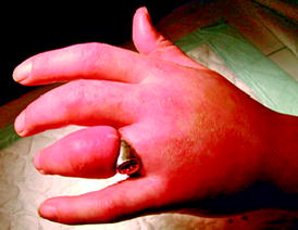
Fig. 6.8
The removal of jewels, such as rings and watches, worn in close proximity or more distally from the lesion should be systematic after an injury (medicolegal implications)
The interview investigates the following:
Diagnostic factors, such as the circumstances of the accident (direct or indirect trauma), immediate functional signs (dislocation often spontaneously reduced by the athlete himself, sensory-motor deficit, functional disability, pain) and the evolution of the patient.
Prognostic factors, such as the time elapsed since the accident and the distal oedema: the greater they are, the more the risk of adhesions and fibrosis is increased, leading to a secondary stiffness. Moreover, it has been shown that whenever the lesion affects the dominant hand, when the patient often performs manual activities, whether related to his profession, to the practice of sports or to the practice of a music instrument, and/or when the initial pain is intense, the risk of secondary complex regional pain syndrome is increased (see sensory and motor representation of the hand, at the level of the cerebral cortex).
Therapeutic factors, such as the professional or athlete patient’s specific, functional needs for the hand, which might influence the clinical management. As more professional activities are required for the accuracy of technical gestures, it becomes more essential that a hand expert’s opinion must be promptly asked to limit the eventual and possibly very debilitating sequelae that could lead to the arrest of the physical activity.
The systematic physical examination, complete (articular, vascular, neurological) and comparative, aims to highlight all positive or negative clinical signs that could guide the diagnosis and indicate the very likely need for further investigation. An uncertain diagnosis is intolerable. Sometimes, when the lesion appears to be complex (open trauma), the patient can be immediately entrusted to a specialised surgeon.
Several settings may be encountered:
In case of wound: Apart from simple cutaneous abrasions, any wound of the hand must be properly explored to make sure that a joint, nerve, vascular or tendon injury that requires debridement and/or repair is not overlooked. In case of consequent loss of substance (digital amputation) or nail avulsion, the intervention of a specialist is required for clinical management. In this case, the wound should be banded and the lost substance placed in a clean towel and bag containing ice, and the patient should be transferred as quickly as possible to a specialised healthcare facility.
In case of osteoarticular or ligament injury: When there is a visible, open fracture or dislocation (wound next to a skeletal deformation), the patient should be immediately referred to a hand surgeon. In case of closed injury, no clinical examination, even if perfectly conducted, would be sufficient to accurately diagnose an osteoarticular or ligament injury. A radiographic assessment should be systematically performed before choosing a treatment option. To avoid any mobilisation susceptible to dislocating a possible avulsion fracture, imaging should always precede the assessment of passive mobility and the assessment of the severity of ligament damage.
The evaluation of active finger mobility is always feasible and can sometimes reveal a clinodactyly, which reflects a disturbed rotation at a fracture site and requires a surgical opinion.
Interphalangeal dislocations, which are easily identifiable, are also common. They should be eligible for first-line radiographic assessment like any other injury. However, they are often managed directly by the athlete or his entourage, ‘blindly’ reduced on the sides of the court by pulling on the finger. The success of this technique, usually conducted in an empirical and reflexive manner, often minimises this injury in sports medicine. However, experience shows that the lack of appropriate postreduction care can cause a much higher rate of sequelae (oedema, pain, instability) than in cases of correct initial management. To avoid these unfortunate pitfalls, it should be kept in mind that prior to an obvious joint distortion, it is necessary to prevent inadvertent, uncontrolled gestures, if not clinically indicated (neurovascular compression, cutaneous threat) and technically controlled (risk of articular incarceration), and to direct the patient to a specialist as soon as possible for appropriate care.
What can the sports physician do while waiting for X–rays and/or immediate transfer to a specialised surgeon?
In case of open injury, cover the wound with a clean towel (preferably sterile) and immobilise the injured limb with a splint.
In case of closed injury, explain to the patient the importance of X-rays for therapeutic options, to improve his compliance to medical decisions.
Relieve pain:
By administering oral analgesics or muscle relaxants, if necessary
By advising the patient, in case of closed injury, to apply ice for 10–15 min after protecting the skin with a cloth and renewing the operation every hour
Secure the injured joint (wrist, finger) with a splint or brace, and clearly indicate to the patient that it is a prediagnosis preliminary treatment.
Control the oedema by elevating the affected limb (arranging arm in a sling, holding the hand higher than the elbow).
What is the sports physician’s role after the X–rays have been performed?
Refer the patient to a hand specialist for:
All injuries who do not need conservative treatments (see corresponding book chapters).
All injuries for which the physician considers that his skills are insufficient to establish a diagnosis and/or implement an appropriate therapy. Indeed, a physician’s lack of experience can lead to excessive, unjustified trauma consisting of the systematic immobilisation of all hand injuries. Yet, as rightfully stated by Moutet, inopportune hand immobilisations are the ‘main providers of stiffness that are often disproportionate to the severity of the initial injury’. In contrast, overconfidence can lead to insufficient measures, preventing complete healing and opening the door to malunions, residual laxity and other secondary complications to deficient care.
In case of sufficient and recognised expertise, consensus treatment guidelines specific to the diagnosed lesion should be applied. For the rehabilitation of an injured hand, the gain in mobility should not be at the cost of the loss of indolence nor at the cost of the loss of stability. Moreover, the intrinsic plus position should be chosen if the immobilisation of the metacarpophalangeal, proximal interphalangeal and distal joints is necessary. Dynamic orthoses should be implemented as soon as possible to protect and direct healing and allow for the preservation of the active motor circuits, particularly in case of associated trunk, plexus or radicular neurological, motor and/or sensory lesion.
Whatever the decision may be, one should explain to the patient the importance of progressive monitoring for early detection of eventual complications and to assess wound healing, which is essential before resuming the activities that caused the trauma.
6.2.2.1 In Case of Tendon Injury
There are two main difficulties:
Make the diagnosis. Indeed, the initial clinical expression of these lesions is often diminished because of moderate pain symptoms and an evoking distortion not always present during the immediate post-traumatic phase. The identification of the lesions is thus difficult if not systematically researched. Moreover, apart from the classic ‘mallet finger’ and ‘jersey finger’, fairly well known by sports physicians, there are other, less common lesions (rupture of the median strip of the extensor, dislocation of the extensor tendons over the metacarpophalangeal joint, etc.) with severe functional consequences when initially neglected. So, when in doubt, the advice of a specialist should be sought.
Establish the severity of the injury: partial or complete rupture? Avulsion of the insertion area? Tendon retraction? These questions usually lead the sports physician to perform a few imaging studies before considering appropriate treatment under specialised monitoring (see relevant chapters).
Key Points
When considering recent hand injuries, the frequency and apparent triviality of some lesions should not overlook rare, and often more serious, diagnoses. Only a thorough knowledge of the pathology and a complete clinical examination can help avoid sometimes inappropriate stereotypical attitudes, which often evolve towards functionally troublesome (oedema, stiffness) or even disabling (laxity, arthritis, etc.) sequelae. This is particularly important for top athletes, because the treatment of these complications, even if entrusted to knowledgeable physicians, rarely leads to complete recovery. The management of an injured hand thus requires a sufficient knowledge to establish an accurate diagnosis of the lesion and to propose the most appropriate treatment.
6.2.3 Old Injuries
The reasons for consultation are diverse: pain, stiffness, feeling of instability and oedema. The sports physician must locate the source of these symptoms during the examination. Two principles will orchestrate his approach [9–18]:
Based on his knowledge of technical sports gestures, he will ask about and investigate injuries associated with the sports practised.
He will remain vigilant and not simply associate the symptoms presented to the overall practice of sport, regardless of the level. All aetiologies should be systematically discussed, including ‘non-sports’ pathologies.
Three situations should be considered: (1) post-traumatic hand, (2) microtraumatic hand as a result of overuse and (3) expression of a more general hand condition (rheumatologic, vascular, neurological, etc.).
6.2.3.1 Post-traumatic Hand
Two situations may arise:
The initial trauma is clearly explained by the patient. The sports physician should then redraw the complete history of the lesion to classify the symptoms reported by the athlete into the following categories: diagnostic error, therapeutic error or therapeutic insufficiency, progressive complications (specify) and sequelae (what kind).
The interview does not reveal any clear trauma. Sometimes the lesion is not very painful (scaphoid fracture) or the onset of complications is delayed (arthritis, pseudoarthritis). In these cases, despite the patient’s claims, the sports physician must nonetheless eliminate the possibility of a formal trauma that was initially underestimated or unnoticed (in the context of multiple trauma or repeated falls).
Additional tests are often helpful to establish a diagnosis. They are inseparable from the physical examination, which guides the prescription (What tests should be performed? How many? How to interpret the results? Pathological aspect or not? etc.).
6.2.3.2 Overused Hand
An Increasingly Common Pathology
The improvement of working conditions and the development of the service industry has gradually led to a sedentarisation of a large part of the population. This development, referred to as modernisation, has allowed for a reduction in physical constraints. However, these constraints were gradually replaced by increasing stress, favoured by technologies requiring ever higher skill levels and faster production rates.
Because physical fatigue has proved to relieve stress, there has been a growing interest in sports among the general population in the past few years, in concert with greater leisure time. Moreover, as young individuals increasingly enter into sports competition, this exposes their immature skeletons to very high stress levels. Finally, because the professionalisation of sports has increased, the resulting financial and psychological implications have led to a much greater physical effort than ever before.
Whatever the context in which they occur, the consequences for hand lesions are hand overuse, purveyor of earlier cartilage and tendon and bone lesions, much more marked in sports practice today than just decades ago. Sports physicians must be aware of these developments so as not to minimise the potential orthopaedic consequences.
Varied Clinical Presentations
Depending on the sport, hands collide (combat sports, diving, etc.), handle technical objects (pads, wrist strap, golf clubs, swords, etc.) or maintain balance (gymnasts, climbers, etc.). Sports physicians should recognise the anatomical elements involved or exposed to these specific gestures in order to identify the conditions of overuse (tendinopathies, stress fractures, canal syndromes, etc.) (see relevant chapters). The higher – quantitatively or qualitatively – the level of practice, the more common the incidence of microtraumas. However, physicians should also search for these traumas in individuals practising as amateurs, because unlike top athletes, their usually more advanced age, combined with a poor mastering of technical gesture(s), can also lead to lesions.
Avoid Overconfidence
The anatomical elements of the hand and wrist are very numerous and can be more or less involved depending on the sport practised. For this reason, the clinical expressions of overuse are varied. Thus, the examination must be rigorous, systematic and comparative to avoid overlooking some of them. As for any joint examination, this should be based on sound knowledge of functional anatomy to give full meaning to the gestures made during the consultation. Despite a more accurate knowledge of microtraumatic diseases, too many athletes still consult a specialised unit for so-called rebel chronic tendinitis.
This catch-all term, improperly used, and with no anatomical or histological basis as demonstrated by numerous studies, often reflects the physician’s incompetence rather than a true diagnosis. These rough approximations can lead to an escalation of complementary investigations, often excessive and inappropriate, which can lead to inadequate and ineffective therapeutic strategies.
Stay updated, free articles. Join our Telegram channel

Full access? Get Clinical Tree


