Malignant Tumors of Bone

Chapter 27 Malignant Tumors of Bone


Osteosarcoma


Osteosarcoma is a tumor characterized by the production of osteoid by malignant cells. It is the most common nonhematologic primary malignancy of bone, accounting for approximately 20% of these malignancies. The incidence is 1 : 3 per 1 million per year. Onset can occur at any age; however, primary high-grade osteosarcoma occurs most commonly in the second decade of life. Parosteal osteosarcoma has a peak incidence in the third and fourth decades, and secondary osteosarcomas (e.g., those that occur in the setting of Paget disease or previous radiation therapy) are more common in older individuals. The incidence is slightly higher in males (with the exception that parosteal osteosarcoma is more common in females). There are no significant differences among races, and genetic factors rarely have been shown to play a role, although osteosarcoma may be more common in patients with the hereditary form of retinoblastoma, Rothmund-Thomson syndrome, and Li-Fraumeni syndrome. All skeletal locations can be affected; however, most primary osteosarcomas occur at the sites of the most rapid bone growth, including the distal femur, the proximal tibia, and the proximal humerus.


Almost all patients with high-grade osteosarcoma report progressive pain (patients with low-grade surface osteosarcomas may report a painless mass) that results from microinfarctions as the invasive tumor cells weaken the involved bone. Pain initially may improve with conservative measures and activity modifications, which can lead to a false sense of security for the patient and the physician. The pain eventually becomes severe if the diagnosis is delayed. Night pain may be an important clue to the true diagnosis; however, only about 25% of patients experience this phenomenon. Patients frequently are misdiagnosed with a more common musculoskeletal problem at the initial visit. The average delay from the onset of symptoms to the correct diagnosis was approximately 15 weeks in one study. This included the sum of the average patient delay of 6 weeks (the time between the onset of symptoms and initial physician encounter) and the average physician delay of 9 weeks (the time from the first visit to the correct diagnosis). The primary reasons for delay on the part of physicians included failure to obtain radiographs at the initial visit and, more important, failure to repeat the radiographs when a patient’s symptoms persisted or worsened.


Although the radiographic appearance of osteosarcoma can vary, plain radiographs are the most valuable tools for making the correct diagnosis. The most common appearance is that of an aggressive lesion in the metaphysis of a long bone. Approximately 10% are primarily diaphyseal, and less than 1% are primarily epiphyseal. Although the lesion can be either predominantly blastic or predominantly lytic, more commonly areas of bone production and bone destruction are present. The lesion usually is quite permeative, and the borders are ill defined. If the tumor has broken through the cortex, a soft tissue mass may be present at the time of diagnosis. Periosteal reaction may take the form of a “Codman triangle,” or it may have a “sunburst” or “hair-on-end” appearance. MRI is the best to measure the extent of the tumor within the bone and in the soft tissue and to determine the relationship of the tumor to nearby anatomical structures. A bone scan should be obtained to look for skeletal metastases, and radiography and CT of the chest should be done to search for pulmonary metastases; the lungs are the most common sites of metastases. These tests should be done before biopsy.


Osteosarcomas are categorized as primary or secondary. Primary osteosarcomas are subcategorized as conventional osteosarcoma, low-grade intramedullary osteosarcoma, parosteal osteosarcoma, periosteal osteosarcoma, high-grade surface osteosarcoma, telangiectatic osteosarcoma, and small cell osteosarcoma.


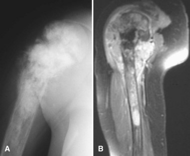
Most osteosarcomas are classified as conventional osteosarcomas (Figs. 27-1 to 27-3) and have a radiographic appearance as previously described. These high-grade tumors begin in an intramedullary location but may break through the cortex and form a soft tissue mass. Histologically, they may be primarily osteoblastic, fibroblastic, or chondroblastic; however, osteoid production from the tumor cells must be shown. The spindle cell component is high grade with hypercellularity, abundant mitotic figures, and marked nuclear pleomorphism.


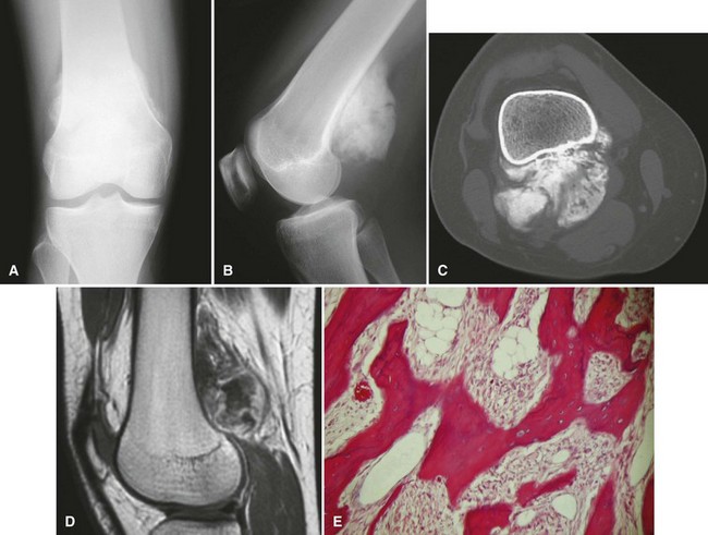
Periosteal osteosarcoma (Fig. 27-4) is an intermediate-grade malignancy that arises on the surface of the bone. The most common locations are the diaphyses of the femur and tibia. It occurs in a slightly older and broader age group. Histological examination of periosteal osteosarcoma shows strands of osteoid-producing spindle cells radiating between lobules of cartilage.


Low-grade intramedullary osteosarcoma is a rare type characterized by an indolent course with relatively benign features on radiograph. In some patients it can be mistaken radiographically and histologically for an osteoblastoma or fibrous dysplasia. As the name implies, it is located in an intramedullary location. If left untreated, it may erode through the cortex very late in the disease process. Microscopically, it consists of slightly atypical spindle cells producing slightly irregular osseous trabeculae.


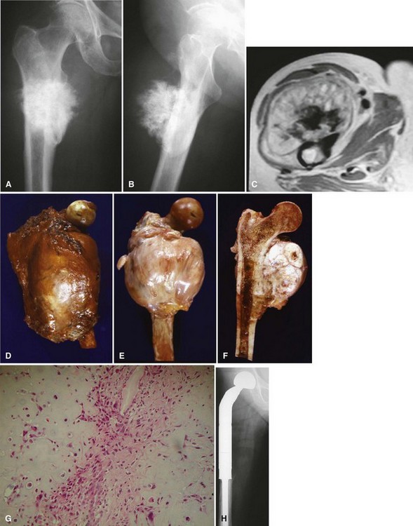
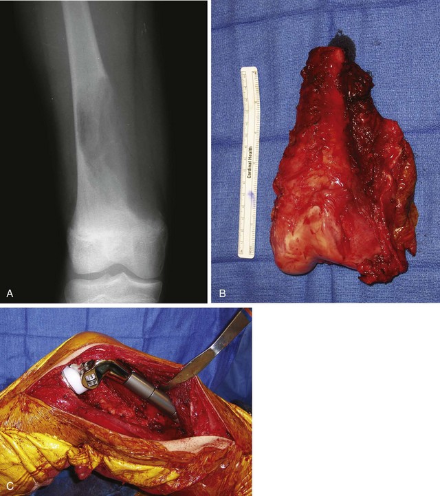
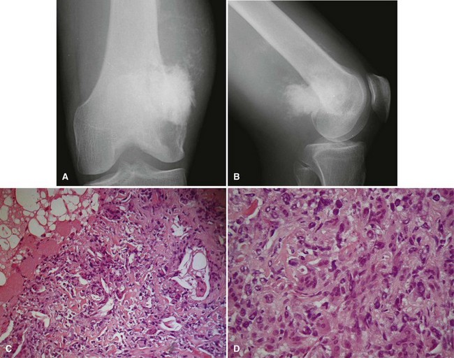
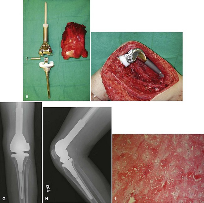
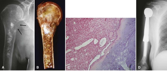
Parosteal osteosarcoma (Fig. 27-5) also is a rare, low-grade malignancy, but it arises on the surface of the bone and invades the medullary cavity only at a late stage. It has a peculiar tendency to occur as a lobulated ossified mass on the posterior aspect of the distal femur. CT may be helpful in differentiating this subtype of osteosarcoma from myositis ossificans or an osteochondroma. The ossification in myositis ossificans is more mature at the periphery of the lesion, whereas the center of a parosteal osteosarcoma is more heavily ossified. Parosteal osteosarcoma can be easily differentiated from an osteochondroma because the CT scan of an osteochondroma shows a medullary cavity containing marrow in continuity with the medullary canal of the involved bone. Microscopically, similar to a low-grade intramedullary osteosarcoma, parosteal osteosarcoma consists of slightly atypical spindle cells producing slightly irregular osseous trabeculae.
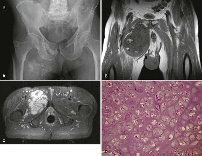


High-grade surface osteosarcoma is the least common type of osteosarcoma. As the name implies, it is an aggressive tumor arising on the outer aspect of the cortex. Radiographs show an invasive lesion with ill-defined borders. Similar to conventional osteosarcoma, the microscopic appearance is that of a high-grade tumor with hypercellularity, mitotic figures, and marked nuclear pleomorphism. In contrast to parosteal osteosarcoma, medullary involvement is common at the time of diagnosis.


Telangiectatic osteosarcoma is a purely lytic lesion. On a radiograph, it can have an invasive appearance or it can have a ballooned appearance similar to that of an aneurysmal bone cyst. Grossly, it resembles a blood-filled cyst with only a very small solid portion. Microscopically, on low power, it most commonly resembles an aneurysmal bone cyst with blood-filled spaces separated by thin septa. On higher-power magnification, however, the cells in the septa appear frankly malignant.


Small cell osteosarcoma, another rare variant, is a high-grade lesion that consists of small blue cells and may resemble Ewing sarcoma or lymphoma. If present in only a small quantity, the osteoid can be difficult to differentiate from the fibrin-like material that may be present in Ewing sarcoma. Cytogenetic and immunohistochemistry studies sometimes are needed to differentiate these lesions.


Secondary osteosarcomas occur at the site of another disease process. They rarely occur in young patients but constitute almost half of the osteosarcomas in patients older than age 50 years. The most common factors associated with secondary osteosarcomas include Paget disease and previous radiation therapy. The incidence of osteosarcoma in Paget disease is approximately 1% and may be higher (5% to 10%) for patients with advanced polyostotic disease. Paget osteosarcoma most commonly occurs in patients in the sixth to eighth decades of life, and the pelvis is the most common location. Radiation-associated osteosarcoma occurs in approximately 1% of patients who have been treated with greater than 2500 cGy and can occur in unusual locations, such as the skull, spine, clavicle, ribs, scapula, and pelvis. Although osteosarcoma is the most common radiation-associated sarcoma, fibrosarcoma and malignant fibrous histiocytoma also are relatively common in this setting. The time to onset of the secondary osteosarcoma averages 10 to 15 years after radiation exposure but may occur 3 years to several decades after treatment. Other conditions that have been reported to be associated with secondary osteosarcomas include fibrous dysplasia, bone infarcts, osteochondromas, chronic osteomyelitis, melorheostosis, and osteogenesis imperfecta; however, secondary osteosarcomas are extremely rare in these settings and a causal relationship has not been established.


Before the advent of multiple-agent chemotherapy, the prognosis for patients with osteosarcoma was dismal. Despite treatment consisting of wide or radical amputation, approximately 80% of patients died as a result of distant metastases, usually within 2 years. With today’s multiple-agent chemotherapy regimens and appropriate surgical treatment, most series report long-term survival of 60% to 75% for patients with high-grade osteosarcoma without metastases at initial presentation and 90% for those with low-grade lesions.


The most important prognostic factor at the time of diagnosis is the tumor stage. Approximately 15% of patients with osteosarcoma have detectable pulmonary metastases at the time of diagnosis. As a group, these patients continue to have a poor prognosis, with less than 20% long-term survival. (Patients with one or a few resectable pulmonary metastases at presentation may have greater than 50% long-term survival, whereas patients with many, large, or unresectable pulmonary metastases have an extremely poor prognosis.) Patients with nonpulmonary metastases (e.g., bone metastases) have an even worse prognosis, with less than 5% long-term survival. Patients with “skip” metastases (i.e., a metastasis within the same bone as the primary tumor or across the joint from the primary tumor) have the same poor prognosis as patients with distant metastases.


The next most important prognostic feature is the grade of the lesion. Low-grade lesions rarely metastasize, and patients with low-grade lesions have a marked survival advantage over patients with high-grade lesions. Size of the primary tumor also seems to be of prognostic significance. Although authors differ on the specific criteria for what constitutes a large or a small tumor, most studies confirm that patients with large tumors have a worse prognosis than patients with smaller tumors. Skeletal location also is thought to be important because patients with more proximal tumors do worse than patients with more distal tumors. Size and location are likely interrelated variables, however, because most proximal tumors are larger at the time of diagnosis than most distal tumors. Paget osteosarcomas continue to have a poor prognosis, with less than 15% long-term survival. Radiation-associated osteosarcomas have been regarded as having a poor prognosis; however, this may be due primarily to their frequent occurrence in unusual locations where resection is difficult. Radiation-associated osteosarcomas in the extremities may have the same prognosis as any other high-grade osteosarcoma. Age at diagnosis and gender do not seem to be of prognostic significance.


As stated earlier, historically, patients with high-grade osteosarcoma were treated with immediate wide or radical amputation. Despite this treatment, 80% of patients with apparently isolated disease died of distant metastases. From this, it can be deduced that most patients with high-grade osteosarcoma have nondetectable micrometastases at presentation. The goal of adjuvant or neoadjuvant chemotherapy is to treat these micrometastases. Currently, at most musculoskeletal oncology centers, the treatment of high-grade osteosarcoma consists of neoadjuvant chemotherapy, wide or radical surgery (resection or amputation), and adjuvant chemotherapy. Pulmonary metastases likewise are resected if possible after neoadjuvant chemotherapy. The histological response of the primary tumor to neoadjuvant chemotherapy has been shown to be a good predictor of long-term survival. Greater than 90% tumor necrosis indicates a very good prognosis. Low-grade osteosarcoma can be treated with wide resection or amputation without chemotherapy.


About 50% of patients with high-grade osteosarcoma have some type of relapse after the initial treatment. About 10% of patients have local recurrence after wide resection or wide amputation. Patients who have a local recurrence have a very poor prognosis and usually are treated with a radical amputation (if cure is the goal) and further chemotherapy. Late pulmonary metastases likewise are treated with surgery and chemotherapy. Poor prognostic factors include rapid relapse after completion of the initial treatment, many (more than eight) pulmonary nodules, large (>3 cm) pulmonary nodules, and unresectable pulmonary nodules. Patients with a few, small, resectable pulmonary nodules that occur late may have a 40% chance of cure with aggressive treatment.


Chondrosarcoma


Chondrosarcoma constitutes about 9% of primary malignancies of bone, an incidence about half that of osteosarcoma. It is the second most common nonhematologic primary malignancy of bone. It occurs over a broad age range, with peaks between 40 and 60 years for primary chondrosarcoma and between 25 and 45 years for secondary chondrosarcoma. Chondrosarcoma can occur in any location; however, most are located in a proximal location such as the pelvis, proximal femur, and proximal humerus. Although chondrosarcomas rarely occur in the hand, they are the most common primary malignancy of bone in this location. Similar to most bone tumors, the incidence is slightly higher among males. Race predilection is not significant.


Clinically, most patients with primary chondrosarcomas report increasing pain. A palpable mass also may be present. Chondrosarcomas frequently are slow growing, and symptoms can be present for several years before a patient seeks medical attention. Pain in the absence of a pathological fracture can be important in helping to differentiate an enchondroma from a low-grade chondrosarcoma. Frequently, patients are referred for evaluation of an asymptomatic cartilaginous lesion discovered as an incidental finding on a bone scan or radiograph obtained for another reason. (The radiographic abnormality usually is the sole reason the patient is referred to the orthopaedic oncologist.) Although an asymptomatic radiographic abnormality is common in a patient with an enchondroma, the diagnosis of chondrosarcoma would be extremely rare in this circumstance. A chondrosarcoma may occur in the area of a treated “enchondroma.” In this circumstance, the original pathology specimen should be reviewed.


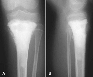
Secondary chondrosarcomas arise at the site of a preexisting benign cartilage lesion. They occur most frequently in the setting of multiple enchondromas and multiple hereditary exostoses. In Ollier disease (multiple enchondromas) the incidence of malignancy (most commonly chondrosarcoma) is approximately 25% by age 40 years, and in patients with Maffucci syndrome (multiple enchondromas with soft tissue hemangiomas) the incidence may be even higher. Although data for osteochondromas are difficult to analyze, the lifetime incidence of secondary chondrosarcoma is estimated to be 5% for patients with multiple hereditary exostoses and approximately 1% for patients with solitary osteochondromas (Fig. 27-6). As discussed in Chapter 25, the true incidence of malignant degeneration of osteochondromas is unknown. Published estimates are likely too high, owing to the effect of referral bias on pathology data at tertiary referral centers. The true prevalence of osteochondromas in the general population is unknown. Whether a solitary benign enchondroma has the potential to give rise to a secondary chondrosarcoma is difficult to determine. If this does occur, the incidence is not high enough to warrant prophylactic treatment of asymptomatic enchondromas. Other conditions that have been reported to be associated with secondary chondrosarcoma include synovial chondromatosis, chondromyxoid fibroma, periosteal chondroma, chondroblastoma, previous radiation treatment, and fibrous dysplasia.


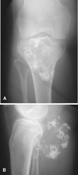
The radiographic appearance of chondrosarcoma frequently is diagnostic (Fig. 27-7). Similar to enchondroma, it is a lesion arising in the medullary cavity with irregular matrix calcification. The pattern of calcification has been described as “punctate,” “popcorn,” or “comma shaped.” Compared with enchondroma, however, chondrosarcoma has a more aggressive appearance with bone destruction, cortical erosions, periosteal reaction, and, rarely, a soft tissue mass. CT can be helpful to show endosteal erosions or other evidence of a destructive lesion and to differentiate benign from malignant cartilage lesions. The site of the lesion also must be considered because lesions in the hand (the most common site for an enchondroma and a rare site for a chondrosarcoma) may appear aggressive and still be diagnosed as benign. The same amount of cortical destruction shown in a pelvic or proximal femoral lesion would be diagnostic of a chondrosarcoma. Finally, the size of the cartilaginous cap of an osteochondroma, as evaluated with CT or MRI, is important in evaluating the possibility of a secondary chondrosarcoma. If the cartilaginous cap is larger than 2 cm in a skeletally mature patient, a secondary chondrosarcoma must be considered.


Histologically, conventional chondrosarcomas are composed of malignant cells with abundant cartilaginous matrix. (If malignant osteoid is present even in small amounts, the diagnosis should be chondroblastic osteosarcoma—a tumor with different prognostic and therapeutic implications.) Differentiating a low-grade chondrosarcoma from an enchondroma can be difficult solely from a biopsy specimen. Factors that favor a malignant diagnosis include hypercellularity, plump nuclei, more than occasional binucleate cells, a permeative pattern, and entrapment of bony trabeculae (Fig. 27-8). As much tissue as possible should be obtained from the biopsy of a borderline lesion. Perhaps in no other circumstance is correlation with the clinical and radiographic findings more important. Lesions in the setting of multiple enchondromas, periosteal chondromas, synovial chondromatosis, and enchondromas of the hand all may appear hypercellular and yet still can be benign. This same appearance in a biopsy specimen taken from a solitary large pelvic lesion with radiographically shown cortical erosions would be diagnostic of a chondrosarcoma.
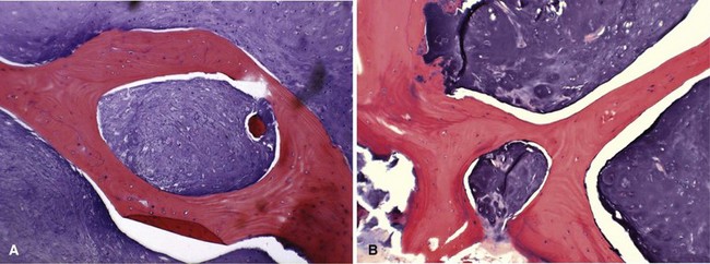


Less common histological subtypes of chondrosarcoma include dedifferentiated chondrosarcoma, clear cell chondrosarcoma, and mesenchymal chondrosarcoma. Together, these subtypes constitute less than 20% of all chondrosarcomas. Histologically, dedifferentiated chondrosarcoma consists of a high-grade sarcoma (most commonly osteosarcoma followed in frequency by fibrosarcoma and malignant fibrous histiocytoma) adjacent to an otherwise typical low-grade chondrosarcoma (Fig. 27-9). The radiographic features of a dedifferentiated chondrosarcoma often show a more aggressive radiolucent area juxtaposed on an otherwise typical chondrosarcoma.


Clear cell chondrosarcoma is a low-grade malignancy. As the name implies, it consists of round cells with abundant clear cytoplasm and distinct cytoplasmic borders with a background of cartilaginous matrix. Multinucleated giant cells usually are apparent. Clear cell chondrosarcoma has a strong tendency to arise in an epiphysis (especially the proximal femur). It may have benign radiographic features and can be confused with chondroblastoma or giant cell tumor.


Mesenchymal chondrosarcoma is a high-grade tumor consisting of small, round blue cells with islands of benign-appearing cartilage. The cellular portions often have a hemangiopericytomatous pattern of growth with “staghorn-like” vessels. Radiographically, mesenchymal chondrosarcoma may look like a conventional chondrosarcoma. More frequently, however, it has a nonspecific, aggressive radiographic appearance.


The treatment of low-grade chondrosarcoma is controversial, with many authors reporting excellent results after extended curettage with the use of intraoperative adjuvant treatments. Extended curettage is considered adequate treatment only for low-grade lesions that are confined within the medullary canal. Those with soft tissue extension should be treated similar to high-grade lesions. The treatment of high-grade chondrosarcoma is wide or radical resection or amputation. Because cartilage is relatively avascular, the cells survive transplantation easily. The local recurrence rate after intraoperative tumor contamination is high. For lesions in an expendable location, primary wide resection without a biopsy may be indicated to decrease the chance of tumor contamination. After wide resection, local recurrence is less than 10% and can be treated with repeat wide resection or wide amputation. Likewise, pulmonary metastases should be treated with surgical resection if possible. Chemotherapy has no role in the treatment of conventional chondrosarcoma but currently is being evaluated for treatment of dedifferentiated and mesenchymal chondrosarcomas. Radiation therapy likewise has a limited role and is used only as a palliative measure for surgically inaccessible lesions.


The prognosis for patients with chondrosarcoma depends mostly on the size, grade, and location of the lesion. If a high-grade lesion cannot be completely resected with wide or radical margins (usually because of its size or location), local recurrence is likely. Patients with low-grade lesions have been reported to have a greater than 90% 10-year survival rate, whereas patients with high-grade conventional chondrosarcoma are reported to have a 20% to 40% 10-year survival rate. The 5-year survival rate is less than 15% for patients with dedifferentiated chondrosarcoma, with most deaths occurring in the first 2 years. Because chondrosarcomas often are slow growing, local recurrences and pulmonary metastases may not be detected until years or decades after the primary procedure. A significant percentage of recurrences show a higher histological grade than the original tumor. Long-term follow-up with regular imaging of the operative site and the chest is imperative so that treatment can be initiated promptly in the event of a recurrence.


Ewing Sarcoma


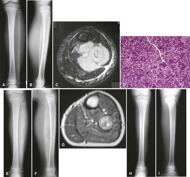
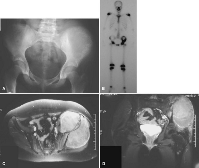
Ewing sarcoma is the third most common nonhematologic primary malignancy of bone, but it is the second most common (after osteosarcoma) in patients younger than 30 years of age and the most common in patients younger than 10 years of age. The incidence is less than 1 per 1 million per year, accounting for about 9% of primary malignancies of bone. Ewing sarcoma has been reported to occur in a wide age range of patients from infants to the elderly, but most occur in patients 5 to 25 years old. The most common locations include the metaphyses of long bones (often with extension into the diaphysis) and the flat bones of the shoulder and pelvic girdles (Figs. 27-10 and 27-11). Rarely, it occurs in the spine or in the small bones of the feet or hands. Similar to most sarcomas of bone, there is a slightly higher incidence in males. Ewing sarcoma is exceedingly rare in individuals of African descent. There are no known predisposing factors.


Pain is an almost universal complaint of patients with Ewing sarcoma. Usually the onset is insidious, and the pain may be of long duration before the patient seeks medical attention. The pain may be only mild and intermittent initially and may respond to initial conservative treatment. The average delay from the onset of symptoms to the diagnosis has been reported to be 34 weeks. The average patient delay in one study was 15 weeks from the onset of symptoms until the first medical appointment, and the average physician delay was 19 weeks from the initial visit to correct diagnosis. These numbers show the importance of radiographs at the initial visit and rechecking them at subsequent visits if the patient continues to have symptoms.


In addition to pain, patients also may have fever, erythema, and swelling, suggesting osteomyelitis. Laboratory studies may reveal an increased white blood cell count, an elevated erythrocyte sedimentation rate, and an elevated C-reactive protein level. To complicate matters further, a needle aspirate of Ewing sarcoma may grossly resemble pus, and the tissue may be sent in its entirety to microbiology and none to pathology. (As a general rule, most biopsy specimens should be sent for culture and pathological analysis.)


Classically, Ewing sarcoma appears radiographically as a destructive lesion in the diaphysis of a long bone with an “onion skin” periosteal reaction. In reality, Ewing sarcoma more often originates in the metaphysis of a long bone but frequently extends for a considerable distance into the diaphysis. Although “skip” metastases (similar to those that occur in osteosarcoma) are not reported in Ewing sarcoma, it is common for a large portion of the bone (or even the entire bone) to be involved. In flat bones, Ewing sarcoma appears as a nonspecific destructive lesion. Regardless of the location, MRI of the entire bone should be ordered to evaluate the full extent of the lesion, which typically extends beyond the abnormality apparent on plain films. MRI also is useful to evaluate the extent of the soft tissue mass, which often is very large. All patients should have a baseline radiograph and CT of the chest because the lung is the most common site of metastases. A bone scan should be performed because bone is the second most common site of metastases. In contrast to other bone sarcomas, a bone marrow aspirate should be obtained as a routine part of the staging of Ewing sarcoma to rule out diffuse systemic disease.


Histologically, Ewing sarcoma consists of small blue cells with very little intercellular matrix. Cytogenetic or immunohistochemical studies often are required to differentiate Ewing sarcoma from other small blue cell tumors. The t(11;22)(q24;q12) is the most common translocation diagnostic of Ewing sarcoma and is present in greater than 90% of cases. Other diagnostic translocations, including t(21;22)(q22;q12) and t(7;22)(p22;q12), also have been identified. Immunohistochemical staining for the CD99 gene product has been reported to be specific for Ewing sarcoma. In addition, Ewing sarcomas usually are periodic acid–Schiff positive (owing to intracellular glycogen) and reticulin negative. This is in contrast to lymphomas, which are periodic acid–Schiff negative and reticulin positive. Lymphomas also stain positive for leukocyte common antigen and other T- and B-cell antigens. Embryonal rhabdomyosarcoma stains positive for desmin, myoglobin, and muscle-specific actins. Hemangiopericytomas stain positive for factor VIII, and small cell metastatic carcinomas and melanomas stain positive for cytokeratin.


The worst prognostic factor is the presence of distant metastases. Even with aggressive treatment, patients with metastases have only a 20% chance of long-term survival. The size of the primary lesion has been shown consistently to be of prognostic significance, although specific parameters have not been firmly established. Location also has been reported to be of prognostic significance, but it is difficult to differentiate the effects of location and size because most proximally located tumors are larger at presentation than distally located tumors. Histological grade is of no prognostic significance because all Ewing sarcomas are considered high grade. Fever, anemia, and elevation of laboratory values (white blood cell count, erythrocyte sedimentation rate, lactate dehydrogenase) have been reported to indicate more extensive disease and a worse prognosis. Older age at presentation (with a cutoff around 12 to 15 years old) and male gender also have been reported to be associated with a worse prognosis. The specific translocation, t(11;22) versus t(21;22), does not seem to affect the clinical course; however, secondary genetic alterations, such as aberrant TP53 expression, may prove to be important. As with osteosarcoma, histological response to neoadjuvant chemotherapy has been shown to be prognostically important. Greater than 90% necrosis after preoperative chemotherapy indicates a good prognosis.


The treatment of Ewing sarcoma must include neoadjuvant or adjuvant chemotherapy, or both, to treat distant metastases that may or may not be readily apparent at the initial staging. Before the use of multiple-agent chemotherapy, long-term survival was less than 10%. Today, most centers report long-term survival rates of 60% to 75%.


Local treatment of the primary lesion is more controversial. Ewing sarcoma is radiosensitive, yet some authors report a decreased rate of local recurrence (<10%) and an increased rate of overall survival with wide resection of the primary tumor. These reports are difficult to interpret, however, because large, central, unresectable tumors often are treated with radiation, whereas smaller, more accessible lesions (which inherently have a better prognosis) are more likely to be treated with surgery. At this time, the choice between surgery and radiation for treatment of the primary lesion must be made on an individual basis. Repeat staging studies should be obtained after neoadjuvant chemotherapy. The repeat radiographs often show increased ossification, and repeat MRI often shows a marked decrease in the soft tissue mass. At this point, if it seems that the lesion can be resected with wide margins with an acceptable functional deficit, surgery should be the treatment of the primary lesion. If wide margins would be difficult to obtain or if the functional deficit resulting from surgery would be unacceptable, radiation of the primary lesion is an acceptable alternative. Radiation also can be used as an adjuvant after a marginal resection or a contaminated wide resection. The treatment plan in each case is most appropriately made after long discussions with the patient and the family. The discussions should include expected function after amputation, limb salvage surgery, or radiation and the inherent short-term and long-term risks involved with each option.


Disease relapse is associated with a poor prognosis despite aggressive treatment of the relapse with further surgery, radiation, and chemotherapy. Patients with local recurrence have been reported to have about a 20% 5-year survival rate, whereas patients who relapse with distant metastases have approximately a 10% 5-year survival rate. As with osteosarcoma, time to relapse has prognostic significance. Patients who relapse within the first year after primary treatment have a worse prognosis than patients who have an extended disease-free interval.


Chordoma


Chordoma is a rare malignant neoplasm that arises from notochord remnants. In the Mayo Clinic series, it accounted for approximately 4% of primary malignancies of bone. Chordoma is the second most common primary malignancy in the spine (behind myeloma) and is the most common primary malignancy of the sacrum. Greater than 50% of chordomas arise in the sacrococcygeal area, and more than 30% arise at the base of the skull. The remainder are dispersed throughout the rest of the spine. Peak incidence for sacrococcygeal chordomas occurs in the fifth to seventh decades, whereas the peak for sphenooccipital lesions is the fourth to sixth decades. Most series show a marked male predominance (3 : 1), especially for sacrococcygeal tumors.


The presenting signs and symptoms vary according to the site of the lesion. Because most chordomas are slow growing, patients frequently have symptoms for more than a year before diagnosis. Patients with tumors in the sphenooccipital region may report headaches or symptoms related to cranial nerve compression. In the spine, symptoms can be caused by nerve root or cord compression. If an anterior mass exists with a cervical spine lesion, the symptoms may be similar to those caused by a retropharyngeal abscess. The most common presenting complaint for patients with sacrococcygeal tumors is low back pain. Bowel and bladder disturbance and sciatic pain also are common with sacral tumors. A palpable mass frequently is present on rectal examination.


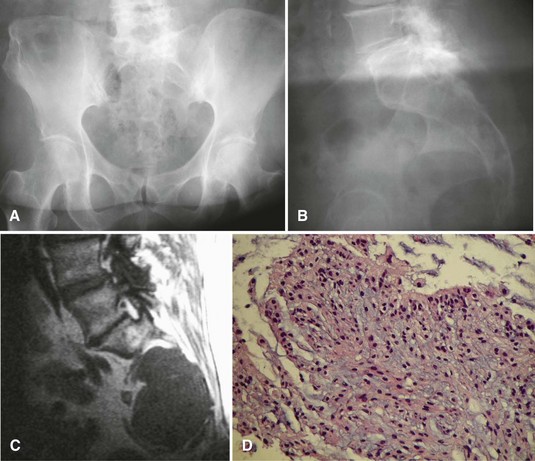
Radiographically, chordomas appear as destructive lesions (Fig. 27-12). They virtually always arise from the midline. Sacrococcygeal lesions often are missed on the initial radiographic examination because of overlying bowel gas. They usually are seen more easily on a lateral view of the sacrum. Likewise, radioisotope accumulation in the bladder can obscure a sacral tumor on a bone scan. More than 50% of chordomas exhibit radiographically detectable calcification. CT may be better for detecting calcification (which may help with the diagnosis), but MRI is better for determining the full extent of the lesion and its relationship to other anatomical structures. A common pitfall in the evaluation of a patient with a chordoma and low back pain is ordering an MRI of only the lumbar spine; this study usually misses a sacrococcygeal chordoma because most arise below S3.


Microscopically, chordoma appears as lobules of cells separated by fibrous bands. The cells usually contain abundant vacuolated cytoplasm (physaliferous cells). The cells usually are arranged in long strands, or “cords,” with a mucinous background. Most chordomas are low grade, although dedifferentiated chordomas exist. These dedifferentiated chordomas contain areas of a high-grade sarcoma—most frequently a malignant fibrous histiocytoma—and behave in a more aggressive manner.


The primary treatment is surgical resection with wide margins, even if this creates a neurological deficit, because progressive growth of the tumor would create a neurological deficit anyway and possibly metastatic disease. Resection that preserves the S3 nerve roots bilaterally results in relatively normal bowel and bladder function, whereas resection above this level results in incremental loss of bowel and bladder function. Resection of bilateral S2 nerve roots results in complete loss of control of bowel and bladder function. If wide margins cannot be obtained or if tumor contamination occurs intraoperatively, radiation may be beneficial. Radiation also may be beneficial for patients in whom resection is not feasible, although a cure is rarely, if ever, achieved in these patients. Chemotherapy is of no proven benefit. Likewise, distant metastases are treated surgically.


The 5-year overall survival rate for patients with chordomas is approximately 60% to 80%, but the survival rate continues to decline with longer follow-up because of late recurrences (25% to 40% 10-year survival). Local recurrences are common because of the difficulty encountered in achieving wide margins. Male gender and younger age at diagnosis have been reported to be associated with a favorable prognosis. A more distal location for sacral lesions also is associated with a better prognosis. Metastases are rare at initial presentation (<5%) but may occur later in 30% to 60%. In addition to the lungs, metastases are common in bone and in unusual locations such as skin, eyelid, brain, liver, and other internal organs.


Adamantinoma


Adamantinoma is a rare neoplasm representing less than 1% of all primary malignancies of bone. Adamantinoma has a wide age distribution, but most patients are in the second or third decade at the time of diagnosis. It has a peculiar predilection for occurring in the tibia (approximately 85%) and may also involve the ipsilateral fibula. It has been postulated that adamantinoma arises from aberrant nests of epithelial cells, which would account for the fact that this tumor primarily occurs in bone that is in a subcutaneous location.


Pain is the most common symptom. Because the lesion is typically slow growing, the pain can be present for many years before the patient seeks medical attention. Because the lesion usually occurs in a subcutaneous location, a palpable mass may be present. Approximately 20% of patients have a pathological fracture.


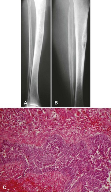
The most common radiographic appearance is that of multiple, sharply demarcated radiolucent lesions in the tibial diaphysis (Fig. 27-13). The radiolucent lesions are separated by areas of dense, sclerotic bone. Although the radiographic appearance is similar to that of osteofibrous dysplasia, adamantinoma usually has a more aggressive appearance. A large portion or even the entire tibia can be involved. Frequently, the fibula also is involved by direct extension of the tumor.


Microscopically, adamantinoma consists of islands of epithelial cells in a fibrous stroma. Some areas of the tumor resemble fibrous dysplasia or osteofibrous dysplasia. (Some authors consider adamantinoma to be a malignant variant of osteofibrous dysplasia.) Nuclear atypia is minimal, and mitotic figures are rare. Immunohistochemical staining usually is positive for cytokeratins and vimentin. It generally is a low-grade lesion, and histological features are not predictive of behavior.


The optimal treatment of adamantinoma is wide resection or amputation. The tumor generally is radioresistant and chemoresistant. Local recurrence occurs in approximately 25% of patients, and amputation should be considered for these patients. Metastases are rare at presentation but may occur later in 30% of patients. Overall survival is approximately 85% at 10 years. Prognosis depends most on the adequacy of the surgical margin. Compared with patients who have marginal or intralesional surgical procedures, patients who have wide or radical procedures have significantly reduced rates of local recurrence and metastases (<10%). Because of the slow-growing nature of this lesion, local recurrence or metastasis may occur very late, reportedly 19 years after the initial treatment. The importance of long-term follow-up must be stressed.


Malignant Vascular Tumors


The terminology used to describe malignant vascular tumors in the literature is confusing. Multiple terms have been used interchangeably, including hemangioendothelioma, hemangioendothelial sarcoma, hemangiosarcoma, angiosarcoma, and others. Although not strictly defined, most authors use the term hemangioendothelioma to describe low-grade malignant vascular tumors and the term angiosarcoma to describe high-grade malignant vascular tumors.


These rare tumors constitute approximately 1% of all primary malignancies of bone. After the first decade, they may occur at any age and in any bone. There is a slight male predominance but no significant race predilection. Stewart-Treves syndrome refers to the occurrence of angiosarcoma in the setting of chronic lymphedema (e.g., in the upper extremity of a patient who has previously undergone a radical mastectomy). Angiosarcomas also have been reported to occur adjacent to orthopaedic implants, although a causal relationship has not been firmly established.


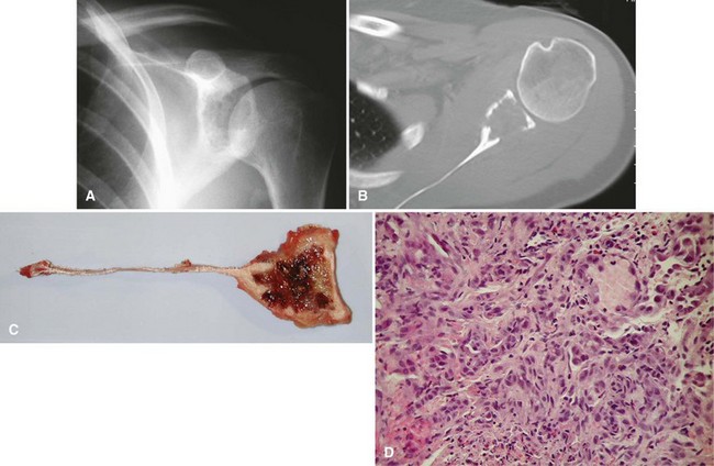
Pain or, more rarely, pathological fracture is the presenting complaint. Duration of symptoms varies depending on the grade of the tumor. The radiographic appearance of this lesion also is correlated with its grade. Low-grade tumors appear as well-demarcated lytic lesions that may or may not have surrounding reactive bone formation (Fig. 27-14). High-grade tumors have a more permeative appearance. Periosteal reaction is unusual. Malignant vascular tumors have a peculiar tendency to be multicentric at presentation regardless of grade. Most commonly, multiple lesions are found within the same bone or within multiple bones of the same extremity.


Microscopically, low-grade tumors show well-formed anastomosing vascular channels lined by plump endothelial cells. Well-differentiated hemangioendotheliomas can be difficult to differentiate from benign hemangiomas. High-grade lesions can be pleomorphic and may appear as an undifferentiated sarcoma or carcinoma. In some extremely pleomorphic lesions, the diagnosis can be made only through immunohistochemistry. Although metastatic carcinoma and malignant vascular tumors may be keratin positive, factor VIII–related antigen, CD31, and CD34 should be positive only in vascular tumors.


Treatment is individualized depending on the clinical situation. Solitary lesions are treated with wide resection if possible. Radiation can be used successfully in the treatment of surgically inaccessible lesions or in the treatment of multiple lesions. For high-grade lesions, adjuvant chemotherapy can be added to the treatment regimen. Prognosis depends most on grade. Patients with low-grade lesions may have better than an 80% chance for long-term survival, whereas patients with high-grade tumors have less than a 20% long-term survival rate.


Malignant Fibrous Histiocytoma and Fibrosarcoma


Although malignant fibrous histiocytoma and fibrosarcoma are described in the literature as being separate entities, the distinction is sometimes arbitrary. The presentation, prognosis, and treatment of these two entities are similar, so they are discussed together.


Malignant fibrous histiocytoma and fibrosarcoma of bone constitute 3% to 5% of primary malignant tumors of bone. Excluding the first decade, they occur at any age with comparable frequency. Males and females are affected equally. There is a slight tendency for the lesion to occur in the distal metaphysis of the femur or the proximal metaphysis of the tibia; however, any bone may be involved. Approximately 25% of these tumors are considered to be secondary to a preexisting bone abnormality. The most commonly reported predisposing conditions include Paget disease, radiation, giant cell tumor, and bone infarction (Fig. 27-15). They also may occur as part of a dedifferentiated chondrosarcoma.


Jun 5, 2016 | Posted by in ORTHOPEDIC | Comments Off on Malignant Tumors of Bone

Full access? Get Clinical Tree

Get Clinical Tree app for offline access