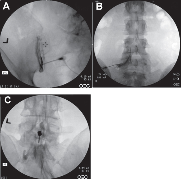
35 Because of their high prevalence and debilitating nature, low back pain and radiculopathy have important social, economic, and clinical implications. Although numerous nonsurgical interventions are available for these conditions, lumbar injection therapies have grown to become a popular conservative treatment option because of their dual role as a therapeutic and diagnostic tool. Lumbar injections may serve as a therapeutic tool by temporarily relieving symptoms in some patients, while also serving as a diagnostic tool to confirm pain etiology. Lumbar injections can be categorized into one of three groups: those that address neural inflammatory symptoms (epidurals and selective nerve blocks), those that address facet joint pain (facet joint blocks and medial branch blocks), and those directed at sacroiliac (SI) joint dysfunction (SI joint blocks). Patients considered for lumbar injections should first give a thorough history and undergo a physical examination. Spinal imaging is typically performed prior to lumbar injections. Imaging, including radiographs, magnetic resonance imaging (MRI), and computed tomographic (CT) myelography, plays a vital role in determining pain etiology and determining the appropriate course of action. As the treating physician searches for pain generators in the spine, fluoroscopically guided, contrast-enhanced diagnostic injections may also assume an important role in identifying the pain generators and directing further therapeutic interventions. Pathology in the SI joint can produce pain in the back, buttocks, and thighs. SI pain has been suggested to make up ~ 15% of low back pain. Because the physical examination is nonspecific, diagnostic injections are crucial to the diagnosis of SI dysfunction. To approach the SI joint, the needle is introduced inferiorly into the diarthrodial portion of the joint (Fig. 35.1A). Epidural steroids help decrease the presence of local inflammatory factors and reduce nerve root irritation, leading to some symptomatic relief in some patients. Some patients even temporarily experience complete symptomatic relief if nerve root irritation is the sole etiology of their low back and leg pain. Some authors have hypothesized that the volume of the injectate is important because it may physically dilute the concentration of local inflammatory mediators, although there is not strong evidence to support this rationale. In general, these injections are most effective for acute or subacute radicular symptoms. The procedure may be done with or without fluoroscopic guidance, although the accuracy of the injection is significantly improved with the use of fluoroscopy. Often, epidural injections are done in a series of two to three, performed 2 to 3 weeks apart, tailored to the patient’s response. Many different types of injections and approaches have been developed utilizing epidural steroid injections, including: 1. Transforaminal epidural steroid injections (Fig. 35.1B), which are performed from a lateral approach so that the injectate can be delivered directly into the neural foramen, anterior to the dural sleeve and around the exiting nerve root. This approach is best suited for unilateral leg symptoms in a specific dermatome that can be targeted to a specific symptomatic nerve root. 2. Translaminar epidural steroid injection (Fig. 35.1C), which is done through a midline approach. This approach is best suited for symptoms involving multiple dermatomes. The term facet joint syndrome describes pain in the back that is derived from degeneration of the facet joint. Both the facet joint and the multifidus muscle are innervated by the medial branch of the dorsal ramus. Each nerve supplies at least two facet joints; therefore, each facet joint may be innervated by more than one nerve. The diagnosis of pain derived from facet joint degeneration depends on a specific clinical presentation and a positive response to diagnostic blocks. Patients whose pain is definitively derived from facet joint degeneration (i.e., consistent positive response to facet or medial branch blocks) may be considered candidates for radiofrequency denervation of medial branches of the dorsal rami. Successful medial branch ablation may prolong the beneficial response to the facet or medial branch blocks. Fig. 35.1 (A) SI arthrogram with the needle in the inferior aspect of the left SI joint. (B) Anteroposterior view of the left L5–S1 transforaminal (nerve root) injection. Shown here is the contrast spread around the proximal takeoff of the nerve root as well as epidural spread at the corresponding level. (C) Translaminar epidural steroid injection with the needle inserted at the L3–L4 interspace. Contrast spread is limited in the caudad direction.
Lumbar Injections and Procedures
![]() Classification
Classification
![]() Workup
Workup
History and Physical Examination
Spinal Imaging
![]() Treatment
Treatment
SI Joint Injections
Epidural Injections
Facet Joints

Stay updated, free articles. Join our Telegram channel

Full access? Get Clinical Tree


