Juvenile Hallux Abducto Valgus Deformity
Donald R. Green
Kieran T. Mahan
Tracy L. Klimaz
An accepted definition of the term juvenile hallux valgus does not exist. However, in 1976, Goldner and Gaines (1) classified the deformity as occurring in an individual of 20 years of less because of the relatively plastic nature of the components of the deformity. Other authors have simply defined the juvenile bunion as manifesting itself during the formative years of life (2,3 and 4). As difficult as this deformity has been to define, it has been a greater challenge for the podiatric physician to treat as there is a lack of uniform guidelines for conservative and surgical care.
AGE OF ONSET
In 1960, Piggott reported on a series of 100 consecutive patients evaluated for hallux valgus deformities in adolescence. Of those interviewed, 57% recalled the onset of their deformity during their adolescent years, 38% did not recall, and only 5% said after the age of 20 (4). Similarly, Hardy and Clapham (5) found that 46% of adult patients remembered the deformity before they were 20 years old.
INCIDENCE
The incidence of hallux abducto valgus in the adolescent population has been reported by various authors to range from 22% to 36% (6,7). Cole reported that 36% of school children between the ages of 8 and 18 displayed mild to severe hallux valgus. Of those, 75% were female (7). In 1981, Helal (8) reported that 92% of his juvenile hallux valgus patients, ranging from 9 to 19 years old, were girls, with 75% occurring bilaterally.
ETIOLOGY
EXTRINSIC FACTORS
Shoe gear was one of the first factors considered in the etiology of the hallux abducto valgus. This was based on the observations that tight and pointed shoes tended to produce a higher incidence of the deformity (9). In addition, the incidence of juvenile hallux valgus has been shown to be higher in the shod population versus those who walk barefoot (10,11). Although decorative and fashionable shoes have been implicated in the progression of the hallux valgus deformity, Hardy and Clapham have suggested that most cases do not appear to be influenced by a history of constricting footwear (5,12). Therefore, shoe gear should be considered an aggravating factor rather than a causative one.
INTRINSIC FACTORS
It has been suggested by several authors that hallux valgus is an inherited deformity. A positive family history for the deformity has been noted in 58% to 80% of the cases (13,14 and 15). Furthermore, significantly earlier ages of onset have also been reported with patients with a positive family history of hallux valgus (16). In 1954, while studying abnormalities of the foot and hand, Johnson stated, “Hallux valgus is inherited through an autosomal dominant trait with incomplete penetrance” (17). In 2007, Pique-Vidal et al (18) examined the pedigree charts from 350 patients and found a family history present in 90% of probands, with vertical transmission affecting some family members across three generations, which is compatible with autosomal dominant inheritance with incomplete penetrance. Race may also play a role in adolescent hallux valgus. Black children are affected five times as often as white children; however, this ratio decreases to 2:1 in adult groups (19).
Several causative factors for the development of the juvenile hallux valgus deformity have been proposed in the literature. In 1996, Schwitalle et al (20) classified the types of juvenile hallux valgus as congenital, neurogenic, and idiopathic types. Root et al (21) also stated that “the causative factor for all hallux abducto valgus deformity is mechanical malfunction of the first metatarsophalangeal joint (MTPJ).” Most authors agree that any factor that will increase subtalar joint pronation and instability of the first ray will enhance the progression of the hallux valgus deformity. Heredity, congenital deformity, metatarsus primus adductus, metatarsus adductus, pes valgus, ankle equinus, intrinsic muscle imbalance, and obesity are among these factors that cause excessive pronation (22). In most cases, it is thought that the etiology of the juvenile hallux valgus deformity is multifactorial, although in some cases one factor may have a dominant role (23).
Despite Hohmann’s (24) statement that “Hallux valgus is always combined with pes planus, and pes planus is always the predisposing factor in hallux valgus,” the association between the two has remained controversial. Hardy and Clapham (5) found the severity of hallux valgus to be correlated with arch height, subcutaneous prominence of the talus, eversion of the calcaneus, and pronation of the foot. Selner et al (22) stated that in general, any proximal deforming force that produces compensation in the foot be excessive pronation of the subtalar join may result in instability of the first ray and promote the development of adolescent hallux valgus. Mann and Coughlin (12) reported that the occurrence of hallux valgus with pes planus is uncommon in patients without neuromuscular disorders. In attempting to resolve this contradiction with other authors Mann and Coughlin state that “A patient with a
pes planus deformity in whom hallux valgus develops will have more rapid progression of the deformity” (25).
pes planus deformity in whom hallux valgus develops will have more rapid progression of the deformity” (25).
Several anatomic variants have been reported to predispose a child to developing a hallux valgus deformity. In 1925, Truslow (26) noted that in hallux abducto valgus, an oblique position of the first metatarsal bone resulting from an anatomic variation was invariably seen. Lapidus believed that an instability of the medial column predisposed the foot to the formation of a bunion deformity. He described the so-called atavistic foot as that which demonstrated a large space between the first and second metatarsals similar to the prehensile great toe of higher primates (27). Mann and Coughlin (12) identified other anatomic factors contributing to the hallux valgus deformity, including rounding of the first metatarsal head, oblique positioning or a curving of the first metatarsocuneiform joint, and lastly the presence of a lateral exostosis or facet at the base of the first metatarsal.
METATARSUS ADDUCTUS
Metatarsus or forefoot adductus is also frequently seen with juvenile hallux valgus and may initiate the development of the deformity due to the compensatory pronation and structural metatarsus primus varus. Failure to recognize the underlying metatarsus adductus may result in complications following surgical correction, especially recurrence (23). In 1994, Pontious et al (28) found approximately 75% of adolescents with hallux abducto valgus also had an abnormally high metatarsus adductus angle (MAA). In a radiographic study of the relationship between metatarsus adductus and hallux valgus Ferrari et al (29) found the prevalence of metatarsus adductus was 55% in subjects with hallux valgus deformity compared with 19% in subjects without the deformity. Many times patients who have metatarsus adductus will appear to have a long first ray due to the transverse plane adduction of the forefoot on the midfoot. It is extremely important to both identify and address the metatarsus adductus in the adolescent with a hallux valgus deformity; missing it could lead to improper procedure selection, under correction, and recurrence.
Hardy and Clapham (5) described the relationship between the length of the first metatarsal and the occurrence of hallux valgus. In their study, they demonstrated an average metatarsal length of +4 mm relative to the second metatarsal in patients with hallux valgus. Those without the deformity, the control group, had an average length of +2 mm compared with the second metatarsal. Therefore, they concluded that those patients with a long first metatarsal will be more likely to develop a more severe hallux valgus.
RADIOGRAPHIC EVALUATION
A full evaluation of the radiographic parameters should be assessed similar to that of the adult hallux valgus deformity. More specific radiographic evaluation of the juvenile hallux valgus deformity involves the evaluation of dorsal-plantar (DP) and lateral x-rays taken in the angle and base of gait. A sesamoid axial view should also be taken. A medial oblique radiograph may be utilized to assess metatarsus primus elevatus deformity. The extent of closure or ossification of the epiphyseal growth plate at the base of the first metatarsal should be evaluated. Patients under the age of 14 to 16 should demonstrate open growth plates on the dorsoplantar view. Occasionally there is a secondary distal epiphyseal growth plate at the head of the first metatarsal (30), the presence of which may alter the selection of procedures and/or timing of surgery (23). The intermetatarsal angle (IMA), hallux abductus angle (HAA), and hallux abductus interphalangeus angle are commonly evaluated angles. Other factors to consider when evaluating the radiographs include metatarsal length, the proximal articular set angle (PASA), and distal articular set angle, sesamoid position, and MAA.
Griffiths and Palladino (31) found that there was a significant relationship between an increased MAA and an increased hallux abductus and PASA. PASA is defined as the relationship between a line connecting the medial and lateral articular margins of the first metatarsal head and the longitudinal axis of the first metatarsal (32). Measurements between 0 and 8 degrees are typically considered normal (32). In 1994, Vittetoe et al (32) found that a clinician measuring the PASA has only a 95% chance of estimating it within 5 degrees of the true angle. A significant increase in the PASA indicates functional adaptation of the first metatarsal due to chronic malpositioning of and abnormal forces on the great toe. In 1986, Amarnek et al measured both pre- and intraoperative PASA and demonstrated that the intraoperative value exceeded the preoperative radiographic measurement by approximately 7 degrees. Final determination of the PASA, therefore, should be made intraoperatively (33).
Metatarsus adductus is evaluated on a weight-bearing DP projection with the subtalar joint placed in a neutral position (34). It is obtained by taking the angular measurement formed between the line bisecting the second metatarsal and the longitudinal line bisection of the lesser tarsus. The normal adult value is between 5 and 17 degrees and is considered pathologic about 20 degrees (29). It is important to determine the accurate IMA in the presence of metatarsus adductus. The true or effective IMA can be defined mathematically by the formula IMA + (MAA — 15) = true IMA, in which IMA is the intermetatarsal angle and MAA is the metatarsus adductus angle (23).
Early observational studies performed by Hardy and Clapham (5) have suggested a relationship between an increased first metatarsal length and the development of hallux valgus. In 2006, McCluney and Tinley (35) demonstrated that a positive first metatarsal protrusion distance is a significant component of juvenile hallux valgus and is an early radiographic feature of the deformity.
In addition to determining angular relationships in the first ray, it is also important to assess the rearfoot for any deformities that may be contributing to the juvenile hallux valgus. These include those that may be associated with a pes planus or equinus deformity (23).
CLINICAL FEATURES
The age of the typical patient with juvenile bunions usually ranges between 11 and 14 years. Some authors have attributed this to the heightened awareness of their bodies during these years. Pain, cosmesis, or the desire to wear certain types of shoe may cause concern for the juvenile patient (22). If the patient is in pain, it is important to ascertain whether the pain is due to activity or shoe wear and if the pain limits the patient’s
function (Fig. 32.1). Despite the presentation and complaint, the evaluation of the adolescent HAV begins with a complete patient history, including the patient’s family history. It is also important to determine the patient’s motivation for seeking treatment as well as their reliability.
function (Fig. 32.1). Despite the presentation and complaint, the evaluation of the adolescent HAV begins with a complete patient history, including the patient’s family history. It is also important to determine the patient’s motivation for seeking treatment as well as their reliability.
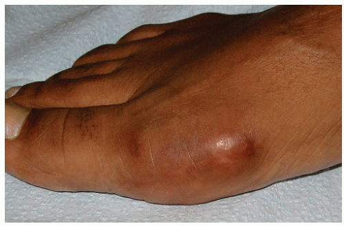 Figure 32.1 Photograph of a painful medial eminence with juvenile hallux abducto valgus. |
The juvenile and adolescent hallux abducto valgus deformity is quite different from the adult form and presents considerations that are quite different from adult hallux valgus surgery (Fig. 32.2). The presence of epiphyses, underlying etiologies, and difficulty with respect to the timing of surgery are among the surgical considerations. While a substantial amount of transverse plane deformity may occur in this population, frontal plane valgus rotation of the hallux is rarely a problem (23). Severe joint degeneration, bursal thickening, prominent medial eminence, and associated digital deformities are rare. However, when such progressive findings are present, this may signify a rapidly progressing deformity with other associated deformities including pes planovalgus, equinus, and metatarsus adductus (23).
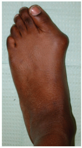 Figure 32.2 Photograph of left foot in an adolescent with juvenile adolescent hallux abducto valgus. |
A complete physical exam, including a biomechanical, orthopaedic, and neurologic examination should be conducted to determine underlying etiologic factors. Neurologic disorders such as cerebral palsy typically produce spasticity or contractures of the Achilles tendon, which may contribute to the hallux valgus deformity (36). The presence of limb-length discrepancy, torsional abnormalities, neuromuscular disease, metatarsus adductus, pes planovalgus, and other factors that contribute to the hallux valgus deformity should be noted (28). It is also important to access the range of motion of the first MTPJ in both the deformed and corrected positions. The presence of a “tracking” or “trackbound” joint indicates deviation of the first MTPJ axis and/or lateral displacement of the sesamoid apparatus. This finding is commonly associated with a significant increase in the PASA (37).
CONSERVATIVE TREATMENT
There has been controversy concerning which patients will require surgery for juvenile hallux valgus and at what age surgery should be performed. Conservative treatment has not been demonstrated to be effective in deterring the progression of the deformity. However, certain conservative measures may be effective if the deformity is mild, minimally painful, and not progressive. Symptoms may respond to wider shoe gear, toe wedges, and bunion pads and shields; however, adolescents may not be inclined to comply with these modifications.
Related posts:

Stay updated, free articles. Join our Telegram channel

Full access? Get Clinical Tree


