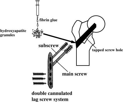
Chapter 19 As the general population ages with increasing life expectancy, an increase in the incidence of proximal femoral fractures is inevitable.1 There is no effective strategy, however, to prevent two major causes of the fracture in the elderly, bone loss and falling down.2,3 Therefore an effective surgical treatment of hip fracture in elderly osteoporotic patients, aiming to restore hip function as early as possible, is needed. Zuckerman et al4 suggested that in elderly patients who are cognitively intact, able to walk, and living at home before the hip fracture, it is necessary to operate and initiate ambulation as soon as possible. In patients with severe osteoporosis, however, postoperative early ambulation following osteosynthesis of the hip fracture is difficult because of bone fragility, and sometimes results in failure of the fixation.5 In such cases, it is necessary to enhance the initial stability of the internal fixation. In the present study, a double-sliding lag screw and plate system (Beak system, 4S Medicals Corp., Saitama, Japan) were used for superior initial stability. The Beak system has greater stiffness in compression and torsion compared to a conventional compression hip screw system (CHS).6 In addition to the new device, hydroxyapatite (HA) granules were used to augment cancellous bone around the lag screws. The purpose of the present report is to introduce preliminary results of osteosynthesis and to present the effect of HA granule augmentation. Osteosynthesis was performed within a week after injury. Under an image intensifier, the fracture was reduced and osteosynthesis was performed using a double cannulated sliding lag screw and plate system (Beak system). Use of the Beak system was approved by the Ministry of Health and Welfare of Japan on Dec. 15, 1994. This system consists of a strong side plate, two cannulated lag screws (main and sub-screws), and cortical screws. The lateral cortex of the femur can be maximally preserved by omitting a barrel structure for the lag screw. The diameter of the main screw was 10 mm at its base with tapered shaft, and the diameter of the subscrew was 7 mm. These two lag screws were both engaged in the femoral head and realized telescoping following the initiation of loading to the fracture site with the locking mechanism after sliding 15 mm in length distally. After drilling and tapping a hole with a guide wire for the main screw, HA granules of 1 to 2 mm in diameter with a porosity of less than 20% (Apaceram, GL1, Pentax, Tokyo, Japan) were packed into the screw hole. Fibrin glue was used to adhere HA granules (Bolheal, Fujisawa Pharmaceutical Corp., Pentax, Tokyo, Japan) prior to packing HA granules into the screw hole. The main screw with a plate was screwed into the pretapped hole, affixing the plate to the femoral shaft. A self-tapping subscrew was then inserted using a guide wire (Fig. 19–1). Patients were encouraged to initiate muscle exercise and ambulation as soon as possible after the operation. Patients were followed for 12 months on average (range 5 to 20 months) with regard to daily activity and X-ray findings. A simple evaluation system was used for grading daily activity of the patient before injury and at the follow-up period as follows: 1. bedridden (point 1) 2. able to walk to bathroom with support (point 2) 3. able to walk in house but not outside the house even with support (point 3) 4. able to walk outside the house with support (point 4) 5. normal activity, walking without support (point 5) FIGURE 19–1 Osteosynthesis procedure using a double cannulated sliding lag screw system and hydroxyapatite (HA) granules. A screw hole is drilled and tapped, then the main screw is inserted following packing of HA granules into the screw hole. Fibrin glue is used to adhere HA granules. The plate is then placed on femoral shaft and compressed by cortical screws, and finally a subscrew is inserted. In all patients, final torque when the main screw was screwed into the hole was improved by augmentation using HA granules and excellent initial stability was achieved. In a separate series of nine patients with proximal femoral fractures (mean age ± SD = 81.9 ± 5.0 years, range 75 to 91 years), the torque of the main lag screw was measured when screwed to a full depth before and after packing HA granules into the tapped hole. The measurement was performed intraoperatively to verify the effect of HA granule augmentation on screw stability. Final torque before and after packing HA granules was compared using a paired t- test. Twenty patients (mean age ± SD = 82.0 ± 5.7 years, range 63 to 88 years) who suffered from proximal femoral fractures caused by minor trauma, such as falling down while walking, were subjected to our treatment protocol. There were 17 intertrochanteric fractures, including one revision case, one intracapsular, and two subtrochanteric fractures in this series. Of the 17 intertrochanteric fractures, six fractures were type I, six were type II, and the other four were type III, according to the classification of Kyle et al.7 X-ray grading of osteoporosis of the contralateral femoral neck (Singh’s index) indicated that seven patients were grade III, 12 patients were grade II, and one patient was grade I.8
INTERNAL FIXATION OF OSTEOPOROTIC
PROXIMAL FEMORAL FRACTURES USING
A NOVEL DEVICE ENHANCED BY HYDROXYAPATITE GRANULES
TESTING OF THE SYSTEM
METHODS
PATIENTS
![]()
Stay updated, free articles. Join our Telegram channel

Full access? Get Clinical Tree


