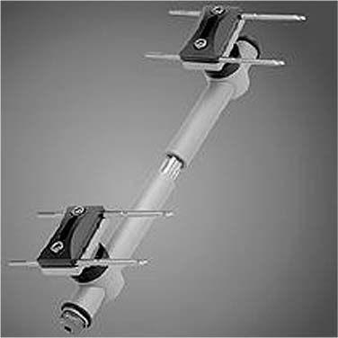
Chapter 14 External fixation is a technique used to manipulate, align, and stabilize osseous fragments by means of pins or wires that connect the bone to a frame outside the body. The history of external fixation of fractures probably begins in the middle of the 19th century with Joseph François Malgaigne1 from France, who developed strapped-on metal points and claws to stabilize displaced fractures, as well as a clamp for the treatment of patellar fractures. Around the turn of the century Parkhill2 in Denver and Lambotte3 in Belgium both built external fixation devices with two unicortical screws in each bone fragment, connected by a clamp outside the skin. The introduction in the 1930s of transfixion pins, the concept of longitudinal distraction, and compression mechanisms led to the more sophisticated devices of Anderson,4 Stader,5 and Hoffmann.6 These devices not only provided a technique to immobilize the bone fragments but also to manipulate and adjust their position during and after application of the fixator, thereby creating a more practical clinical technique. The Russian surgeon Ilizarov and coworkers7 developed highly complex ring fixators for the correction of limb length discrepancies, malalignments, and segmental transport after corticotomy. In the 1970s De Bastiani and colleagues8,9 introduced the concept of dynamic axial fixation with a fixator frame that had a telescopic part, allowing the bone fragments to slide toward each other, thereby facilitating bone union. The fixator pins constitute the link between the external frame and the bone. Their diameter varies between approximately 2 and 6 mm with the strength and rigidity increasing with their diameter.10 Half pins have a terminally threaded segment that reaches just beyond the far cortex of the bone. In contrast, transfixion pins have a centrally threaded segment and can be connected to the fixator frame at both ends of the pin because the pin passes entirely through the limb. Transfixion pins therefore pose a greater threat to muscles, tendons, nerves, and vessels. Recently the concept of “pinless” external fixation has been reintroduced with pointed clamps that penetrate the cortex only minimally, similarly to a reduction forceps.11 Tensioned wires are used to connect bone fragments to a fixator with a ring construction. Purchase can be increased by an “olive,” a bulbous protuberance that anchors the wire to one cortex of the bone. Clamps link the fixator pins (or wires) to the connecting rod. Simple clamps connect individual pins, whereas other fixator systems use clamps to connect a cluster of two or three parallel pins. The clamps are attached to the connecting rods, which are often made of stainless steel, aluminum, or carbon fiber. The construction of the frame can either be simple, with each pin connected directly to the rods, or modular, with clusters of pins held in a clamp that is connected to the rest of the frame (Fig. 14–1). Modular frames often allow pin placement before complete reduction of the fracture, thereby enhancing the possibility of adjusting alignment and length afterward. Uniplanar frame constructions have all pins and rods in the same plane, although the frame can be unilateral using half pins or bilateral using transfixion pins. Biplanar frames have pins entering the bone at different angles to each other (most often 90 degrees) and can also be built in a unilateral or bilateral mode.12 Ring fixators consist of wires and half pins connected to rings around the bone, which are subsequently attached to the fixator rods. The combination of a half pin frame with parts of a ring fixator results in a hybrid frame. FIGURE 14–1 Example of a modular, unilateral external fixation device (Synthes, Paoli, PA, USA). With increasing clinical experience, the biomechanical properties of the external fixation devices have become the subject of research. The stability of the fixator, determined primarily by the properties of the pins and by the frame geometry, has been the main concern. The pins are the weakest component of the system but the stability of the construction is increased by a greater diameter and number of pins, preferably spread over the entire fragment length.13–15 Introducing the pin shaft into the proximal cortex has been said to double the stiffness of most pins, provide tighter pin-bone fit, decreased soft tissue irritation, and reduced stress at the pin-bone interface.16,17 Others instead have associated this technique with an increased rate of pin loosening and therefore recommend the use of larger root-diameter pins with bicortical threads for better purchase and decreased loosening characteristics.18 In a study comparing the healing pattern of osteotomies fixed with four-pin versus six-pin unilateral external fixation the axial, torsional, and bending stiffness of the four-pin configuration was 50 to 70% of that of the six-pin configuration. Bone porosity and pin loosening were significantly increased in the four-pin configuration.19 This suggests that a lower stiffness of the external fixator (i.e., a smaller number of pins) increases the risk of later pin-bone interface problems. Initially bending preload was advised to increase purchase in bone and to achieve compression at the fracture site,20 but Hyldahl21 demonstrated that radial preload is preferable to bending preload because resorption around the pin is reduced. Radial preload can be achieved by first drilling a pilot hole slightly smaller than the core diameter of the pin or by using pins with a conical, tapered thread to produce radial preload as the pin is introduced. However, insertion of fixator pins with misfits of greater than 0.4 mm can result in significant microscopic structural damage to the bone surrounding the pin.22 Biplanar frames increase bending and torsional stiffness as compared to uniplanar, unilateral frames.17,23 However, complex multiplanar configurations presently are rarely indicated since stronger unilateral frames with increased pin diameter have been designed.18,24 In addition, a smaller distance between the bone and the fixator frame is an effective method to increase strength: decreasing the distance between the bone and the rod by 1 cm can increase the frame stiffness by 40%.13,15,17,25 To allow image intensifier and X-ray views through the fixator and to reduce the weight of the construction, carbon rods are frequently applied. Although the carbon rods are stiffer than stainless steel rods, the bending stiffness of an external fixator construction with carbon rods is approximately 85% of the stiffness of standard frames. This is most likely due to the clamps being less effective in connecting the carbon fiber rods rigidly to the metal fixator pins.26 The stability of the frame together with the type of fracture, the accuracy of reduction, and the amount and type of stresses occurring at the bone ends are the most important factors determining the mechanical circumstances for fracture healing. These stresses are dictated by functional activity and loading at the fracture site. The amount of loading and the presence of a significant fracture gap resulting in oscillating fracture motion under loading are the most important factors in promoting callus formation.27 However, although less rigid configurations lead to earlier periosteal callus formation, there is an increased stress at the pin-bone interface and increased cortical porosity, both leading to an increased rate of pin loosening.19,23,25 Clinically, the amount of motion at the fracture site and thus the mode of fracture healing depend on the frame and pin configuration as well as on the type of fracture, the reduction, the amount of physiologic loading, and the performance of the pin-bone interface.15 Although in use for more than a century, external fixators have been applied with little consistency, with variable results, and with a high rate of complications.28 Among the properties that distinguish external fixators, other fixation methods are: stabilization at a distance from the injury, a greater versatility in accommodating a variety of bone and soft tissue lesions, ability to stabilize injuries extending across joints, and adjustability of alignment after application of the device. In the presence of marked osteoporosis, external fixation may provide a method of obtaining adequate stabilization when effective internal fixation might be precluded by the degree of comminution or osteoporosis. Clinical indications include the following18,29: • Severe open injuries with massive associated soft tissue injury. Once (repeated) debridements have resulted in sufficient soft tissue coverage without contamination, external fixation can be converted to internal fixation techniques. • Severe periarticular injuries, particularly in the proximal and distal tibia, when soft tissue conditions preclude emergent lengthy operative procedures for open reduction and internal fixation. External fixation in severe fractures around the distal articular surface of the tibia can provide time for soft tissue healing with subsequent internal fixation. This sequence does not have to lead to inferior results compared to primary internal fixation.30,31 • Unstable and intraarticular distal radial fractures. This type of fracture is one of the few periarticular fractures that are not amenable to open reduction, anatomic reconstruction of the articular surface, and internal fixation. Specifically, in the presence of severe osteoporosis the purchase of screws in the metaphyseal area and in small fragments can be a problem. In these cases, realignment of the fracture fragments may be secured by the application of distraction across the adjacent joint, a technique based on the principle of ligamentotaxis.32 External fixation provides a method of neutralization and reduction maintenance for these frequently encountered fractures. To overcome some of the disadvantages of joint immobilization, articulating “dynamic” external fixation devices that permit early functional treatment have been developed.33–36 • Unstable and severely polytraumatized patients. With the increasing aging of the population, elderly polytraumatized patients will probably be seen more frequently. In patients with injuries such as severe head injuries, pulmonary contusion, or hypothermia, after lifesaving measures have been taken, external fixators can be applied rapidly, providing fracture stability in expectation of improvement of the patients’ general condition.
EXTERNAL FIXATION IN
OSTEOPOROTIC BONE
FIXATOR DESIGN
BIOMECHANICAL ASPECTS
INDICATIONS
Related posts:
 Norian SRS Resorbable Cement for Augmentation of Internal Fixation of Hip Fractures
Basic Principles and Techniques of Internal Fixation in Osteoporotic Bone
An Injectable Cementing Screw for Fixation in Osteoporotic Bone
Improving the Distal Fixation of Intramedullary Nails in Osteoporotic Bone
Internal Fixation of Osteoporotic Long Bone
Enhanced Stability of External Fixation with Hydroxyapatite-Coated Pins
Norian SRS Resorbable Cement for Augmentation of Internal Fixation of Hip Fractures
Basic Principles and Techniques of Internal Fixation in Osteoporotic Bone
An Injectable Cementing Screw for Fixation in Osteoporotic Bone
Improving the Distal Fixation of Intramedullary Nails in Osteoporotic Bone
Internal Fixation of Osteoporotic Long Bone
Enhanced Stability of External Fixation with Hydroxyapatite-Coated Pins
Stay updated, free articles. Join our Telegram channel

Full access? Get Clinical Tree


