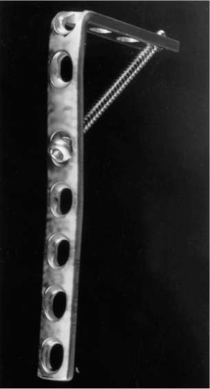
Chapter 24 Osteoporosis is thought to affect 15 to 20 million people in the United States. A significant proportion of these will be females affected by type 1 or perimenopausal osteoporosis, which has the effect of reducing the amount of cancellous bone in the axial and appendicular skeleton while leaving the cortical bone relatively unaffected. Localized osteoporosis affecting metaphyseal bone is also seen in infiltrative conditions such as leukemia, multiple myeloma, and metastatic bone disease. These pathological conditions predispose individuals to predominantly metaphyseal fractures (e.g., distal radius, vertebrae, proximal humerus, proximal and distal tibia). Such fractures may be difficult to treat because of comminution, joint proximity, reduced metaphyseal bone volume, and malignant infiltration. Nonunion of such fractures makes treatment more challenging because of increased local osteopenia and joint stiffness following prolonged immobilization. Also, surgical exposure is restricted to that used in previous attempts at internal fixation, and the overall quality of the soft tissue envelope may be compromised. There may be technical difficulties with internal fixation because of preexisting deformity, and infection may be present. Fortunately this situation is uncommon because of the enhanced vascularity and the large cross-sectional area available for healing in the metaphysis. The theoretical resistance to screw pullout is described in the equation: where F is the pullout force, L is the length of engagement of the screw, P is the pitch, C is the outer circumference of the screw, S is the shear strength of the bone, and G is a geometric factor and accounts for the volume of bone into which the screw has purchase.1 Patients with poor bone stock will have reduced S values and a reduced absolute volume of bone behind the pitch of the screw (G). Various authors have confirmed poor pullout strengths of screws in osteopenic bone,2–4 thus standard fixation techniques used to achieve secure fixation and allow early functional recovery may be found wanting. The blade plate is a rigid device providing a broad surface area of contact within metaphyseal bone. It is therefore capable of resisting considerable torsion and bending moments. Its use has been described in the treatment of fractures and nonunions in the proximal tibia, proximal humerus, and the subtrochanteric and supracondylar regions of the femur.5–8 Biomechanical studies have confirmed improved loads to failure of blade plates over conventional screw-plate devices.9,10 The use of an angled-blade plate device rather than a screw-plate device is recommended as the bone is impacted by the blade rather than removed by the reaming process required for screw insertion, which would be undesirable in bone that is already of poor quality. Despite the advantages of blade plates, failure by pullout is seen when satisfactory fixation to protect from varus or valgus strain cannot be achieved. This is seen in patients with particularly poor bone stock. In this situation the use of an interlocked angled-blade plate may be considered. This is a technique of blade plate fixation that allows extra stability to be achieved without the need for extra soft tissue dissection or fixation hardware. Preoperative planning with radiographs and templates is essential to decide on the appropriate plate length and shape. There should be at least 10 holes in the plate to allow a minimum fixation of six cortices in the diaphyseal fragment and two holes to sit within the weak metaphyseal bone (Fig. 24–1). A 3.5 mm dynamic compression plate (DCP) is suitable for the humerus and a 4.5 mm DCP is adequate for the proximal tibia. The plate is prebent to the planned acute angle in an industrial vice. The angle of the bend of the plate will determine the number of interlocking screws that can be inserted (i.e., more than a right angle usually allows two screws to be successfully interlocked) (Fig. 24–2). The plate-to-plate interlocking screws are tested before sterilization of the implant and the appropriate screw lengths determined. FIGURE 24–1 The customized blade plate demonstrating the interlocking screw in place prior to sterilization. The fracture is approached through the appropriate exposure with minimal soft tissue dissection. The proximal humerus is exposed through a deltopectoral approach. The proximal tibia is approached through a midline extensile approach. This ensures future access for joint athroplasty is not compromised. The distal tibia is approached anteriorly.
INTERLOCKED BLADE PLATES IN
THE TREATMENT OF OSTEOPOROTIC
METAPHYSEAL FRACTURES

OPERATIVE TECHNIQUE
Stay updated, free articles. Join our Telegram channel

Full access? Get Clinical Tree


