Fig. 9.1
The angular relationship between the trochlear (upper line) and transcondylar (lower line) axes in transverse (a) and coronal (b) planes (c): spheres fitted to lateral and medial facets of the trochlea are illustrated
The trochlear groove can be simplified as a circle, which is positioned laterally to both the anatomical and mechanical axes. The trochlear axis defines a new distal femoral axis between the centres of the spheres fitted to the trochlear facets.
As for the patellar geometry, an anthropometric study did not show any racial differences in patellar dimensions. Length and width varied from 47 to 58 and 51 to 57 mm [19]. It has also been shown that the thickness of the patella can be approximated as half of its width [31].
The patellar thickness can be approximated as half of its width.
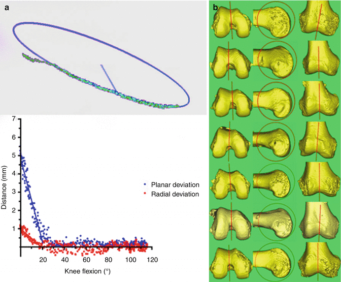
Despite a large number of in vitro and in vivo studies, the description of patellar motion relative to the femur has been not unambiguously clarified [35]. This is due to the different methods employed, making comparison difficult. The use of different coordinate systems is another factor resulting in variable results. Imaging studies tend to describe the motion of the patella relative to the femoral groove [18, 49, 50, 57, 65]. It is relatively straightforward to identify the groove and measure this on cross-sectional 2D images from CT or MRI. However, with three-dimensional tracking studies, there are difficulties in defining the groove axis and plane of reference as the trochlear groove is curved and different definitions of this axis have been used [11]. An in vitro study by our team has simplified the description of patellar tracking by linking it to the 3D geometry of the distal femur [32]. Once the frame of reference had been aligned to the trochlear axis, there was minimum medial-lateral translation of the patella. The patella tracked in a straight line in the coronal plane. The exception to this was close to full extension, when the patella translated laterally as it disengaged from the trochlear groove (Fig. 9.2).


Fig. 9.2
(a) The moving landmark of the patellar centre during knee flexion for one knee is visualised, and a best-fit circle is constructed to these points. The deviation from this circle is plotted against the knee flexion angle for two flexion-extension cycles. A line fitted to the graph crossed the x-axis at the flexion angle at which the path of the centre of the patella deviated from the circle. (b) The circle fitted to the path of the patella is illustrated in transverse, sagittal and coronal views
In early knee flexion, the patella moved medially to engage with the trochlear groove. The trajectory of the centre of the patella then became circular and uniplanar at 16º ± 3.9° of knee flexion; on average, this was after the distal patella but before the centre of the patellar median ridge entered the trochlear groove. The distal articular surface of the patella entered the trochlea at 6º ± 2.8° knee flexion and the centre of the articular surface entered it at 22º ± 5.3° of flexion.
In early knee flexion the patella moves medially to engage with the trochlear groove. The trajectory of the centre of the patella then became circular and uniplanar at 16º ± 3.9° of knee flexion (after the distal patella but before the centre of the patellar median ridge enters the trochlear groove). The distal articular surface of the patella enters the trochlea at 6º ± 2.8° knee flexion and the centre of the articular surface at 22º ± 5.3° of flexion.
9.2 Component Design
9.2.1 Patellar Component
There are five major types of patellar component designs: dome shaped, modified dome shaped, anatomically shaped, cylindrical shaped and mobile bearing [38] (Fig. 9.3). Each of them shows specific pros and cons. It has been shown that the most important design feature is how well it matches the femoral component through the flexion cycle [14, 16, 48, 68, 69, 72].


Fig. 9.3
The most commonly used patellar designs are biconvex, round and oval such as shown here for the GII/Legion® knee system (Smith & Nephew, Switzerland)
The patellofemoral contact areas in modified dome-shaped, anatomic and cylindrical patellar components are higher compared to the domed design [46, 63, 70], but at the same time less forgiving for malpositioning or malalignment [37].
Metal-backed patellar components are not in common use anymore, as implant-related complications such as metal-metal contact and metal wear represent severe problems for the knee joint.
Major types of patellar component designs are dome shaped, modified dome shaped, anatomically shaped, cylindrical shaped and mobile bearing.
9.2.2 Femoral Component
The main determinant within the patellofemoral mechanism is said to be the design of the prosthetic groove. The importance of femoral component design on patellofemoral behaviour has been shown by comparing arthroplasty designs with distinct differences in trochlear geometry. Theiss et al. [46] showed a 14-fold decrease in patellar-related complications using a patellar-friendly design with an extended anterior flange and a deeper and wider trochlear groove. The authors concluded that more proximal capture of the patella in a deeper groove with more gradual proximal-to-distal transition appeared advantageous in reducing patellar morbidity. Whiteside et al. also demonstrated that deepening and distal extension of the trochlear groove improved patellar tracking [56].
The design of the femoral component is even more important if the patella is not resurfaced. It has been shown that the native patella remodels to the shape of the prosthetic trochlea with a less stress contouring needed with an anatomical design [37].
Femoral component design has significant influence on patellofemoral tracking. The most important factors are a patellar-friendly design with an extended anterior flange and a deeper and wider trochlear groove.
9.3 Surgical Technique
It has been shown that the major factors affecting patellar tracking after TKR are patellar component medialisation, patellar resection angle and femoral component rotation [3].
9.3.1 Femoral and Tibial Components
It is well recognised that prosthetic patellar maltracking can result from soft tissue imbalance and component malalignment of the tibia and femur. External rotation of the femoral component has been shown to improve patellar tracking and decrease the need for lateral retinacular release [36, 3]. Although combined internal rotation of the tibial and femoral component has been linked to increased incidence of anterior knee pain [6], tibial component rotation alone does not seem to affect patellar tracking significantly [47, 51, 55].
Prosthetic patellar maltracking can result from soft tissue imbalance and component malalignment of the tibia and femur. In particular, combined internal rotation of the tibial and femoral component has been linked to increased incidence of anterior knee pain. Tibial component rotation alone does not seem to affect patellar tracking significantly.
9.3.2 Patellar Component
Resurfacing the patella in TKR remains controversial [5–7, 9, 22, 37, 53]. But for those cases with patellar resurfacing, surgeons focus on patellar resection angle (PRA), patellar medialisation and patellar thickness.
Biomechanical studies have shown that the patella improves the effective extension function of the quadriceps muscle by increasing the moment arm of the patellar tendon. It also centralises the converging forces of the quadriceps muscle and transmits the tension around the femur to the patellar tendon [2, 25, 28]. A reduced patellar thickness may reduce the efficiency of the extensor mechanism since it plays a central role in quadriceps biomechanics. In addition, a thin patella due to overresection has been shown to lead to high strains, which may contribute to early failure [41, 61].
Equally, establishing the appropriate thickness of the patellar implant-bone composite during TKR is important to restore the patellofemoral joint and to balance its soft tissues [26]. The range of motion of the knee can be affected by an increased thickness of the patellar prosthesis-bone composite after TKR [8]. Other detrimental effects of overstuffing the patellofemoral joint have also been shown in laboratory studies [27, 34, 67].
The primary goal during TKR is to match the native patellar thickness with the patellar implant-bone composite. An increased thickness may lead to patellofemoral overstuffing. A reduced patellar thickness may reduce the efficiency of the extensor mechanism and increase the risk of early failure and fracture.
Most surgeons try to achieve a symmetric patellar cut (with equal medial and lateral thickness). Surgeons tend to avoid overstuffing the patellofemoral joint by resecting the amount of bone that corresponds to the thickness of the patellar implant. However, often the thickness of the patella has been affected by disease. Other bony and soft tissue landmarks have been described to achieve the correct depth of patellar resection. This includes the lateral facet subchondral bone [30, 58], at the level of the posterior limit of the quadriceps tendon and the patellar tendon attachment [39, 40, 43] and the capsular attachment to the patella [23]. An anatomical study showed that the thickness of the patella was half its width [31]. This can be used to avoid overstuffing the patella in cases where the patella is excavated and there is no median ridge present.
References
1.
2.
Aglietti P, Buzzi R, Insall JN. Disorders of the patellofemoral joint. In: Insall JN, editor. Surgery of the knee. New York: Churchill Livingstone; 1993. p. 246–51.
3.
Anglin C, Brimacombe JM, Hodgson AJ, Masri BA, Greidanus NV, Tonetti J, Wilson DR. Determinants of patellar tracking in total knee arthroplasty. Clin Biomech (Bristol, Avon). 2008;23(7):900–10.CrossRef
4.
5.
6.
Barrack RL, Bertot AJ, Wolfe MW, Waldman DA, Milicic M, Myers L. Patellar resurfacing in total knee arthroplasty. A prospective, randomized, double-blind study with five to seven years of follow-up. J Bone Joint Surg Am. 2001;83-A(9):1376–81.PubMed
7.
Barrack RL, Wolfe MW. Patellar resurfacing in total knee arthroplasty. J Am Acad Orthop Surg. 2000;8(2):75–82.PubMed
8.


