Fig. 62.1
Lateral X-ray showing an interprosthetic fracture between a dynamic hip screw (DHS) and a nailed cementoplasty performed for a previous periprosthetic fracture around the knee
In summary, it can be concluded that locking plates and intramedullary nails provide sufficient stability and at the end of the day each surgeon should use an implant with which he or she is familiar.
If surgeons opt for a locking plate system, they must familiarise themselves with the biomechanical factors that influence fracture motion and hence fracture healing. We described the concept of dynamic plate osteosynthesis in detail in a review article [18], and it is partly repeated in the following section.
Locking plates and intramedullary nails provide sufficient stability for distal femoral fractures.
62.2 Dynamic Plate Osteosynthesis
Both biological and mechanical factors have to be met for optimal fracture healing. In particular, the size of the fracture gap and the amount of fracture motion are important for fracture healing. Aro and Chao described the principles underlying bone healing [1]. The authors distinguished between osteonal and non-osteonal bone healing. In the case of the osteonal fracture healing, abundant callus formation is observed. No primary healing of the cortex is observed and remodelling processes are slow. An example of this type of fracture healing is cast immobilisation, where the fracture gap and the initial motion between the fragments are large. Abundant callus is needed to allow sufficient stability for remodelling and bone healing. In a mechanically stable situation, as is the case of a rigid osteosynthesis, primary osteonal fracture healing will take place. Regenerating osteones will migrate directly from one fragment through the fracture gap to the opposite fragment. No callus will be seen.
This kind of fracture healing is only possible in rigid plate osteosynthesis when the fragments are in direct contact (interfragmentary compression) or when the fracture gap is very small. In less rigid constructs, micro-motion at the fracture site occurs. Fracture healing will be initiated by periosteal and endostal callus formation, followed by osteonal fracture healing. This type of fracture healing is called secondary osteonal fracture healing. This is the preferred kind of fracture healing due to the faster remodelling potential. Hence, it is important for a surgeon to know to what extent he can influence the amount of fracture motion and consecutively the speed of fracture healing.
A biomechanical study has been performed to investigate the surgical factors that influence the amount of fracture motion for locking plates [19]. As in conventional plating technique, the most important factor is the bridging length, e.g. the placement of the screws near the fracture site. Omitting three to five plate holes at the fracture gap and avoiding lag screws, especially through the plate, allows sufficient micro-motion and therefore faster bone healing. Another factor is the choice of the plate material. A titanium plate is twice as elastic as a steel plate and therefore allows more micro-motion with the same plate configuration. With locking systems, there is a risk of generating a more rigid construct, resulting in a higher risk for non-union. Hence, for successful plating, we recommend the following principles in the diaphyseal and metaphyseal region:
1.
Use a long plate with few screws.
2.
Overlap a proximal implant (hip stem, intramedullary nail, plate) by 4–6 cm.
3.
Maintain sufficient bridging length. In general, three or five plate holes should be omitted at the fracture site. This allows sufficient fracture motion. If a lag screw is indicated for technical reasons, for example, in the case of a simple spiral fracture, do not place it through a plate hole. We recommend the use of a small fragment (3.5) screw outside the plate.
4.
Treat the periosteum with as much care as possible. Never strip the periosteum off the bone. Keep fragments covered with muscle. Use minimally invasive techniques with aiming devices when feasible.
5.
Consider the fact that a steel plate is twice as rigid as a titanium plate for the same construct.
For plating of fractures in the diaphyseal and metaphyseal region, use of a long plate with few screws, overlapping with a proximal implant by 4–6 cm and omitting of at least three to five plate holes at fracture site and careful treatment of periosteum, is recommended.
62.3 Assessment and General Principles
The overall goal of treatment of any periprosthetic fracture should be to achieve union of the fracture whilst maintaining a well-functioning and well-aligned prosthesis. The decision-making is often complex and takes into account the pre-fracture status of the TKR. A thorough history should be obtained. The patient’s comorbidities and mobility should be assessed. If possible, previous radiographs are examined to ensure that there were no signs of wear or loosening of the TKR prior to the fracture. In addition, the stability of the knee needs to be carefully assessed.
Operative treatment is planned meticulously. Details of the implants are obtained, particularly the nature of the femoral component, as passage of an intramedullary nail may not be possible with certain designs. The bone stock around the implant is assessed. Occasionally, a prosthesis that is judged to be well fixed on radiographs may be found to be loose intraoperatively. If open reduction and internal fixation is performed, revision TKR should be available. The surgeon should be capable to perform both open reduction and internal fixation or revision knee replacement.
62.4 Treatment Options
62.4.1 Femoral Fractures
Historically, fractures have been treated conservatively; however, this can only be recommended for minor or undisplaced fractures in very frail patients. Fractures can be slow to unite, and prolonged periods of immobility must be avoided to prevent intractable stiffness and poor functional outcome. The most frequent methods of treatment for Rorabeck type 1 and 2 fractures are intramedullary nailing and lateral locking plating (see Chap. 13). There are no good randomised studies investigating the superiority of either method. A systematic review of 415 patients found that both intramedullary nails and locking plating were superior to nonoperative treatment or traditional plating [11]. There was no significant difference between intramedullary nails and locked plating.
Intramedullary nailing does not disturb the fracture envelope and therefore does not devitalise fracture fragments. It may be difficult or impossible in low supracondylar fractures due to the lack of bone stock, in which locking screws cannot safely be placed. It is important to achieve an accurate reduction, which may be difficult to achieve by closed methods. In such cases, the fracture site is opened either via a small lateral approach or through the original midline approach. This leads to soft tissue stripping, but the benefits of accurate reduction outweigh the risks of such exposure.
The majority of modern condylar designs allow passage of the nail through the notch; however, occasionally closed box, posterior stabilised designs are encountered. The use of a diamond-tipped burr to create an entry point in the box has been described [13]. This technique is not recommended due to the metallic debris that could compromise the life of the polyethylene. Furthermore, there are enough other options that a nail can be avoided in this situation. Curral and colleagues presented a useful paper detailing the dimensions of the intercondylar area of condylar knee designs and its compatibility with retrograde nailing [6].
The development of locking plating systems has revolutionised the treatment of such fractures. Many systems allow a minimally invasive approach with radiolucent jigs. The soft tissue envelope and blood supply are therefore less traumatised [9]. This and the superior cut-out strength probably explain the superior results seen over conventional plating systems. One publication demonstrated that they can also be used in very low supracondylar fractures [20]. However, they do not define “very low” in terms of distance from the joint. Screws may need to be unicortical, in case of a stemmed TKR component. Newer designs such as the NCB (Zimmer, Warsaw, Indiana) and polyaxial locked plate (Depuy, Warsaw, Indiana) provide further options as the screws can be placed at a variable angle and still locked into the plate [8]. There are now also specific plates designed for periprosthetic fractures, which allow more screw options around a well-fixed stem and also provide a more stable fixation compared to conventional locking plates (Case 2).
A serious problem concerning distal femur fractures, including periprosthetic fractures, remains the incidence of delayed union, non-union and implant failures [7]. Disturbed blood supply of the distal femoral region and biomechanical factors such as construct stiffness have been blamed for the high complication rate of these fractures. The surgical principles to reduce implant stiffness of locking plates have been published [19]. A new development is the concept of far cortical locking, whereby the near drill hole is over-drilled in a similar fashion to a lag screw. This reduces the stiffness of the construct and may lead to more significant callus formation through increased micromotion [3, 4]. As yet, the evidence for this technique is limited to biomechanical and animal models.
If the femoral component is loose or there is insufficient bone stock, the femoral component will need to be revised. The amount of bone loss will dictate which implant should be used but not invariably a distal femoral replacement may be required. In such cases, a hinged replacement is preferred to achieve stability. It is therefore likely that both components will have to be revised. The complication and reoperation rate following such procedures is high [14, 17].
For Rorabeck type 1 and 2 fractures, a polyaxial locking plate system is recommended. A minimally invasive approach is used to allow accurate reduction of the fracture with limited interfragmentary fixation. The plate is then used in a bridging mode, with small stab incisions proximally to place screws. If the prosthesis appears well fixed, then a lateral approach is used; if there is any concern that conversion to a revision prosthesis is required, then a midline approach is performed. For type 3 fractures, a revision TKR is used. If the bone quality is sufficient, then metaphyseal sleeves and cementless stems are used to bypass the fracture. It is important to note that an intramedullary stem does not provide sufficient rotational control of the fracture, so a supplementary plate may be required. The use of cement to secure the stems is avoided to prevent compromising fracture healing.
62.4.2 Tibial Fractures
There is a relative paucity of literature concerning the treatment of tibial periprosthetic fractures compared to femoral fractures. The majority of tibial fractures around the tibial plateau are associated with a loose or failing TKR. These fractures require revision TKR. Although it is possible to treat such fractures nonoperatively in a brace, a significant number will fail this treatment and require revision [10]. The Felix classification does offer a useful guide for treatment; however, it is more complex than the Backstein classification, which splits fractures into type 1 and type 2 depending on whether there is sufficient proximal bone to allow fixation [2, 10] (see Chap. 13). If the prosthesis is stable and the fracture distal to the prosthesis, then standard plating techniques can be used. Fractures of the tibial tubercle are rare; if undisplaced, they can be treated with splinting but displaced fractures require operative stabilisation to restore the integrity of the extensor mechanism [10].
62.4.3 Patella Fractures
Periprosthetic patella fractures are often the most challenging of all periprosthetic fractures. Results of internal fixation are poor and fraught with a high complication rate [5, 15]. A systematic review found an infection rate of 19 % and a non-union rate of 92 % with internal fixation with tension band wiring or cerclage wiring [5].
There is consensus that wherever possible, fractures should be treated conservatively with observation or splinting [5, 15].
If the extensor mechanism is disrupted, then surgical treatment is usually required. Given the poor track record of open reduction and internal fixation (ORIF), consideration should be given to excision of small distal or proximal bone fragments and direct repair of tendon to bone [5].
If the patella component is loose, then surgical treatment will be required to remove it. The surgical decision about whether to reimplant a component depends on the available bone stock; a minimum residual width of 10 mm is suggested. Occasionally, total patellectomy will need to be performed. Whilst the results may be acceptable, the knee should be carefully assessed for axial and rotatory stability, as occasionally revision of the tibial and femoral components will be required [16].
62.5 Case Examples and Suggested Techniques
The following section presents some common clinical situations and discusses the options for treatment. A method is described, but rather than represent the “best option,” we hope that this section will provide a stimulus for thought and help in the more difficult cases that present themselves.
62.5.1 Case 1: Simple Spiral Fracture
An 80-year-old patient with previously well-functioning TKR who presented after a simple fall (Figs. 62.2 and 62.3).
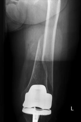
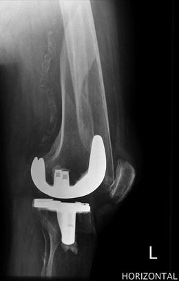
Fig. 62.2
AP X-ray showing simple spiral fracture above TKR
Fig. 62.3
The lateral X-ray shows that the implant appears well fixed
62.5.1.1 Issues
Osteoporotic bone
TKR well fixed
Simple spiral fracture which needs some bone contact
Calcified femoral vessel
Related posts:
 Diagnosis of Periprosthetic Joint Infection After Total Knee Replacement
How Can Preoperative Planning Prevent Occurrence of a Painful Total Knee Replacement?
Causes and Diagnosis of Aseptic Loosening After Total Knee Replacement
Discussion to Chap. 16: Mid-Flexion Instability After TKR Due to Femoral Malrotation
Specific Orthopaedic Imaging Analysis Software: Clinical Benefit for TKR Revision Surgeon
Constrained Condylar Total Knee Replacement
Diagnosis of Periprosthetic Joint Infection After Total Knee Replacement
How Can Preoperative Planning Prevent Occurrence of a Painful Total Knee Replacement?
Causes and Diagnosis of Aseptic Loosening After Total Knee Replacement
Discussion to Chap. 16: Mid-Flexion Instability After TKR Due to Femoral Malrotation
Specific Orthopaedic Imaging Analysis Software: Clinical Benefit for TKR Revision Surgeon
Constrained Condylar Total Knee Replacement
Stay updated, free articles. Join our Telegram channel

Full access? Get Clinical Tree


