Femur
Plane
Landmark
Alignment
Femoral bone
Coronal and sagittal plane
Intramedullary canal
Varus/valgus alignment, flexion, and extension
Distal femoral condylar line
Coronal plane
Distal medial and lateral femoral condyle
Varus/valgus alignment
Posterior condylar line
Transversal plane
Posterior medial and lateral femoral condyle
Internal/external rotation
Anatomical epicondylar line
Transversal plane
Medial and lateral epicondylar tubercle
Internal/external rotation
Surgical epicondylar line
Transversal plane
Medial epicondylar sulcus and lateral epicondylar tubercle
Internal/external rotation
Whiteside’s line
Transversal plane
Center of the trochlea
Internal/external rotation
Distance between the medial epicondyle and the joint line
Coronal plane
Medial epicondyle and distal femoral condyle
Joint line level
Anterior component reference
Sagittal plane
Anterior cortical bone of the metaphyseal bone
AP sizing of the femoral component
Varus/valgus alignment and the mediolateral alignment of TKR components are determined in the coronal plane.
The femoral component is generally aligned in 6° of valgus to the anatomical axis of the femur. The tibial tray is generally placed in 90° to the diaphysis or mechanical axis of the tibia.
Intramedullary alignment for component placement is recommended in revision TKR on both the femur and tibia. It allows correct TKR component placement in the coronal and sagittal plane, and severe bone defects, especially on the femoral side, do not compromise the technique. Longer intramedullary stems will guarantee that the components are well aligned to the diaphysis of the bone. The femoral metaphysis typically shows a trumpet-like shape, and shorter stems might run not exactly in line with the diaphysis of the bone (Figs. 40.1 and 40.2). This may cause varus or valgus position of the femoral TKR component. Oswald et al. reported a difference of 1.5° between the angle of the femoral mechanical axis and either the distal femoral anatomical axis or central anatomical axis [19]. The angle between the anatomical axis and the mechanical axis of the femur is widely influenced by femoral bowing. Femoral bowing can be defined as the offset between the center of the femoral head and the anatomical axis and showed a range between 6.8 and −0.4 cm [12].
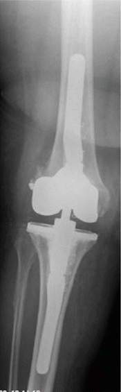
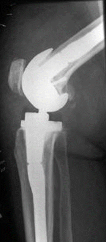
Fig. 40.1
Anterior-posterior radiographs after TKR revision surgery. A CCK prosthesis was implanted. It shows that the femoral stem is not in line with the diaphysis of the femur. The femoral component is placed more laterally and in valgus position
Fig. 40.2
In the lateral view, the components are placed correctly. The offset stem on the tibial site moves the tibial plateau more anteriorly as anatomically necessary
Intramedullary alignment may create a risk of fat embolism or increased bleeding from the femoral canal. Extramedullary alignment does not bear these kinds of risks. However, the extramedullary alignment technique showed 10 % more outliers in the coronal plane compared to the intramedullary alignment [2]. In contrast, others reported similar overall accuracy of extramedullary and intramedullary femoral alignment [14].
Offset stems are frequently required on both the femur and tibia due to the orientation mismatch of metaphysis and diaphysis [1]. When using straight stems, a lateralized femoral component TKR placement is more likely to occur.
Femoral component sizing in primary TKR in the sagittal plane can be based on either anterior or posterior referencing technique (see Chap. 15) [6]. The posterior femoral condyles are partially missing when revision TKR is performed. The anterior femoral cortex provides guidance for correct component placement in the sagittal plane. Sometimes, the component needs to be placed more anteriorly in order to avoid notching at the anterior femoral cortex or posteriorly in order to avoid overstuffing of the patellofemoral compartment. A higher posterior condylar offset was seen when offset stems were used in comparison to straight stems [4]. In general, the femoral component needs to be placed slightly posterior and medially in regard to the distal femur because of the mismatch between the diaphysis and the epiphysis of the femur.
Severe notching might compromise the femoral component fixation. Biomechanical studies have shown that the intramedullary stems will improve component stability in cases with severe femoral anterior notching [5].
Various anatomical landmarks have been described to serve as references for correct component placement in the transversal plane. Several landmarks are missing in type 3 bony defects. The posterior femoral condyle is one of the most frequently used landmarks for correct component placement in rotation. However, no difference in accuracy was found when comparing the posterior condylar line with other commonly used landmarks such as the Whiteside’s line or the transepicondylar line [23]. The trochlea and thus the Whiteside’s line and the posterior femoral condyles are missing in revision TKR. These landmarks are neither available for correct component placement in rotation nor for component sizing.
The transepicondylar line is, in the majority of revision cases, still preserved. This line matches very closely the flexion and extension axis of the knee. The medial and lateral epicondyles serve as a reliable landmark in revision TKR. The femoral component is rotated parallel to the surgical transepicondylar line, a line drawn between the medial intertubercular sulcus and the lateral epicondyle [27]. The reliability of the transepicondylar line has been studied, and a variation of 15 mm in the anteroposterior direction and of 19 mm in the proximal distal direction is reported [25]. This variation in anterior-posterior orientation might have a significant impact on femoral component rotation and the variation in the proximal to distal direction in varus/valgus alignment.
When most of the femoral bony landmarks are missing, such as in severe Type III defects, TKR trial components might be helpful in order to identify the correct component position [10] (Figs. 40.3 and 40.4). Sometimes, it also might be helpful to use the previous component for indentifying the rotational position. Two 1.4 mm K-wires might be placed in the proximal metaphysis in order to mark the flexion and extension axis prior to component removal.
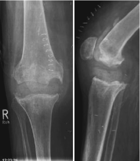
Figs. 40.3 and 40.4
Grade III bony defect on both the tibial and femoral side. The medial and lateral epicondyles can still serve as references for correct femoral TKR component alignment
Intramedullary alignment is considered to be the most reliable for correct varus/valgus TKR component placement. The transepicondylar line serves as a reliable reference line for component placement in rotation.
40.2 Anatomical Landmarks for Tibial Component Placement
Several anatomical landmarks are available (Table 40.2). Bone defects require regularly stemmed implants in order to take load from the tibial plateau. Intramedullary alignment might be used in primary and revision TKR. Long-leg X-rays should be available prior to surgery in order to detect bowing of the tibia. No difference in tibial component alignment has been reported between intramedullary or extramedullary alignment [26]. Others reported an increase of 1.3 ± 1.4° of varus alignment and 4 ± 2.1° of posterior slope when the intramedullary technique was used, while the extramedullary technique resulted 1.5 ± 1.8° of valgus and 1.8 ± 1.1° of posterior slope in regard to the natural anatomy [17].
Table 40.2
Anatomical tibial landmarks and reference lines for primary and revision TKR
Tibia | Plane | Landmark | Alignment |
|---|---|---|---|
Tibial bone | Coronal plane | Intramedullary tibial canal | Varus/valgus alignment, posterior slope |
Center of the upper ankle joint | Coronal plane | Center between the medial and lateral malleolus | Varus/valgus alignment |
Second metatarsal line | Coronal plane | Second metatarsal bone | Varus/valgus alignment |
Tibial bone | Sagittal plane | Anterior cortex of the tibial diaphysis | Posterior slope |
Medial or lateral tibial plateau | Sagittal plane | Medial or lateral joint surface | Posterior slope |
Tibial tubercle | Transverse plane | Medial 1/3 of the tibial tubercle | Tibial component rotation |
Posterior cortical line bone of the tibia | Transverse plane | Posterior cortical bone of the tibia | Tibial component rotation |
Anterior border of the tibia plateau | Transverse plane | Anterior cortical bone of the tibia | Tibial component rotation |
Akagi’s line | Transverse plane
Related posts: Diagnosis of Periprosthetic Joint Infection After Total Knee Replacement
How Can Preoperative Planning Prevent Occurrence of a Painful Total Knee Replacement?
Causes and Diagnosis of Aseptic Loosening After Total Knee Replacement
Discussion to Chap. 16: Mid-Flexion Instability After TKR Due to Femoral Malrotation
Specific Orthopaedic Imaging Analysis Software: Clinical Benefit for TKR Revision Surgeon
Constrained Condylar Total Knee Replacement Diagnosis of Periprosthetic Joint Infection After Total Knee Replacement
How Can Preoperative Planning Prevent Occurrence of a Painful Total Knee Replacement?
Causes and Diagnosis of Aseptic Loosening After Total Knee Replacement
Discussion to Chap. 16: Mid-Flexion Instability After TKR Due to Femoral Malrotation
Specific Orthopaedic Imaging Analysis Software: Clinical Benefit for TKR Revision Surgeon
Constrained Condylar Total Knee Replacement
Stay updated, free articles. Join our Telegram channel
Full access? Get Clinical Tree
 Get Clinical Tree app for offline access

|