Fig. 1
Radiographs (anterior-posterior and lateral-a and long leg view-b) of the left knee showed a primary cruciate retaining mobile-bearing TKR. No signs of loosening or infection. The tibial component was well positioned. No over- or undersizing of the TKR components. A patella infera was seen which was attributed to a proximalisation of the joint line. In addition, there was a significantly flexed femoral TKR component

Fig. 2
99mTc-HDP-SPECT/CT images of the left knee showed no increased tracer uptake around the femoral and tibial TKR component indicating a well-fixed TKR and no infection. There was markedly increased bone tracer uptake of the patella indicating an increased loading of the patella due to a patella infera
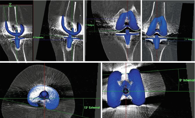
Fig. 3
Measurements of the TKR component position using a customised and validated software showed – 12° flexion (left) and 5° internal rotation (middle) of the femoral TKR component and 5° posterior slope of the tibial TKR component (left up) and 12° external tibial rotation (left bottom)
Questions
1.
What is your differential diagnosis now?
2.
What are your next steps in diagnostics?
99mTc-HDP-SPECT/CT confirmed that there was no loosening or infection. Hyperflexion of the femoral component of the TKR on the left side (12°) with a consecutive ventral, over-shielding, and patellar stress at the upper pole of the left patella baja (Figs. 2 and 3).
Questions
1.
What is your diagnosis and proposed treatment?
2.
 Diagnosis of Periprosthetic Joint Infection After Total Knee Replacement
How Can Preoperative Planning Prevent Occurrence of a Painful Total Knee Replacement?
Causes and Diagnosis of Aseptic Loosening After Total Knee Replacement
Instability of Total Knee Replacement
Discussion to Chap. 53: Persistent Anterior Knee Pain After Total Knee Replacement
Treatment of Instability After Total Knee Replacement
Diagnosis of Periprosthetic Joint Infection After Total Knee Replacement
How Can Preoperative Planning Prevent Occurrence of a Painful Total Knee Replacement?
Causes and Diagnosis of Aseptic Loosening After Total Knee Replacement
Instability of Total Knee Replacement
Discussion to Chap. 53: Persistent Anterior Knee Pain After Total Knee Replacement
Treatment of Instability After Total Knee Replacement


How would you address the femoral component malposition?
Related posts:
Diagnosis of Periprosthetic Joint Infection After Total Knee Replacement
How Can Preoperative Planning Prevent Occurrence of a Painful Total Knee Replacement?
Causes and Diagnosis of Aseptic Loosening After Total Knee Replacement
Instability of Total Knee Replacement
Discussion to Chap. 53: Persistent Anterior Knee Pain After Total Knee Replacement
Treatment of Instability After Total Knee Replacement
Stay updated, free articles. Join our Telegram channel
Full access? Get Clinical Tree
