Fig. 10.1
Photograph of a chronically exposed TKR in a subject with rheumatoid arthritis that failed multiple irrigation and debridements, extensor mechanism reconstructions, and a medial gastrocnemius flap
10.1 Important Anatomical Knowledge for Normal Wound Healing
In contrast to the skin circulation of the thigh proximal to the knee, there is no underlying muscle or intermuscular septae anterior to the knee to provide direct arterial perforating vessels to the skin. The majority of the blood supply to the skin is derived from terminal branches of the peripatellar anastomotic arterial ring [2]. The medial and lateral superior geniculate arteries, the supreme geniculate artery, the anterior tibial recurrent artery, and a branch of the profunda femoris artery contribute to the peripatellar anastomotic arterial ring [3] (Fig. 10.2). The subcutaneous fascia carries the majority of the vascularity to the skin. These subcutaneous perforators are delicate vessels and can be disrupted by excessive traction.
Fig. 10.2
Diagram demonstrating the peripatellar arterial anastomotic ring (SG supreme genicular artery, LSG lateral superior genicular artery, MSG medial superior genicular artery, LIG lateral inferior genicular artery, MIG medial inferior genicular artery, ATR anterior tibial recurrent artery)
The majority of blood supply to the skin comes from terminal branches of the peripatellar anastomotic arterial ring.
Normal wound healing consists of an inflammatory stage, a proliferative stage, and a wound maturation stage [4]. The inflammatory stage involves an influx of platelets, polymorphonuclear leukocytes, lymphocytes, and macrophages. During this stage, vascular permeability is increased by the release of histamine. The local cellular response is fortified by the release of cytokines, platelet-derived growth factors, the complement cascade, and prostaglandins. Fibroblasts, the major source of collagen production, migrate to the wound during the proliferative stage. They produce a ground substance that acts as a matrix for the collagen. The wound maturation stage continues for several months during which the collagen fibrils become more structured and organized [5]. The first weeks of wound healing are when the most rapid gains in wound strength occur [6].
10.2 Patient Risk Factors
A critical aspect of the preoperative plan for TKR is identifying patients at risk for wound complications. A thorough medical history and physical exam help identify risk factors associated with poor wound healing. These risk factors include obesity [7–10], poor nutrition [4, 11–14], tobacco usage [15–17], rheumatoid arthritis [18], and poorly controlled diabetes mellitus [19–23]. Preoperative recognition and then treatment of these high risk patients can potentially decrease the risk of a wound complication.
Typical patient risk factors for wound healing after TKR are obesity, poor nutritional state, smoking, rheumatoid arthritis, and poorly controlled diabetes mellitus.
Many patients desiring total joint replacement are obese [10]. Obese patients often require vigorous soft-tissue retraction to achieve adequate exposure. This can lead to disruption of the delicate cutaneous vascular perforators. Multiple studies suggest that morbid obesity is associated with an increased rate of deep infection in primary TKR [7, 9]. Parvizi et al. [8] found that morbidly obese patients treated with bariatric surgery do well following total joint arthroplasty with an acceptable complication rate. Morbidly obese patients should be clearly and directly counseled prior to surgery regarding their increased risk of wound complications.
Despite the epidemic of obesity, many of these patients are poorly nourished. Forty-two percent of 129 patients undergoing elective orthopedic procedures had evidence of clinical or subclinical malnutrition [13]. Malnutrition is associated with poor wound healing and the early local inflammatory response is dependent on adequate nutrition [4]. Serum albumin levels less than 3.5 g/dL and total lymphocyte counts of less than 1,500 cells/mm3 are associated with wound complications [11–14]. Nutritional intervention prior to surgery may help improve wound healing potential in subjects who are malnourished [12].
Uncontrolled diabetes mellitus is also associated with poor wound healing and subsequent infection [20]. Normal nerve, vascular, and immune function are altered by poor diabetic control [23]. In a review of 59 TKRs in 40 patients with diabetes mellitus, England et al. [19] found an infection rate of 7 % and a revision rate of 10 % at an average follow-up period of 4.3 years. Twelve percent of these TKRs developed wound complications. Multiple studies suggest good glycemic control reverses the negative effects of glycosylated hemoglobin [21, 22]. Unfortunately, hemoglobin A1c, which is a serologic marker for the average systemic glucose concentration over 1–3 months, has not been shown to be predictive of infection risk in total joint replacements [20].
Patients with rheumatoid arthritis undergoing TKR should be carefully investigated preoperatively. In particular their medication such as MTX, corticosteroids, TNF alpha blockers, or leflunomide should be carefully managed. Consultation with the rheumatologist and general practitioner is advocated.
Progressive symmetric synovitis and joint destruction are the hallmarks of rheumatoid arthritis. Disease-modifying antirheumatic drugs (DMARDs) have revolutionized the care of rheumatoid arthritis and when started early can mitigate musculoskeletal damage [18]. However, the use of DMARDs and corticosteroids may be associated with wound healing complications [24, 25]. Methotrexate, leflunomide, and the TNF alpha inhibiting agents need to be carefully managed during the perioperative period. Because there is limited information in the literature regarding perioperative management of DMARDs, consultation with the patient’s rheumatologist or internist becomes critical when trying to manage these medications [18]. Stopping these medications for a period of time preoperatively and postoperatively is often recommended. Corticosteroids hinder macrophage migration, wound contracture, fibrin synthesis, and the formation and ingrowth of blood vessels. Chronic corticosteroid use decreases collagenase clearance and reduces wound tensile strength. It is important to consult the appropriate services for guidance when managing these medications.
Tobacco smoking has long been associated with the development of postoperative wound complications [15–17]. Smoking causes peripheral vasoconstriction through the adrenergic effects of nicotine. Moller et al. [17] found that smoking was the single most important risk factor for the development of postoperative complications related to wound healing and the need for postoperative intensive care in 811 consecutive patients who underwent hip or knee arthroplasty. Craig et al. [15] found that cessation of smoking, even at the time of surgical preparation, obviated much of the noxious effect of smoking and increased survival of hamster skin flaps. Smoking cessation should be a part of the preoperative counseling.
Smoking cessation should be a part of the preoperative counseling.
10.3 Surgical Risk Factors
Surgeons can optimize wound healing potential through meticulous surgical technique. Because the blood supply to the skin around the knee is precarious, gentle tissue handling is mandatory to avoid disruption of fasciocutaneous vascular perforators supplying the skin. Other operative factors that directly impact wound healing are the location of the incision and the management of soft-tissue flaps.
Careful preoperative planning minimizes wound complications. All surgical scars around the knee should be diligently studied at the time of initial consultation. Prior operative reports should be reviewed to determine the soft-tissue dissection utilized in previous operative procedures [26]. A plastic surgeon consultation may be valuable in cases complicated by multiple incisions, burns, or previous radiation therapy. Complex wounds at risk for soft complications have been successfully treated with soft-tissue expansion [27] or prophylactic flap coverage [28].
The anterior midline skin incision is the least disruptive approach to the arterial network. Large lateral based flaps should be avoided.
Medial peripatellar incisions necessitate the creation of large laterally based flaps. Because the lateral wound edge is more hypoxic than the medial edge, it is desirable to avoid large laterally based flaps [29]. Placing the incision slightly lateral in obese patients helps with patella eversion because it decreases the soft-tissue bulk. Minimally invasive techniques tend to increase the skin tension. Slightly longer skin incisions decrease excessive skin tension and thereby help protect the subcutaneous fasciocutaenous perforators. When skin flaps are necessary due to previous incisions, subfascial dissection is critical to avoid disruption of the cutaneous blood supply.
To avoid the compromised vascularity associated with narrow skin bridges, previous surgical incision should be incorporated into the new skin incision whenever possible. Placement of a new incision adjacent to a previous incision leaving a narrow skin bridge risks skin necrosis (Fig. 10.3). This phenomenon can innocently occur if previous skin incisions are not marked out with an indelible ink marker before the limb is prepped. Iodine-based prep solutions and impregnated draping can make precise identification of previous incisions difficult and result in creation of a new incision with an undesirable narrow skin bridge. If only part of the previous incision is to be used, try to maximize the angle of intersection (at least 60°) between the new and old scar. Because wide scars typically have thin subcutaneous tissue and damage to the underlying dermal plexus, it is wise to excise them. Short medial or lateral peripatellar incisions often present from previous open meniscectomy procedures typically can be ignored. Transverse incisions can be crossed at a 90° angle [30].
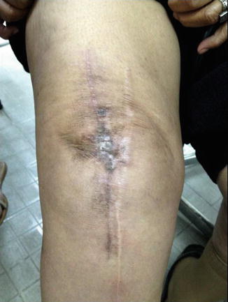
Fig. 10.3
Photograph of a knee with two longitudinal incisions and a narrow skin bridge which resulted in necrosis requiring debridement and secondary closure
Whenever possible incorporate previous skin incisions into your new approach. Avoid narrow skin bridges at all circumstances. If only part of the previous skin incision can be used, maximize the angle of intersection (>60°). Transverse incisions should be crossed at 90° angle.
Because the predominant blood supply is medial, in cases of multiple extended longitudinal incisions, the most lateral incision should be used [31]. Consider a new incision if the scar is too far from midline. The minimum distance between an old and the new incision should be from 2.5 to 8 cm [32].
Minimum distance between a previous and new skin incision should be at least 2.5 cm.
Because lateral patellar releases may compromise wound healing, they should be performed only if necessary. Johnson et al. [33] found that division of the superolateral geniculate vessels during a lateral retinacular release procedure results in an increased incidence of wound discoloration (14 %) and superficial wound infection (38 %). They also noted a reduction in lateral wound edge skin oxygen levels following lateral releases.
Related posts:
 Diagnosis of Periprosthetic Joint Infection After Total Knee Replacement
Measured Resection and Gap Balancing Technique in TKR
Material Failure of Total Knee Replacement
Instability of Total Knee Replacement
Discussion to Chap. 53: Persistent Anterior Knee Pain After Total Knee Replacement
Treatment of Instability After Total Knee Replacement
Diagnosis of Periprosthetic Joint Infection After Total Knee Replacement
Measured Resection and Gap Balancing Technique in TKR
Material Failure of Total Knee Replacement
Instability of Total Knee Replacement
Discussion to Chap. 53: Persistent Anterior Knee Pain After Total Knee Replacement
Treatment of Instability After Total Knee Replacement
Stay updated, free articles. Join our Telegram channel

Full access? Get Clinical Tree


