Fig. 35.1
(a, b) The C-sign. This term reflects the shape of the hand when a patient describes deep interior hip pain. The hand is cupped above the greater trochanter with the thumb posterior and the fingers gripping deep into the anterior groin (Reprinted with permission J. W. Thomas Byrd, MD)
Special attention should be paid to abdominal-based pain, as athletic pubalgia (AP) can often be present in the setting of hip pathology. Hammoud has shown that athletes with symptomatic FAI also have a high incidence of AP with 32 % of the patients in their series having undergone surgical treatment for both FAI and athletic pubalgia [14]. Patients with AP will often complain of pain near the pubic tubercle at the insertion of the conjoint tendon of the rectus abdominis musculature. Pain may also be present in the groin along the course of the adductor musculature, making an accurate distinction between the true causes of groin pain even more difficult. Patients with AP will often complain of pain with sudden activities such as sneezing, coughing, sprinting, kicking, and sit-ups, but rarely have pain at rest [15, 16].
Physical examination of the hip starts with an evaluation of the patient’s gait pattern. Specifically, patients may oftentimes present with an antalgic gait pattern secondary to the inflammation and pain they are experiencing in their hip. With an antalgic gait pattern, the stance phase of gait is abnormally shortened relative to the swing phase. A Trendelenburg gait may also be present as a result of abductor weakness. During the stance phase, normal abductor strength is necessary to maintain a level pelvis. To compensate, the trunk lurches to the weakened side, reducing the forces required of the abductors.
The patient is then laid supine on the exam table in a pair of shorts, with their shoes and socks off. Leg lengths should also be measured from the anterior superior iliac spine down to the medial malleolus. Leg length discrepancies are not uncommon and can result in an altered gait pattern. The resting position of the feet can provide clues to the capsular status of the hip joint. Patients with hip pathology may develop a contracted hip capsule due to synovial inflammation or capsular laxity due to labral tearing and the loss of the suction-seal effect of the labrum which results in repeated capsular overload and stretching [17, 18]. This is best appreciated by comparing the resting position of the foot in the affected limb versus that of the unaffected limb. In the setting of a contracted hip capsule, the foot will sit internally rotated compared to the contralateral side. In a hip with capsular laxity, the opposite holds true as the foot sits externally rotated compared to the contralateral unaffected limb. The dial test can also be used to assess capsular laxity. This test is performed with the patient lying supine on the examination table with the limb in the neutral position. The limb is then internally rotated and then released, allowing passive external rotation. A positive test occurs when the patient’s limb passively externally rotates beyond 45° and lacks a solid end point [19].
Range of motion testing is then performed with the patient in the supine position. Flexion, extension, abduction, adduction, internal rotation, and external rotation are all assessed. It is important, especially when testing hip flexion, to place a hand on the anterior superior iliac spine to allow the examiner to detect pelvic rotation when assessing range of motion. Once the pelvis starts to rotate or lift off the table, true isolated hip motion has ceased. This is the equivalent of assessing true glenohumeral motion in the shoulder by eliminating scapulothoracic motion. To determine true hip joint motion, compensatory pelvic motion must be eliminated to obtain an accurate measure. In the setting of FAI, hip flexion and internal rotation are often decreased due to a mechanical block to motion. A global loss of motion is not uncommon due to the synovial hypertrophy and inflammation that result from labral pathology and the impingement-mediated process. Excessive joint mobility and motion is often present in patients with hip dysplasia, as the normal bony constraints are no longer present, allowing for supraphysiologic motion to occur. Patients without dysplasia may develop capsular laxity as a result of the loss of the normal stabilizing suction-seal effect of the labrum. This results in capsular overload and eventual capsular stretching with plastic deformation. This most commonly involves the anterior capsular and iliofemoral ligament, resulting in an increase in hip extension and external rotation. Femoral version can be predicted from the sum total of internal and external range of motion. Increased internal rotation with a loss of external rotation is seen with increased femoral anteversion. Loss of internal rotation with an increase of external rotation is seen with femoral retroversion. One special note about adhesive capsulitis, occasionally encountered in middle-aged females: a characteristic feature is disproportionate painful restriction of external rotation compared to internal rotation [20].
Several examination maneuvers exist to detect labral pathology. The log roll test is simply a passive internal and external rotation test of the hip with the patient in the resting supine position, and a positive test is specific for intra-articular irritation of the hip joint. The flexion, adduction, internal rotation (FADDIR) test engages the femoral neck against the anterior rim of the acetabulum and recreates labral impingement [21]. With the patient in the supine position, the hip is maximally flexed, internally rotated, and adducted. Recreation of groin-based pain is considered a positive test for labral pathology. The flexion, abduction, external rotation (FABER) test is used to detect lateral labral pathology and also to stress the anterior capsulolabral complex. With the patient supine, the hip is brought into 90° of flexion, abduction, and external rotation so that the foot sits on the contralateral knee. The height of the knee from the examination table is then measured and compared to the contralateral side. This provides some insight into the integrity of the iliofemoral ligament and anterior capsular structures. This also provides the maximum amount of stress to the lateral labrum and can help detect labral pathology [22]. The Stinchfield test is used to detect iliopsoas irritation and possible iliopsoas impingement against the labrum [23]. With the patient lying supine, a straight leg raise is actively performed to 45°, at which time active downward pressure is applied to the thigh, isolating the iliopsoas complex. Recreation of pain or weakness indicates a positive test. The posterior rim impingement test is performed by bringing the leg into extension and external rotation, allowing for engagement of the femoral neck against the posterior acetabular rim. This is often performed by bringing the patient’s leg off the end or side of the table to allow for full hip extension to occur.
With the patient in the lateral position, the palpation of the trochanteric facets can be performed [24]. This allows for the detection of possible abductor pathology which, in the setting of labral pathology, is often a true gluteus medius or minimus tendinitis. Passive adduction tests are also performed in the lateral position to detect tightness of the iliotibial band, gluteus medius, and gluteus maximus. Each test is performed by allowing passive adduction of the limb. With the knee in full extension, contracture of the iliotibial band can be detected. With the knee flexed to 90°, contracture of the gluteus medius can be detected. With the knee in full extension and the hip forward flexed, gluteus maximus contracture can be detected [23].
Diagnostic studies to diagnose labral pathology include plain radiographs and MRI. Plain radiographs should always include a well-entered AP pelvis [25] and, variably, a frog lateral [26], a cross-table lateral, and a false-profile view [27]. An appropriate AP pelvis x-ray is indicated by the coccyx being centered over the pubic symphysis, with the tip of the coccyx 1–2 cm from the pubic symphysis [28]. This allows for standardization and correction of pelvic tilt and inclination which can alter the acetabular orientation and version [29]. The lateral center-edge angle of Wiberg, the Tonnis angle, and the acetabular version are measured from the AP pelvis radiograph. A crossover sign is indicative of acetabular retroversion and indicates possible pincer impingement. A normal lateral center-edge angle is between 25° and 40°, with less than 25° representing dysplasia and greater than 40° representing acetabular over-coverage [12]. The cross-table lateral and frog radiographs allow for evaluation of the head-neck offset and measurement of the alpha angle to determine if cam impingement exists. Normal head-neck offset measures approximately 10 mm, with anything less suggesting cam impingement [30]. A normal alpha angle measures approximately 50° or less, and anything greater than that may indicate cam impingement [31]. The false-profile view allows for assessment of the anterior center-edge angle and gives an assessment of anterior acetabular coverage. It also allows for visualization of the anterior-inferior iliac spine morphology, as this may serve as a source of extra-articular impingement [5, 32]. Lastly, in cases of suspected degenerative disease, the false profile can be very helpful to detect anterior or posterior joint space narrowing that may be missed in an AP projection.
If labral pathology is suspected, conventional magnetic resonance imaging (MRI) or MRI combined with arthrography (MRA) may be useful. Both demonstrate greater sensitivity at detecting labral lesions than accompanying articular damage [33]. MRA reports better sensitivity, but eliminates the ability to assess for the presence of an effusion and may obscure subchondral or soft tissue edema [34, 35]. A negative study should not preclude consideration of arthroscopic treatment and evaluation when there is a high clinical suspicion of a labral lesion, as even MRA has a poor negative-predictive value and cannot be used to rule out a labral lesion [34, 36]. X-rays are used as more of a screening tool, but once the diagnosis of FAI with labral pathology has been established and arthroscopy has been proposed, then a CT scan with 3D reconstructions can be very helpful in planning the surgical correction. This allows for a more accurate assessment of the osseous anatomy of the hip [37, 38]. Axial cuts through the distal femoral condyles are made to allow for accurate measurement of the overall femoral version, which is a vital piece of information needed for appropriate surgical treatment. The 3D reconstructions also allow for identification of cam lesions that may be missed on the multiple planes of the CT scan, as oftentimes the cam can be subtle and missed with standard CT-scan cuts. The 3D reconstructed views provide excellent visualization and detail of the AIIS. Overall, the 3D-reconstructed CT scan is the most vital imaging study used in formulating an arthroscopic plan, as it allows for an accurate identification of impingement lesions, bony resection planes, and age in overall capsular and soft tissue management intraoperatively.
35.3 Treatment Strategy
Treatment of acetabular labral tears normally starts with an effort to identify and modulate offending activities and may include a course of nonoperative trial of physical therapy, anti-inflammatory medications, and, oftentimes, an injection into the femoroacetabular joint. Joint injections are done using ultrasound guidance in the office but can also be done using fluoroscopic guidance [39]. Physical therapy should focus on core-muscle strengthening, correction of abnormal muscle firing patterns, and abductor strengthening to provide stability to the hip joint in cases of capsular laxity. Oftentimes, athletes will elect nonoperative treatment in an attempt to “play out their season” and elect for operative treatment in the off-season.
Operative treatment consists of arthroscopic or open correction of the impingement process with labral refixation or reconstruction. Several studies have shown that the results of labral repair are superior to simple labral debridement [40–42]. In the event that labral repair is not viable, labral reconstruction has been proposed. Currently, reconstruction is most clearly indicated for symptoms associated with a deficient labrum, often associated with previous surgical debridement [43]. Keep in mind that there may often be multiple pain generators, and it can be difficult to ascribe symptoms solely to a labral deficiency. Although there were limitations of their methodology, Matsuda and Domb have reported results of labral reconstruction similar to those of labral repair and outperforming those of simple labral debridement [44, 45]. Open treatment consists of surgical dislocation of the hip, which was developed by Ganz and has been used by others to address labral pathology [42, 46–49]. Our current preferred method is to address labral pathology utilizing arthroscopic treatment in an outpatient setting with the patient in the supine position [50, 51].
35.3.1 Labral Debridement
Labrectomy is infrequently necessary. Even comminuted and degenerative tears can demonstrate successful outcomes with restoration. However, occasionally, simple debridement may be deemed the best option for some patients: elderly patients, patients with limited capacity to comply with necessary postoperative precautions, and some tears with limited healing capacity. Debridement alone does not necessarily result in uniformly poor results [52]. However, debridement must be carried out in a thoughtful fashion, as there is compelling evidence that excessive labral removal can result in accelerated degenerative changes [42]. The goal of labral debridement is to remove the damaged tissue, preserve as much healthy labrum as possible, and create a stable transition zone, lessening the likelihood of persistent symptoms or propagating further tearing (Fig. 35.2) [53].
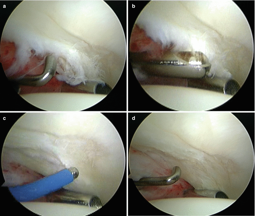
Fig. 35.2
Arthroscopic view of a right hip from the anterior portal. (a) A fragmented labral tear with degeneration within its substance is identified. (b) Debridement is initiated with the power shaver. (c) A portion of the comminuted labral tear is conservatively stabilized with a radio-frequency probe. (d) The damaged portion has been removed, preserving the healthy substance of the labrum (Reprinted with permission J. W. Thomas Byrd, MD)
35.3.2 Surgical Technique for Arthroscopic Repair/Refixation
The majority of labral tears are amenable to restoration and preservation. Mostly commonly, labral tears requiring treatment are seen in conjunction with pincer impingement and require concomitant acetabuloplasty. Thus, the labrum is mobilized as necessary with sharp dissection, shavers, and radio-frequency devices to expose the pincer lesion that must be resected. If the chondrolabral junction is intact, it is preferable to preserve its integrity, but adequate mobilization for proper correction of the pincer lesion takes precedent over the goal of preserving the chondrolabral junction.
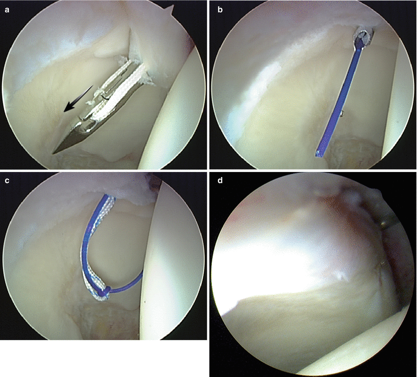
Most labral tears begin anteriorly and extend laterally. Thus, most of the work is done with the arthroscope in the anterolateral position as a viewing portal and the anterior position as the working portal with a large-diameter disposable cannula (Fig. 35.3) [51, 53]. The anchors are placed on the capsular side of the labrum as close as possible to the articular rim but always being careful to avoid perforating the articular surface. If the anchors are placed too far away from the rim, it does not restore the labral anatomy, and it is unlikely that it is restoring its function. The anchors are placed and tied starting from medial to lateral.
Fig. 35.3
(a) For this right hip, three standard portals are utilized for routine arthroscopy, including the anterior (A), anterolateral (AL), and posterolateral (PL). A large-diameter disposable cannula has been placed anteriorly for suture management. The anchor delivery system (arrow) has been placed distally midway between the anterior and anterolateral portals. (b) Schematic illustrates the drill sleeve placed against the acetabular rim (Reprinted with permission J. W. Thomas Byrd, MD)
Most anchors are placed from a percutaneous distally based site which easily allows placement from the 3 o’clock position anteriorly to well posterior to the 12 o’clock position laterally on a right hip [54]. Occasionally, a far medial anchor is placed from the anterior portal, giving a better direction to avoid the anchor perforating the medial pelvic cortex, possibly irritating the iliopsoas tendon.
When drilling, the articular surface of the acetabulum is carefully observed. Any signs of articular motion with the drilling process indicate that it could be violating the subchondral surface and necessitates redrilling further away. Anchors are spaced at 8–10 mm intervals for adequate security of the restored labrum.
Less commonly, for tears extending well posterior to the 12 o’clock position, the arthroscope can be switched to the anterior portal, and the anchors can be placed from the anterolateral portal. In this position, fluoroscopy helps to assure that, with drilling, there is adequate diversion from the articular surface. Fluoroscopy does not help for anteriorly based anchors because the direction of placement is more in the plane of the x-ray beam (Fig. 35.4).
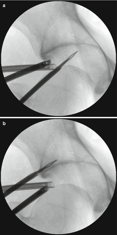
Fig. 35.4
(a) Fluoroscopic image of a right hip as drilling is performed for an anteriorly based anchor. Since the direction of placement is partly in the plane of the image, it is not helpful for assuring proper placement. (b) AP fluoroscopic image of a right hip with the anchor drill hole being placed laterally. Since this is perpendicular to the x-ray beam, the image is helpful in assuring that the anchor will diverge from the articular surface (Reprinted with permission J. W. Thomas Byrd, MD)
Anchor placement is consistent from one case to the next, but suture management is variable and dictated by the size, morphology, and pattern of labral tearing. If the chondrolabral junction is intact, a simple suture can be used through the lateral margin of the labrum, rolling it up against the rim and recreating the labral structure (Fig. 35.5). For a large labrum, or one in which the chondrolabral junction has been disrupted, a modified single-limb mattress suture creating a labral base fixation can restore the chondrolabral junction and the normal anatomy of the labrum (Fig. 35.6) [55]. For a small labrum, or one in which the quality of the labral tissue is poor, a simple loop suture may be necessary in order to restore adequate labral tissue to the acetabular rim (Fig. 35.7).