Category 1: Visceral causes
Inguinal hernia
Other abdominal hernias
Testicular torsion
Category 2: Hip-associated causes
Acetabular labral tear and femoroacetabular impingement
Osteoarthritis
Snapping hip syndrome and iliopsoas tendinopathy
Avascular necrosis
Iliotibial band syndrome
Category 3: Pubic symphyseal causes
Rectus abdominis tear
Adductor muscle–tendon dysfunction
Rectus abdominis–adductor longus aponeurosis tear
Osteitis pubis
Category 4: Infectious causes
Septic arthritis
Osteomyelitis
Category 5: Pelvic inflammatory disease
Prostatitis
Epididymitis and orchitis
Herpes infection
Category 6: Inflammatory causes
Endometriosis
Inflammatory bowel disease
Pelvic inflammatory disease
Category 7: Traumatic causes
Stress fracture
Tendon avulsion
Muscle contusion
Baseball pitcher–hockey goalie syndrome
Category 8: Developmental causes
Apophysitis
Growth plate stress injury or fracture
Legg–Calvé–Perthes disease
Developmental dysplasia
Slipped capital femoral epiphysis
Category 9: Neurologic causes
Nerve entrapment syndromes (e.g., ilioinguinal nerve)
Referred pain
Sacroiliitis
Sciatic entrapment (piriformis syndrome)
Hamstring strain
Knee pain
Category 10: Neoplastic causes
Testicular carcinoma
Osteoid osteoma
8.3 Injury Mechanisms and Predisposing Factors
Intrinsic and extrinsic factors may predispose the athlete to the groin pain syndrome. Among the intrinsic factors, those receiving the greater consensus in literature [1, 10, 32, 53–59] are as follows:
1.
Hip and/or sacrum–iliac joint diseases
2.
Lower limbs asymmetry
3.
Lumbar hyperlordosis
4.
Functional imbalance between abdominal and adductor muscles, with a weakness of the abdominal muscles compared to the adductors leading to their excessive stiffness or a weakness of both muscular groups, leading to a reactive contracture of adductor muscles
5.
Excessive hamstring stiffness
6.
Adductor weakness
7.
Previous injury
It is important to remember that some authors [60] proposed as intrinsic cause a core muscular weakness or a delayed onset of transversus abdominal muscle recruitment.
Furthermore, there is an ongoing debate in literature regarding the age and/or sport experience as risk factors for groin injury [60–62].
Regarding the inadequacy of pitch surfaces, we must make some important clarifications. A parameter which we must carefully assess is represented by the interaction, in terms of mechanical constraint, between the pitch and the shoe. An interesting data in this regard comes from the American National Football League (NFL), which shows that abductor tendinopathy would increase by 27 % on the artificial turf pitches when compared to natural turf pitches [66], although these data do not find further confirmation in the literature [67, 68]. Also some natural grass surfaces may be a risk factor for the onset of abductor tendinopathy. The association of hot climates and some types of grass having a particularly strong and deep root system creates an excessive constraint between the shoe and the ground. Conversely, other types of grass with an insufficient radical apparatus, if used in cold climates, would not be able to create a sufficient mechanical constraint between the foot and the playing surface. Both situations could represent a risk factor for onset of adductor tendinopathy especially in athletes with pelvic instability [66].
One of the sports where groin pain is most frequent is football [69]. Many technical movements in football may favor the onset of the injury: jumps, dribbling, cutting movements in general, and tackles performed sliding with abducted leg and adductor muscle contracted. These are factors that cause high stress on the pubic symphysis, triggering a synergic mechanism between adductors and abdominal muscles [43]. Moreover, shooting and running performed on irregular surfaces represent other intense and abnormal functional stress factors [70].
In this context, it is important to consider the Maigne theory [71], based on the functional imbalance of the football players’ column biomechanics. Specifically, this theory argues that football players are playing in a constant hyperlordotic gait which creates a conflict at the dorsal-lumbar spine level between the vertebral joints and genito-abdominal nerves, responsible for the groin region sensitive innervations. This theory could justify the high incidence of groin pain in football reported by different authors [72, 73].
There is no strong evidence in the literature supporting a causal association for any extrinsic or intrinsic risk factors and groin pain syndrome onset. In effect, the majority of the studies are based on conjecture, expert opinion, or case series.
Athletes affected by groin pain syndrome would most likely be subjected to a combination of excessive muscular contractions by abdominal and adductor muscles. Torsion and impact causing bone stress can occur during running, violent movement performed with poor muscle control (such as sprint, shoots, tackles, change of direction), and mechanical constraints especially of torsion type at the pubic symphysis level [12, 32, 63, 65, 74]. The majority of authors agree that during normal activity, the abdominal and adductor muscles have an antagonistic but biomechanically balanced function. In the case of groin pain, there is no more muscle balance between the adductors and abdominals, with the adductor muscles being too powerful and the abdominals too weak or with adductors being extremely stiff thus producing an abnormal tension in the pelvis with a negative impact on the pubis [19, 23, 36, 44, 57, 75, 76]. Finally, quadriceps muscle hypertonia would further aggravate this functional imbalance [76].
It is important to underline the rectus abdominis and adductor longus origin from a common aponeurosis insertion at the periosteum of the anterior aspect of the pubic body and their antagonist function during rotation and extension [77].
Moreover, we must remember that also a force ratio less than 80 % between the adductor and abductor muscles has been identified as a potential groin pain risk factor [45]. Other authors found that the same deficit between the extensor and the flexor trunk muscle force ratio could induce groin pain [16]. Finally, other studies [1] include poor proprioception among the predisposing factors. However, our therapeutic experience does not allow us to share this hypothesis; in effect, both static proprioception management and dynamic proprioception management reflect an extremely multifactorial control mode which makes it difficult to provide evidence in this specific field.
It is important to remember that six of the seven adductor muscles1are innervated by the obturator nerve and that their origin is in close proximity to the pubis. This allows them biomechanically to act in open kinetic chain as hip adductors and have an important stabilizing role in the closed kinetic chain. Not surprisingly, athletes affected by groin pain syndrome generally have significant concentric muscle strength in the lower limb muscles while simultaneously presenting with a deficit of postural muscle strength [1, 45].
8.4 Clinical and Diagnostic Examination
Symptoms of groin pain syndrome are bilateral in 12 % of cases, affecting the adductor region in 40 % of the cases and the perineal area only in 6 % of the cases [14]. The onset of reported groin pain syndrome symptoms is insidious in 2/3 of the patients and acute in 1/3 [14]. The groin pain clinical framework is characterized by subjective and objective symptomatology.
Subjective symptoms are mainly identified in pain and functional deficit [78, 79]. The intensity of pain has highly significant variability and can range from a mere annoyance to acute pain. The intensity of which can even affect the patient’s normal daily life activity, such as walking, dressing, and getting out of bed or car, and sometimes even preventing sleep. The painful event can occur during competition and/or training. It can already be present prior to exercise and disappear during warm-up, reappearing later during activity or appearing after the exercise, while cooling down, or even the morning after. In extreme cases, symptoms can effectively preclude performance. Pain may radiate outward and extend along the adductor and/or abdominal muscles in the direction of the perineum and the genitals. This generates possible diagnostic errors [79]. The functional deficit is obviously correlated with pain intensity.
From an objective point of view, the patient can complain of pain at palpation, resisted contraction, and during stretching. In addition, clinical examination is based on several muscle tests based both on active contractions and on passive and active muscle stretching [80–83]. Moreover, in this context, it is important to observe how the patient moves, walks, and undresses [84].
8.4.1 Imaging
Radiological investigations can help in groin pain syndrome diagnosis. Pelvic X-rays highlighting the pubic symphysis are always advisable to rule out possible bone erosion, pubic branch dissymmetry, osteoarthritis (also frequent in young subjects), hip joint pathology, and especially tumors or avulsion fractures [85–87]. It is important to emphasize how through a dynamic X-ray made in alternating monopodalic support, the so-called flamingo views (Fig. 8.1), when a vertical offset greater than 3 mm between the pubic horizontal branches is found, we can make the diagnosis of symphysis instability [44, 88, 89]. Musculoskeletal ultrasound (US) finds its indication in inguinal hernia suspicion. It can highlight edema areas, hematomas (in the case of muscle–tendon tears), myxoid degeneration areas, chondral metaplasia or metaplastic calcification, and fibrosis [30, 90] with the advantage of having the possibility of being carried out in dynamic conditions. This highlights musculofascial movements and, in particular, inguinal bulging (inguinal canal posterior wall weakness). However, US currently falls short in the identification of inflammatory and degenerative bone processes.
Fig. 8.1
A double-stance X-ray (a) compared to a dynamic flamingo view X-ray (b) made in alternating single-stance support (in this case in single right stance). The subject, a 25-year-old professional football player, shows a vertical offset greater than 3 mm between the pubic horizontal branch that allows us to make the diagnosis of symphysis instability
Nuclear bone scan is a highly sensitive but nonspecific tool. Every type of symphysis bone lesion of traumatic, tumoral, or infectious etiology would lead to an increased uptake activity at the symphysis level [30, 91, 92]. However, a previous uptake that normalizes after conservative treatment is an important factor which may play a role in making a decision for possible return to sports activity [91, 93, 94].
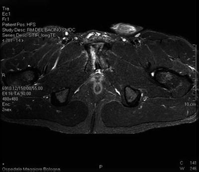
Magnetic resonance imaging (MRI) is considered the gold standard examination providing detailed information concerning both bone and insertion structures [8, 30, 86, 89]. An MRI groin pain-specific protocol should include sequences covering the entire bony pelvis as well as higher-resolution sequences dedicated to the pubic symphysis region. A relatively outperformed model like a 1.5 T MRI unit is an adequate instrument to generate high-quality images of the pelvis, while a 3 T scanner can offer indubitable advantages in signal and resolution but is also prone to generate more imaging artifacts [95]. Images must be acquired in standard coronal, sagittal, and axial planes; however, it is important to underline that coronal oblique imaging plane performed along the anterior margin of the iliac crest is a very important sequence for optimal assessment of the rectus abdominis/adductor longus common aponeurosis at the pubic level [96]. Some authors proposed the use of intravenous contrast, but its use generally adds little in the identification of lesions, and a non-contrast protocol at 1.5 T can be considered standard [52].
One of the most important advantages in the use of MRI for the assessment of patients affected by groin pain is its high sensitivity for a wide array of both musculoskeletal and visceral lesions that may concur to the symptomatology. In effect, it is not uncommon to discover an unsuspected lesion with pelvic MRI. For these reasons, it is important to include in MRI protocol several large field of view sequences covering the entire bony and visceral pelvis even if there is a strong suspicion for a simple pubic symphysis lesion. In fact, it is not uncommon that the groin pain is caused by bursitis, benign and malignant soft tissue tumors in various locations around the pelvis, visceral pelvis sources such endometriosis and inflammatory bowel disease, osseous injuries such stress fracture, primary osseous tumor such as osteoid osteoma, or scarring and fibrosis related to prior herniorrhaphy. With a deep MRI evaluation protocol, the majority of these lesions should be observed or at least suspected [97].
8.5 Rehabilitation and Treatment Strategy
8.5.1 Type of Exercise and the Progression of Work Plan
Concerning the type of exercise, the study with the strongest evidence considers strengthening exercise as the main component of the work plan [80, 98, 99]. Target muscles involved are the adductor, abductor, hip flexor, and deep and superficial abdomen muscles. The progression begins with isometric contractions and continues with concentric and eccentric exercises, reaching the functional standing position. This is to be as similar as to those required by the athlete’s specific sport activity during the last stage of the rehabilitation protocol. Isokinetic exercises should also be present throughout the protocol. Holmich et al. [80] used a predetermined graduated exercise protocol, while many researches adopt the following criteria for exercise progression:
1.
Absence of pain during exercise
2.
Full acquisition of functional control
3.
Ability of performing functional exercise or a predetermined number of repetitions
The available evidence suggests that strengthening exercise represents an important component in an effective work plan. However, variability between the different protocols in terms of the muscle concerned does not allow for a conclusion to be reached on the specific target muscle group [80, 98, 99]. Conversely, research shows a uniformity of exercise progression from the isometric modality to be completed by sport-specific functional standing positions.
8.5.2 The Intensity, the Frequency, and the Duration of Exercise
To the best of our knowledge, only one reliable study may be found in the available literature providing enough detail concerning intervention frequency and duration of exercise [80]. This study suggests a work plan of 90 min of strengthening exercises for the hip and abdominal muscles to be performed three times per week for an overall duration of 8–12 weeks. According to this research, the outcome is good, allowing the athlete to return to sport activities without groin pain.
The duration of conservative treatment is between a minimum of 2–3 weeks [14] and a maximum of 6 months generally [100]. The majority of authors agree on a duration of around 6 months [23, 92, 94, 101–104]. In summary, it is clear that the variation in duration of rehabilitation work plans used reflects the variation in the severity and multifactorial characteristics of groin pain.
8.5.3 Therapeutic Interventions
In essence, the majority of studies report the use of one or more co-interventions, from manipulation techniques and massages [92, 102–104] to anti-inflammatory [18, 98, 100, 101, 105] and corticosteroid medication [58, 106, 107]. Some studies included jogging, running, and cycling as co-interventions [56, 98, 99, 104]. Furthermore, some studies underline the importance of physiotherapist-supervised exercise programs [56, 99, 102].
8.5.4 Surgical Treatment
As previously discussed, groin pain syndrome may be caused by several pathologies responding to conservative therapy. However, if conservative therapy fails, then a surgical option must be considered. In this final section, we will briefly describe the most common diseases requiring such treatment.
8.5.5 Inguinal Hernia
Athletes are susceptible to inguinal (direct and indirect) hernias like the general population and sometimes even more, especially in sports like weightlifting. However, in athletes, direct hernias are more frequent [108]. Real-time dynamic US during a provocative maneuver, such as Valsalva, may help visualize a subtle hernia possibly causing symptoms only during sport activity and otherwise difficult to detect. The risk of complications such as bowel incarceration and strangulation is not an issue in this case; it is impossible to participate in sports due to pain. This is why in most cases posterior wall weakness of the inguinal canal are surgically repaired [109].
Even though surgical treatment is successful in the large majority of cases, one should bear in mind the possibility of surgical complications and, in some cases, the inability to achieve prior levels of athletic performance [52]. It has been proposed that this variability in surgical repair outcome is occasionally due to the increasing stabilization of the pubic region because of progressive fibrosis [52]. However, patients with inguinal hernia have little chance of success with conservative treatment [52, 110]. After herniorrhaphy, an average of 87 % of the athletes have a positive outcome and are able to return to full and unrestricted athletic activity in 4 weeks or less [29, 110, 111].
8.5.6 Sports Hernia
Sports hernia, also known as sportsman’s hernia, athletic hernia, and incipient hernia, represents a difficult clinical problem [112].
The diagnosis of sports hernia is formulated when no inguinal hernia is found but there is persistent inguinal pain during sports activity. The symptoms resemble an hernia and are present only during sport. We must also point out that some authors underline that sports hernia is often associated with femoroacetabular dysplasia and/or femoroacetabular impingement [113].
There also is no hernia present on physical examination and ultrasound, hence the term sports hernia (Fig. 8.2). Sports hernias rarely improve without surgery [11, 114–118], and surgical repair should be considered when conservative treatment over a period of 6–8 weeks has failed. Careful examination has to additionally exclude other potential pain sources [112, 119].
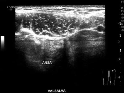
Fig. 8.2
Left inguinal ultrasound in a 27-year-old professional footballer that shows a modest pre-hernial area with about 8 mm of intestinal loop in correspondence to the weak zone. This situation is pathognomonic for sports hernia
Some authors propose laparoscopic repair with prosthetic mesh [120, 121]. This “tension-free” technique involves placing prosthetic material suitably shaped, non-absorbable, and biocompatible. This acts as mechanical reinforcement of the abdominal wall [120, 121]. However, the mesh has no elasticity and creates more scar tissue, and mesh-related complications can occur years after surgery. Another laparoscopic method used for the treatment of sport hernias is inguinal release procedure [122]. After laparoscopic repair, the recovery before full return to competition is generally between 2 and 8 weeks [110, 115, 119, 123–128].
Some authors prefer open surgical inguinal repair: Shouldice repair, Maloney darn, or Bassini with or without adductor longus tenotomy or only the “minimal repair” of the weak area of transversalis fascia [14, 129, 130]. In a meta-analysis study [119], the authors found that the period of time to return to sport is on average 17.7 weeks for patients who underwent open approaches and 6.1 weeks for laparoscopic repairs. Several authors underline mesh-related complications such as infections with chronic groin infection and fistula formation. These complications sometimes require mesh removal [131] or cause mesh migration and penetration into the bladder or bowel [132, 133]. In addition, a foreign body reaction with decrease of arterial perfusion and testicular temperature [134] accompanied by secondary azoospermia may occur [134, 135].
It is interesting to note that Muschaweck et al. [112, 130] after previously utilizing the Shouldice repair under local anesthesia for years, in 2000 developed a new surgical technique called the “minimal repair technique.” The aim of this surgical intervention was to stabilize the posterior wall by a tension-free suture without the use of a prosthetic mesh and by repairing only the weak spot of the transversalis fascia. The authors chose to avoid the use of a prosthetic mesh to allow the athlete’s full elasticity and muscle sliding between the abdominal muscles after surgery [112]. According to some authors, opinions regarding this technique apart from avoiding prosthetic mesh insertion have several advantages. These include not requiring general anesthesia, less traumatization, and a lower risk of severe complications. The authors underline a quicker resumption of sports activity following this surgical technique compared to the laparoscopic or open surgery with mesh insertion. They report that on average their patients resumed moderate training after 7 days and felt complete relief of pain after 14 days. Return to full activity was achieved after 18.5 days [112].
8.5.7 Adductor Tendinopathy
With the increase of knowledge of the pubic symphysis’ complex anatomy, the incidence of isolated adductor tension lesion has seemingly decreased [96]. In any case, adductor tendinopathy is one of the most common causes of groin pain syndrome in athletes in athletes and is most often associated with either rectus abdominis/adductor longus aponeurosis lesions or midline pubic plate lesions (i.e., lesions originate at the midline of the pubis and propagate either unilaterally or bilaterally, also called “midline core muscle injuries”). One of the main causes of groin pain syndrome is the imbalance between the abdominal and hip adductor muscles, with the abdominals too weak or the adductors too strong [5]. Adductor tendinopathy is frequently related to an adductor longus overuse or to its aponeurotic injury [136]. A vast majority of patients respond positively to conservative treatment, both in the case of overuse tendinopathy or in muscle–tendon injury. There are not many scientific papers on failed conservative treatment on chronic adductor-related groin pain [137]. Adductor tenotomy is proposed for cases nonresponsive to conservative treatment [5, 136–139]. The criteria for surgery is a history of long-standing (ranging from 3 to 48 months according to various authors) and of distinct pain at the origin of the adductor longus muscle, refractory to conservative treatment. The operation is performed by releasing the anterior ligamentous fibers of the adductor longus while keeping the fleshy part of the muscle intact on the deep aspect, thus minimizing the loss of adductor strength after surgery and constituting a template for future regrowth of the tendon. In the patients undergoing tenotomy, there is an average of 10 % postoperative strength reduction which does not result in any obvious functional or speed limitation because other muscles in the adductor group, namely, adductor brevis, adductor magnus, and pectineus, take over adductor longus function [140]. In the reported studies [129, 136, 137], the subjects returned to competitive sport after 19.8 weeks (range 27–14 weeks). The cited studies report that following surgery, 70.6 % of the subjects (range 90–62 %) performed sport activities at the same level, 24 % (range 32–9 %) performed sports activities at a reduced level, and 5 % had to stop sport activities altogether. It is interesting to note that some authors associate the adductor tendon release to a pelvic floor repair [45, 141].
Surgically treated adductor acute tears are rarely described in scientific literature. We could find only one study [138] reporting three cases of acute proximal adductor longus insertional tear repaired with anchor sutures and followed by postoperative rehabilitation. The patients followed in this study resumed their full sport ability after 5, 6, and 7 months, respectively.
8.5.8 Osteitis Pubis
Osteitis pubis is a common medical problem in soccer players, long-distance runners, and hockey players. In terms of etiology, the main risk factor is believed to be pubic symphysis instability [52]. This causes a chronic, repetitive shear and an imbalanced tensile stress of the muscles inserted on the pubic symphysis. This biomechanical alteration can cause an inflammatory response with osteitis and periostitis.
Normally, from a radiological point of view, into the MRI pubic symphysis evaluation, any subchondral bone marrow edema, bony sclerosis, or cystic or osteophytic formation is termed osteitis pubis. This type of assessment is not entirely correct. In effect, a true active osteitis pubis should include at least an element of subchondral bone marrow edema (often asymmetric) spanning the pubis joint anterior to posterior on axial fat-suppressed sequence (Fig. 8.3). This bone marrow edema extending into the anteroposterior totality of pubic rami should be distinguished from sub-entheseal marrow edema at pubic tubercle level sometimes present in a rectus abdominis and/or adductor longus tendinopathy without osteitis pubis at the symphysis [96, 142]. It is in any case important to note that osteitis pubis is strongly associated with rectus abdominis and/or adductor longus tendinopathy [96]. Osteitis pubis is normally a “self-limiting” disease and requires a lengthy treatment of 12-month duration on average [105]. The management is initially conservative with physical rehabilitation, NSAID, and/or steroid injections. The historical surgical treatment options were symphysis curettage and arthrodesis and are now abandoned by the majority of surgeons. This is due to the lack of results and frequent side effects. In most cases, adductor tenotomy/surgical abdominal strengthening is reserved for the subjects with symptoms nonresponsive to conservative treatment [52, 107].