Fig. 23.1
Coracoclavicular ligaments: conoid ligament – located medial, coned or triangular in shape. Runs from the posterior medial aspect of the coracoid process to the posterior conoid tubercle in the clavicle. Responsible for restraining superior–inferior displacements. Trapezoid ligament – located lateral, quadrilateral in shape. Runs from the coracoid process shaft oblique and superior–lateral to the anterior–lateral clavicle trapezoid ridge. Responsible for resisting compressive forces and lateral displacement of the clavicle
Indirect mechanisms of ACJ injuries are rare. A fall on to the adducted arm leads to a pushing force of the humeral head upward against the inferior aspect of the acromion. The resulting forces create a wide spectrum of ACJ injuries including inferior displacement of the clavicle beneath the coracoid process (type VI dislocation according to the classification of Rockwood [12]).
23.3 Clinical and Diagnostic Examination
A detailed history including the mechanism of injury, location and duration of pain, and associated symptoms is vital to diagnosing an AC joint injury.
An accurate clinical evaluation may be difficult and painful in the acute setting. It is important to examine the patient in a sitting or standing position, allowing the weight of the injured arm to exaggerate any deformity. Pain may be variable in nature, given the AC joints’ dual innervation from the suprascapular nerve and the lateral pectoral nerve [13]; however, the diagnosis is often clinched with a visible or palpable defect noted at the AC joint. Tenderness directly at the ACJ is the main symptom with visually evident step formation between the acromion and the distal clavicle end in complete ACJ dislocations. A comparison to the unaffected contralateral ACJ should be drawn due to a sometimes physiological prominent distal clavicle end on both shoulders. A key part of the clinical assessment represents the testing of horizontal instability. Hereby, the distal clavicle is shifted posteriorly with the acromion fixed by the other hand. An increased posterior translation in comparison to the unaffected side indicates a horizontal component of ACJ instability. Discomfort is often exacerbated with range of motion of the shoulder and with loading of the joint with the crossarm adduction test, which is performed by forward elevating the arm to 90° with arm adduction. Assessment of the AC joint for stability after an acute injury may be difficult secondary to guarding; however, for subacute and chronic injuries, this should be attempted. The Paxinos test (thumb pressure at the posterior AC joint) combined with a positive bone scan has been found to predict AC joint pathology with a high degree of confidence. Pain localized to the acromioclavicular joint or “on top” is diagnostic of acromioclavicular joint abnormality, whereas pain or painful clicking described as “inside” the shoulder is considered indicative of labral abnormality. However, the sensitivity of this test for AC pathology is only 41 %, with a specificity of 94 % [14]. A simple shoulder shrug may be helpful in determining if the deltotrapezial fascia has been separated from the clavicle [5]. Reduction of the AC joint can also be tested by stabilizing the clavicle in one hand and with the other hand placing an upward force on the ipsilateral elbow and assessing the joint for visible or palpable incongruity. Additionally, a thorough neurovascular assessment of the upper extremity including the cervical spine should be performed. Suspicion for other associated injuries, such as clavicle, coracoid, and rib fractures, should be raised with higher injury mechanisms [15].
They were initially graded I through III based on radiographic displacement and the degree of ligamentous damage [16, 17]. Rockwood later added types IV through VI to the classification system. The rising type correlates with greater displacement and higher levels of ligamentous injury [5].
Type I: This typically low-energy injury involves a sprain to the AC ligaments only. The CC ligaments are spared by the absorption of the impact by the AC ligaments. With the AC and CC ligaments intact, radiographic imaging appears normal.
Type II: As the energy imparted to the shoulder is increased, the AC joint capsule and ligaments are ruptured, and the distal clavicle is thereby rendered unstable in the horizontal plane. The CC ligaments remain intact, and there may be slight elevation of the clavicle on radiographs; however, the displacement is less than 100 % of the diameter of the distal clavicle, and the radiographic CC distance is increased by less than 20 %.
Type III: This higher-energy injury represents a complete disruption of both the AC and CC ligaments, which leads to complete dislocation of the AC joint. The insertion of the deltotrapezial fascia remains intact. Radiographs demonstrate displacement of the clavicle greater than 100 % of the diameter of the distal clavicle, and the radiographic CC distance is increased by 20–100 % [5].
Type IV: This injury involves a complete rupture of the AC and CC ligaments with posterior displacement of the distal clavicle into the trapezius fascia. It is important in this setting to evaluate the SC joint as concomitant anterior dislocation can occur.
Type V: This higher-energy variant of a type III injury represents a complete disruption of both the AC and CC ligaments, which leads to complete dislocation of the AC joint. The deltotrapezial fascia is stripped from its attachment to the clavicle. Radiographs demonstrate displacement of the clavicle greater than 300 % of the diameter of the distal clavicle, and the radiographic CC distance is increased by 100–300 %.
Type VI: This rare injury involves inferior displacement of the clavicle either subacromial or subcoracoid behind the conjoined tendon. The mechanism involves severe hyperabduction and external rotation of the arm combined with scapular retraction. It results from high-energy trauma, and neurovascular impairment is often present prior to reduction [15].
Confirmation of AC joint injury involves a complete radiographic shoulder series, which is essential in the analysis and classification of these injuries. Anteroposterior, scapular Y, and axillary views are obtained. These radiographs serve to provide information regarding the nearby glenohumeral joint and can rule out coexisting pathology. As standard anteroposterior (a.p.) projection, the Zanca view [18] (Fig. 23.4) has been established with the x-ray beam tilted for 10° in a caudocranial direction completed by a panorama stress view with a weight of 10 kg hanging on both wrists. Different methods have been employed for weight bearing. Detection of an increased AC and/or CC distance is indicative of ACJ dislocation. The normal ACJ width in the frontal plane (Zanca view) measures 1–3 mm and decreases with age. An ACJ width >7 mm in men and 6 mm in women is found to be pathologic (Fig. 23.2). The introduction of Rockwood’s classification necessitated a second plane to detect posterior dislocation of the distal clavicle. Routinely, at most radiologic departments, an axillary view with the patient in a sitting, supine, or standing position is performed [19].
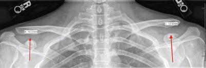
Fig. 23.2
Zanca view is the most accurate view to visualize the AC joint. This view is achieved by tilting the x-ray beam 10–15° cephalad and using one-half of the standard penetrance. Because of the significant variation in AC joint anatomy from one side to another, a bilateral Zanca view is recommended to visualize both AC joints on a single x-ray cassette while maintaining the same orientation of the x-ray beam. By visualizing both AC joints on the same cassette, the CC distance can be compared from side to side, pre- and postoperatively. The two arrows show the two acromion-clavicular joints. On the right is possible to observe a healthy clavicle. On the left an acromioclavicular dislocation
Magnetic resonance (MR) imaging represents a sensitive diagnostic tool in evaluation of ACJ disorders. Assessment of the stabilizing soft tissue structures involving the AC ligaments, CC ligaments, and delto-trapezoidal fascia is possible in a reliable manner, and its results can change the clinical grading of dislocation [20, 21].
MR imaging is also useful if surgery is considered to identify accompanying pathologies and again to identify underestimated injuries.
T1-weighted imaging best demonstrates the CC ligaments, and fat-suppressed proton density-weighted or T2-weighted imaging best demonstrates the ligamentous disruption, when surrounded by blood or fluid [20].
In the author’s clinical practice, a panorama stress view and axillary dynamic radiological evaluation represent the basic imaging tools, on which a therapeutic decision can be made in almost all cases. MRI is used only in selected cases, where associated glenohumeral soft tissue injuries are assumed.
23.4 Treatment Strategy
The goals of treatment for AC injuries are achieving painless range of motion of the shoulder, obtaining full strength, and exhibiting no limitation in activity. The treatment strategy varies according to the classification of the lesion.
Rockwood Type I
Sprains or partial tears at the ACJ are beyond all doubt treated nonoperatively [3, 5, 7, 17, 22]. Joint stability is maintained and ligament healing will occur in virtually all cases. Conservative therapy in terms of “skilful neglect” seems to be appropriate and sufficient. Occasionally, symptoms may appear between 6 months and 5 years, the 90 % are insignificant, reasonably well tolerated [38] and resolve within 12 months [23].
Rockwood Type II
General treatment recommendations are nonoperative for type II injuries [33, 5, 7, 22–24]. Similar to type I injuries, in most cases, symptoms disappear within 12 months [23]. Reasons for persistent complaints are residual instability, tearing of the intraarticular disk, articular cartilage injuries, residual joint incongruity, osteolysis of the lateral clavicle, and weakness [3, 5]. In case of type II injuries it’s possible to observe an increased anteroposterior translation in terms of horizontal stability. This may be a further explanation for persistent symptoms due to mis-/underdiagnosis of the initial injury degree.
Rockwood Type III
Operative treatment of grade III injuries results in a better cosmetic outcome but greater duration of sick leave compared to nonoperative management. No difference regarding strength, pain, throwing ability, and incidence of acromioclavicular joint osteoarthritis has been observed between the treatments. Current treatment recommendations favor surgical treatment in young patients with physically demanding occupations or sporting activities. The current scientific evidence seems to show rather a cosmetic advantage of surgical treatment than a functional one [3, 5, 6].
Rockwood Type IV
There is consensus in the literature that the treatment of type IV injuries should be surgical [3, 5]. The argument to treat even the most inactive patients, was the extremely high pain level, considering only closed reduction as sufficient therapeutic measure [7]. When considering the complete disruption of the AC ligaments and the detachment of the delto-trapezoidal insertion, closed reduction alone is not deemed to be sufficient, requiring surgical stabilization. Surgical treatment should focus on ACJ reduction, AC ligament fixation, and reconstruction of the delto-trapezoidal fascia. Obviously if the CC ligament complex is involved, its pathology has to be addressed as well.
Rockwood Type V
All stabilizing anatomical structures, including the CC and AC ligaments and the delto-trapezoidal fascia, are disrupted. The treatment should be operative [7] with reconstruction of the stabilizing structures including the delto-trapezoidal fascia. Thus, open procedures may be of advantage as compared to arthroscopic techniques, which usually fail to address adequately reconstruction of the delto-trapezoidal fascia.
Rockwood Type VI
This type of ACJ dislocation is quite rare and has been reported only in case reports [3, 15, 24, 25]. The treatment is always operative with reduction of the distal clavicle end and ACJ stabilization. Closed reduction may be difficult due to entrapment of the distal clavicle posterior to the conjoint tendon.
Related posts:

Stay updated, free articles. Join our Telegram channel

Full access? Get Clinical Tree


