Foot Disorders


Foot Disorders


Eric L. Kolodin

Thomas Vitale

Lynn H. Gerber


INTRODUCTION

Foot pain and problems are common among people of all ages. The prevalence has been reported as 15% to 40%, depending on the demographic distribution of the patient population (1, 2, 3). Foot disorders in the younger age group are frequently the result of trauma or congenital anomalies, and require a different approach to management than the elderly adult. The published literature on foot pain in the adult/aged population is quite extensive and indicates that it affects between 20% and 30% of community-dwelling elders (4). Some studies have shown that foot pain is associated with decreased ability to perform activities of daily living (5), problems with balance and gait (6, 7, 8) and increased risk of falls. Others have not found that to be the case (9). In fact, they reported that while there are a significant number of foot problems facing the elderly, only plantar fasciitis and a pes cavus foot profile are associated with functional foot problems. Another study has shown that elders with rheumatoid arthritis and who have significant foot pain are more likely to report back, hip, hand, and wrist pain. They also have pes planus and loss of motion in the ankle joint (10).

While it is useful to understand the issues of prevalence of foot pain and problems, and their associations with function, these must be understood within the context of the specific population (e.g., diagnostic group, age, etc.) of interest.


EXAMINATION


Physical Examination

The foot examination begins with an assessment of all the segments of the lower extremity. This should include measures of muscle strength, range of motion (ROM), alignment, stride characteristics, neurological examination (e.g., sensation, vibration and proprioception), vascular status, skin and nails, and review of shoe selection and wear patterns.


ANATOMY

Examination of the foot is challenging because of its complex anatomy, its biomechanical properties, and its function. This section attempts to describe important anatomic information that will enable the clinician to understand the affected anatomic site, but the reader is referred to books on anatomy, which present a more in-depth review (11, 12, 13).

Each foot is composed of 26 bones and 55 joints with muscle attachments that have their origin either within the foot or from the anatomical structures above the ankle. It is easier, therefore, to divide the foot into sections: the forefoot, midfoot, and rearfoot-ankle complex. This simplifies the examination process and provides the foundation necessary to understand biomechanical principles. The forefoot consists of 5 metatarsals and 14 phalanges constituting the five digits. The midfoot includes three cuneiform, cuboid, and navicular. The rearfoot-ankle complex is composed of the talus, calcaneus, and distal ends of the tibia and fibula (Fig. 38-1). These three divisions form a tenuous interrelationship that can easily be disrupted, resulting in dysfunction and pathology.

The hallux contains two phalanges, and the remaining four digits contain three each. The hallux has one joint: the interphalangeal joint. The lesser digits form the proximal and distal interphalangeal joints. There is a tendency for the intermediate and distal phalanges of the fifth toe to be fused together. The first metatarsal head distally articulates on the plantar surface with the tibial and fibula sesamoid bones, which are within the flexor hallucis brevis tendon. There are three cuneiforms that articulate with the base of metatarsals one through three. The cuboid articulates with the fourth and fifth metatarsal bases. The five metatarsals, along with the three cuneiforms and cuboid, form Lisfranc’s joint. Injury to this joint often is missed at initial presentation with significant associated morbidity. The keystone of this joint is the second metatarsal cuneiform articulation. The base of the fifth metatarsal, along with the lateral styloid process, serves as the insertion site of the peroneal brevis tendon. Injuries to the base of the fifth metatarsal and the styloid process are common, especially after inversion injuries of the ankle. The configuration of the articulation between the metatarsals, cuneiforms, and cuboid helps form the transverse metatarsal arch. Proximally, the cuneiforms articulate with the navicular, and laterally, the cuboid articulates with the navicular and lateral cuneiform. Medially, on the navicular, there is an enlarged tuberosity that serves as part of the insertion points of the tibialis posterior tendon. The navicular and cuboid proximally articulate with the talus and calcaneus, respectively, forming the midtarsal joint. The calcaneus is the largest bone of the foot. It functions as a major weight-bearing bone and serves as the insertion site posteriorly for the Achilles tendon. Many of the intrinsic muscles
arise from its plantar tubercles. The calcaneus is composed primarily of cancellous bone. Its dorsal surface articulates with the talus through three articulating surfaces—anterior, middle, and posterior—called facets. This forms the subtalar joint. A tunnel forms across the middle facet that begins laterally as the sinus tarsi and ends medially as the tarsal canal. Medially, the calcaneus forms a projection called the sustentaculum tali that provides a groove for the flexor hallucis longus tendon. The talus acts as a torque converter that connects the leg to the foot. It is almost completely covered with cartilage and has no muscular or tendinous attachments. Anatomically, it is divided into the head, neck, and body. The body, through its superior trochlear surface, the talar dome, forms the ankle joint with the distal extensions of the fibula and tibia, termed malleoli. The trochlear surface is wider anteriorly, which provides increased stability of the ankle in dorsiflexion. Plantar flexion of the ankle results in articulation, with the narrower portion of the posterior trochlear surface, resulting in intrinsic instability associated with the common ankle sprain. Fractures of the talus at its neck are associated with increased frequency of avascular necrosis owing to its tenuous but generous blood supply. Fractures of the talar body often result in posttraumatic arthritis. Posteriorly, the talus forms two tubercles—medial and lateral. The lateral tubercle has been called Steida’s process. Sometimes, the lateral tubercle fails to ossify completely; it is then termed the os trigonum.


FIGURE 38-1. Osseous anatomy of the foot.

Blood supply to the foot is the result of branches from the popliteal artery. The popliteal artery divides into the anterior tibial, posterior tibial, and peroneal arteries. The branches of these arteries are noted in Table 38-1. The venous drainage system of the foot is divided into deep and superficial segments. The lesser saphenous and greater saphenous veins form the superficial system, whereas the deep venous arch provides the deep drainage.


TABLE 38.1 Branches of the Anterior Tibial, Posterior Tibial, and Peroneal Arteries


Artery


Branches


Anterior tibial


Anterior medial and lateral malleolar


Medial and lateral tarsal Dorsal pedis


Arcuate, first dorsal and plantar metatarsal


Posterior tibial


Posterior medial malleolar


Medial and lateral plantar


Peroneal


Posterior lateral malleolar


Perforating peroneal


Motor and sensory nerves to the foot arise from branches of the sciatic nerve. The sciatic nerve divides into the tibial nerve, common peroneal nerve, and sural nerve. The common peroneal nerve divides into the superficial and deep peroneal nerves. They provide sensory input to the dorsal aspect of the foot and digits. The sural nerve innervates the lateral aspect of the foot. The saphenous nerve innervates the medial aspect of the foot and is a branch of the femoral nerve. The motor component of the peroneal nerve innervates the peroneus longus, peroneus brevis, extensor hallucis longus, extensor digitorum longus and brevis, tibialis anterior, and peroneus tertius. The tibial nerve divides into the medial and lateral plantar nerves. They provide sensory and motor innervation to the plantar aspect of the foot.

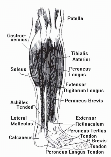
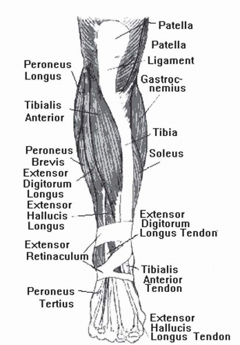
The muscles of the foot are divided into extrinsic and intrinsic groups (Figs. 38-2 and 38-3). The extrinsic muscles arise in the leg and insert into the foot and are held in place by various retinacula as they enter the foot. They are divided into four compartments: superficial posterior, deep posterior, lateral, and anterior (Table 38-2). The intrinsic muscles of the foot are divided into dorsal and plantar groups. The plantar muscles are divided into four layers, with the first layer being the most posterior or superficial and the fourth layer the most anterior or deep (Table 38-3).


Functional Anatomy

The foot is divided into three functional units or parts: the hindfoot (talus and calcaneus and its attachments) provides the foot with stability; the midfoot (navicular, cuboid, cuneiforms, Lisfranc’s and Chopart’s joints) provides both sagittal and frontal plane motion; and the forefoot (metatarsals and phalanges) permits push-off. The ligamentous structures establish and preserve the longitudinal arch, the intermetatarsal transverse arch, the position of the sesamoids, and stability of the ankle mortise.


FIGURE 38-2. Lateral view of the intrinsic and extrinsic muscles of the foot.

The subtalar joint is composed of the talus and calcaneus. The bones articulate at the posterior, middle, and anterior facets. The joint is responsible for triplane motion, primarily pronation and supination. There is 20 degrees of inversion to 10 degrees of eversion to the subtalar joint. In closed kinetic chain pronation, the calcaneus everts, with adduction
and plantar flexion of the talus. The opposite is true for closed kinetic supination (Fig. 38-4).


TABLE 38.2 Extrinsic Muscles of the Foot


Extrinsic


Muscle Groups


Muscles


Function


Superficial posterior


Gastrocnemius


Plantar flexion


Soleus


Plantar flexion


Deep posterior


Tibialis posterior


Plantar flexion


Flexor digitorum longus


Plantar flexion


Flexor hallucis longus


Plantar flexion


Lateral


Peroneus longus


Eversion


Peroneus brevis


Eversion


Anterior


Tibialis anterior


Dorsiflexion


Extensor hallucis longus


Dorsiflexion


Extensor digitorum longus


Dorsiflexion


Peroneus tertius


Dorsiflexion


FIGURE 38-3. Anterior view of the intrinsic and extrinsic muscles of the foot.


TABLE 38.3 Intrinsic Muscles of the Foot


Intrinsic


Muscle


Groups


Muscle


Function


Dorsal


Extensor digitorum brevis


Extension of toes


Extensor hallucis brevis


Extension of hallux


Plantar


Layer 1


Abductor hallucis


Abduction of hallux


Flexor digitorum brevis


Flexion of toes


Abductor digiti minimi pedis


Abduction of fifth toe


Layer 2


Quadratus plantae


Helps in flexion of toes


Lumbricles (4)


Flexion MTPJ/ extension IPJ


Tendons of long flexors


Layer 3


Flexor hallucis brevis


Flexion of hallux


Adductor hallucis


Adduction of hallux


Flexor digiti minimi brevis


Flexion of fifth toe


Layer 4


Plantar interossei


Adduction of toes


Dorsal interossei


Abduction of toes


Tendons of peroneal longus and tibialis posterior


IPJ, interphalangeal joint; MTPJ, metatarsal phalangeal joint.


FIGURE 38-4. Abducted stance with “too-many-toes sign.”

The position of the subtalar joint dictates the position of the midtarsal joint. When the subtalar joint supinates, the arch raises the forefoot, and the foot becomes more rigid. The midtarsal joint “locks.” When there is subtalar joint pronation, the midtarsal “unlocks” and becomes less rigid. The foot becomes a mobile adaptor, allowing for increased motion to take place to accommodate for varying terrain.

The midtarsal joint is made up of the calcaneocuboid and talonavicular joints. The midtarsal joint has two axes of motion: the longitudinal and oblique axes. The longitudinal axis inverts and everts the forefoot while pronation or supination occurs. The oblique axis allows for adduction, abduction, plantar flexion, and dorsiflexion of the foot.


Physical Examination of the Foot

ROM assessment of the lower extremity is important because absence of normal range has functionally important sequelae. For example, a tight Achilles tendon, which limits ankle dorsiflexion, creates a force anterior to the axis of the ankle, which results in pronation, or collapse of the medial column. Limitation of hallux dorsiflexion to less than 25 degrees creates an impediment to forefoot push-off.

When testing ROM for the ankle, there should be at least 10 degrees of dorsiflexion with 45 degrees of plantar flexion. When testing the range of dorsiflexion, the examiner should hold the foot medially. This keeps the midtarsal joint locked, and a more accurate measurement can be achieved. Dorsiflexion is measured with the knee extended and flexed. When the knee is flexed, there is more dorsiflexion of the ankle because of the “unlocking” of the gastrocsoleus complex. The ankle motion should be smooth and unrestricted. The subtalar joint should measure 20 degrees of inversion and 10 degrees of eversion. This can be measured with the patient in the prone position. The heel is bisected in half, and the examiner can then use a tractograph and invert and evert the foot for the measurement. The metatarsophalangeal joints, the distal and proximal interphalangeal joints of the lesser toes, and the interphalangeal joint of the hallux should have smooth and unrestricted dorsiflexion and plantar flexion.

Muscle strength testing provides important information about whether the foot is likely to be supported in stance (static) and dynamic phases of gait.

Support of the foot in stance comes from the ligaments and plantar fascia, which stabilize the longitudinal arch and the intrinsic muscles of the foot. The creation and establishment of the longitudinal arch in the dynamic phase depend on the posterior tibialis and intrinsic and extrinsic muscles of the foot. The gastrocsoleus provides propulsion through sagittal plane motion at the ankle. Weakness in these structures may help explain an apropulsive gait or instability during stance.

Determination of static alignment of the foot provides important information about how the foot will contact the ground. The preferred position is that the foot should be parallel to the floor. If ligamentous structures are intact, ROM abnormality or muscle imbalance is likely to explain alignment problems. Examination demonstrates possible forefoot varus or valgus deformity. This is performed with the patient in the prone position with the subtalar joint neutral.

The feet should be evaluated in the standing position, and the arch height is evaluated as flat, low, normal, or high, based on the navicular position. The angle of the talus measured radiographically may be used to assess arch height. Bisecting the Achilles tendon will demonstrate whether the calcaneus is everted or inverted with respect to the midline. A varus rearfoot generally causes pressure to the posterosuperior lateral aspect of the calcaneus. A bone spur can develop in this area and cause pain. This is known as a Haglund’s deformity. Severe calcaneoeversion can cause a lateral impingement and pain to palpation below the lateral malleolus.

The vascular exam will evaluate peripheral pulses in the foot. Dorsally, there is a dorsalis pedis pulse, and medially a posterior tibial pulse. If one cannot feel a pulse and there is questionable vascular compromise, the physician can order further vascular testing such as noninvasive arterial Doppler. This exam would be beneficial when dealing with lower extremity ulcerations. Capillary filling time is tested and should be within 2 seconds. Press the distal tip of the digit and determine how fast the digit fills up with blood. Another area of testing would be for dependent rubor. Dangle the foot off the examining table and see if the foot becomes more red or purple. If this occurs, there may be pathology to the venous system of the lower extremity. Any varicosities should also be noted to the lower extremities, as well as chronic swelling. Patients with brawny edema from venous stasis disease may develop ulcerations to the medial lower leg or ankle.

The neurological exam will test deep tendon reflexes, ankle clonus, Babinski’s sign, sharp and dull discrimination, as well as vibratory sense and proprioception. Patients who are neuropathic have diminished sensation, vibratory sense,
and lack of proprioception. It is useful to test for protective sensation using Semmes-Weinstein filaments. Those who cannot feel the touch of a nylon filament 5 mm in diameter are at significant risk for ulceration. They are at risk for ulcerations because they lack sensation to the foot and do not feel the pressure caused by a foreign body, minor cut, abrasions from poorly fitted shoes, or thickened calluses.

Examination of the skin indicates where pressure points are. Callosities are in areas of high pressure and often need to be relieved.


DIAGNOSTIC TESTING

The proper selection of imaging techniques enhances the ability to diagnose foot disorders. When dealing with foot pain, one should be knowledgeable about selecting from among plain radiographs, computerized tomography, magnetic resonant imaging (MRI), and ultrasound. The application of diagnostic ultrasound has offered clinicians and clinical investigators opportunities to increase the sensitivity and specificity of patient evaluation and assessment of treatment outcomes.

A good review of which diagnostic tools are best applied to which clinical conditions is available for the reader (14). A summary table of recommendations (see Table 38-1) is given.

Ultrasound has recently received attention because of its usefulness in clinical settings. Sonography has been shown to provide valuable information about soft tissue, including muscle, fascia, tendon, blood vessels, and nerves. Ultrasound is convenient, low cost, and low risk and may be particularly useful in helping to diagnose and follow problems associated with plantar fasciitis and posterior tibialis tendon dysfunction, significant clinical problems (14, 15).


BIOMECHANICS

Foot motion depends on intrinsic and extrinsic muscles that are divided into four compartments (see Table 38-2). These muscles produce sagittal, frontal, and transverse plane motion.

To understand foot biomechanics, the physician must be proficient in the terms used to describe the motion and position of the foot. A list of terms commonly used follows:

Abduction: movement of the foot away from the midline of the body

Adduction: movement of the foot toward the midline of the body

Eversion: turning inward of the feet with the soles pointing away from each other

Inversion: turning inward of the feet with the soles pointing toward each other

Dorsiflexion: motion whereby the distal end of the foot moves toward the tibia

Plantar flexion: motion occurring to the foot whereby the distal end of the foot moves away from the tibia

Pronation: triplane motion of the foot in the direction of abduction, eversion, and dorsiflexion

Supination: triplane motion of the foot in the direction of adduction, inversion, and plantar flexion

Subtalar joint: point at which the normal foot is neither inverted nor everted

Neutral position: from the floor during stance; clinically, whereby the posterior aspect of the calcaneus lines up with the lower one third of the leg

Forefoot varus: inversion to the plantar surface of the forefoot with the subtalar joint neutral and the midtarsal joint pronated; this is a congenital bony deformity

Forefoot valgus: eversion to the plantar surface of the forefoot when the subtalar joint is neutral and the midtarsal joint is pronated; the plane of the metatarsals are everted with the first metatarsal lower than the fifth metatarsal; the first metatarsal may be flexible or rigid; this is a congenital bony deformity

Rearfoot varus: inverted position of the sagittal plane and the plantar surface of the calcaneus to the weight-bearing surface with the subtalar joint in the neutral position and the body standing in normal base of gait (10 degrees to 15 degrees of abduction)

Rearfoot valgus: the sagittal plane of the posterior and plantar surface of the calcaneus are everted to the weight-bearing surface when the subtalar joint is in neutral position while the body is standing in base of gait

Often, with increased subtalar joint pronation, the foot becomes hypermobile, and increased stress and strain are placed on the lower extremities. A functional valgus occurs at the knee, causing increased stress to the medial aspect of the knee. Hyperpronation causes increased strain on the plantar fascia, causing a pull on the origin of the fascia of the calcaneus. Patients are predisposed to bunions, hammertoes, and postural fatigue with increased subtalar joint pronation. Posterior tibial and Achilles tendinitis can occur with a hypermobile foot because of the increased stretching to the tendon.

A supinated foot or high-arched foot is much more rigid. This type of foot structure predisposes the patient to lateral ankle sprains, lateral Achilles tendinitis, claw toes (flexion deformity at the proximal and distal interphalangeal joints), increased incidence of avulsion fractures to the base of the fifth metatarsal, peroneal subluxation, metatarsalgia, and sesamoid pain or fractures.


MECHANICAL DYSFUNCTION


Posterior Tibialis Tendinitis


Anatomy and Pathomechanics

The posterior tibial tendon has its origin from the lateral posterior aspect of the tibia, medial aspect of the body of the fibula, and interosseous membrane of the leg (16). The tendon courses under the medial malleolus and into the foot, with
most of the tendon inserting into the navicular tuberosity. The remaining smaller slips of the tendon insert into all tarsal bones except the talus.

The posterior tibial tendon is a plantar flexor and invertor of the foot and a strong supinator of the subtalar and midtarsal joints. The tendon dynamically supports the medial longitudinal arch (17). The peroneus brevis tendon is the antagonistic tendon to the posterior tibial tendon (18), which is an adductor at the level of the midtarsal joint (19).

When there is weakness of the posterior tibial tendon, the foot becomes more abducted because of the overpowering effect of the peroneus brevis (20). When the posterior tibial tendon weakens, the midtarsal joint does not lock, causing increased pronation at the subtalar joint. In time, the medial longitudinal arch collapses, and there is increased shock at the rearfoot, causing a decrease in the propulsive activity of the foot (19).


Etiology

Posterior tibial tendon ruptures may be traumatic or degenerative in nature. Acute traumatic ruptures are not common, but may occur from puncture wounds, lacerations to the medial ankle (21), and severe eversion ankle sprains (22). Most cases of dysfunction are degenerative (16), such as inflammatory arthritis (23). Diabetes mellitus, obesity, and prior local steroid exposure have been known to cause a degenerative process to the tendon (14). Tendon hypovascularity at the midportion of the tendon just distal to the medial malleolus has been known to cause ruptures to the posterior tibial tendon (14). Abnormal biomechanical forces (equinus, pes valgus) may cause chronic tenosynovitis and weakening of the posterior tibial tendon (15).

A classification system for dysfunctional posterior tibial tendon etiologies has been formulated by Mueller and is based on four categories (19). Type I is a direct rupture due to direct injury to the tendon. Type II is a result of a systemic disease such as diabetes or inflammatory arthritis. Type III is idiopathic or possibly degenerative. Type IV is functional and may be related to severe pronation or stretching of the tendon.


Clinical Findings

The clinical concerns of the patient may depend on the chronicity of the deformity. In the acute stage, there is swelling and pain along the course of the posterior tibial tendon. The patient may experience the symptoms as an ankle injury (16). In the chronic stage, the patient notices a change in the appearance of the foot. There may be a collapse of the arch, or “too many toes” sign, whereby the toes are pointing outward as the patient is seen from behind (see Fig. 38-4). The patient’s gait becomes increasingly apropulsive, with limited heel lift and toe off. There is excessive wear on the medial aspect of the last of the shoe. The patient demonstrates little or no inversion power with resistance. There may be a palpable gap in the tendon itself. It is our opinion that the tear is most noticeable just proximal and posterior to the medial malleolus. Jahss described tears occurring about 1½ in. proximal to the navicular (24). In the advanced chronic patient, there may be pain of the lateral foot and ankle, specifically in the sinus tarsi. There may be lateral ankle impingement with involvement of the calcaneofibular ligament, creating an ankle valgus deformity. A long-standing valgus deformity of the ankle can produce a valgus force on the knees, creating pain and deformity to the knees. In the chronic patient, there might be crepitus, with painful ROM of the ankle and subtalar joint. There may be arthritic changes noted on x-ray to the ankle or subtalar joint.


Diagnostic Imaging

Standard radiographs do not demonstrate a tendon tear but distinguish any adaptive changes to the subtalar or ankle joints. Standard weight-bearing dorsoplantar, oblique, and lateral x-rays should be ordered in patients displaying pain and weakness in the posterior tibial tendon. In long-standing deformities, there are arthritic changes in the subtalar and ankle joints. MRI is one of the most accurate tests that can depict the extent of pathology of the tendon (25). CT can depict the condition of the tendon; however, the definition of tendon outline is not as clear. CT scans can be helpful in identifying osseous deformities such as degenerative arthritis and subtalar joint dislocations. MRI aids the practitioner with excellent resolution of the image regarding tendon outline, synovial fluid, and vertical splits in the tendon. Axial and sagittal images demonstrate tendon pathology well (26). The axial view helps delineate tendon girth and shape, fluid accumulation, and signal changes within the tendon itself (27). Sagittal images allow the practitioner to view the extent of the pathology longitudinally. Coronal images are not as helpful in delineating posterior tibial pathology (16). Ultrasound is useful for describing the course of the tendon and can identify whether there is a tear, tendinitis, tendinosis, and peritendinitis (28, 29).

In the axial view, the tendon is normally round or oval and is twice the size of the flexor digitorum longus tendon. In the normal tendon, there should be no accumulation of fluid surrounding the tendon (26). A tear on the axial image indicates mixed signal intensities, with the size of the tendon increasing or decreasing, depending on the extent of the tear.


Treatment

Conservative treatment is reserved in those patients who are older or who lead sedentary lives. Conservative modalities include removable walking casts (Cam walkers), custom foot orthotics (to control poor biomechanics), braces, ankle-foot orthotics, orthopedic shoes with medial heel wedges, and compression stockings (16). The goal for conservative treatment is to reduce the pain and inflammation and to delay the progression of the deformity. If conservative treatment fails, surgical options should be explored. Surgery in the early phase before significant tear may include a synovectomy to the tendon sheath. After a tear, tendon transfers (flexor digitorum longus) or an isolated fusion of the subtalar joint or triple arthrodesis (fusion of subtalar and midtarsal joints) may be required.


Achilles Tendinitis


Anatomy

The Achilles tendon is composed of the gastrocnemius and soleus muscles (triceps surae). It forms a spiral configuration that inserts into the central one third of the posterior calcaneus. A retrocalcaneal bursa can be found between the calcaneus and Achilles tendon. The Achilles tendon is covered by a paratenon. This paratenon is a loose elastic tissue that stretches with the Achilles tendon. The central layer of the paratenon (mesotenon) is responsible for the tendon’s blood supply. The action of the tendon is to decelerate and stabilize the foot, and then accelerate the foot during the gait cycle (16).


Mechanism of Injury

Poor foot and ankle mechanics and overuse can predispose the patient to injury. A fixed forefoot equinus results in compensation at the ankle joint. The ankle compensates with dorsiflexion, allowing the forefoot and rearfoot to remain on the ground during the midstance phase of gait. This may result in an overuse injury to the tendon. Compensation at the subtalar joint for any forefoot to rearfoot imbalance places strain on the Achilles tendon. If the patient has a high forefoot varus deformity, the subtalar joint may compensate by everting the calcaneus (hyperpronation) and placing more load on the medial side of the Achilles tendon. A rigid plantar-flexed first metatarsal or cavovarus places strain on the lateral (hypersupination) side of the Achilles tendon (16).


Physical Findings and Treatment

There are three distinct areas of pathology to the Achilles tendon: the insertion site of the tendon into the posterior one third of the calcaneus with or without bone involvement, the midportion (vulnerable zone) with peritendinitis or partial or total rupture, and the myotendinous junction (30). Most injuries are related to overuse or poor foot biomechanics. The patients will give a history of increased activity or intensity to their workouts or beginning an exercise program after a period of inactivity.

Insertional Achilles tendonitis may result from a bony abnormality to the calcaneus. Patients describe their Achilles as stiff in the morning. A bony prominence to the posterior superior lateral aspect of the calcaneus (Haglund’s deformity) or insertional calcification may cause pain to the insertion site. This is quite evident in certain shoes rubbing on the area. Surgical management of this problem would be the last resort.

The patient may begin home treatment consisting of rest, ice, anti-inflammatory medication, night splints, deep transverse friction massage, and a ¼-in. heel lift to the shoes. Shoes with a lower heel counter or sneakers with a U-shaped Achilles notch to the back of the heel counter might help reduce the inflammation to the area. Stretching and strengthening exercises for the posterior leg and hamstring muscles will allow the Achilles tendon to function more efficiently. An ankle dorsiflexion stretch held for 30 seconds 10 times twice a day may help with the symptoms of pain. Home strengthening exercises using TheraBand or toe raises can be beneficial as well.

Physical therapy might be helpful in cases in which the patient is not improving on his or her own along with a removable walking cast (Cam walker). Therapy may include ultrasound, iontophoresis, deep transverse friction massage, and stretching and strengthening exercises. It is our recommendation not to inject cortisone into the Achilles tendon. The crystals of the cortisone may weaken and rupture the tendon.

Midportion pain to the tendon presents with fusiform swelling. This area is vulnerable to rupture because of the diminished blood supply. MRI is helpful in determining the extent of injury to the area. Treatment includes physical therapy (deep transverse friction massage, ultrasound, stretching exercises, and ice). Cast immobilization may be necessary in the beginning to help rest the area. We prefer removable walking casts such as a Cam walker. Home exercises for the patient should include stretching the Achilles, with ice and massage to the area.

Myotendinous junction injuries are not serious and heal readily with rest and physical therapy. In the more acute or chronic cases, a walking cast may be needed for pain relief. This type of injury heals within 4 to 6 weeks.


Peroneal Tendinitis


Anatomy

The peroneus brevis originates from the lateral distal two thirds of the fibula and inserts into the lateral base of the fifth metatarsal. The peroneus brevis is the strongest abductor of the foot as well as a flexor to the ankle and everter of the foot (31). The muscle stabilizes the lateral segment of the foot, specifically at the calcaneocuboid joint.

The peroneus longus muscle originates at the proximal two thirds of the lateral fibula. The tendon passes inferior to the lateral malleolus and under the cuboid and courses medially under the foot to insert into the lateral plantar base of the first metatarsal and first cuneiform. The peroneus longus is a plantar flexor and everter of the foot. The tendon at its insertion helps stabilize the first metatarsal.


Mechanism of Injury

Injuries to the tendon may occur traumatically or insidiously from poor foot biomechanics. Forced inversion sprains may cause pain to the peroneal tendons. A patient who has a supinated foot is predisposed to this type of injury because there is increased stress to the tendons on the lateral ankle.


Clinical Findings and Treatment

Pain to the peroneal tendons may be present inferior to the lateral malleolus, at the peroneal tubercle, or at the peroneal groove under the cuboid where the peroneus longus courses medially to insert at the lateral base of the first metatarsal and first cuneiform. Peroneus brevis tendonitis may also be present at its insertion point on the lateral base of the fifth metatarsal. Passive dorsiflexion of the first metatarsal may be helpful in determining tendonitis of the peroneus longus. Passive inversion of the calcaneus with adduction of the forefoot is a good way of testing for peroneus brevis tendinitis.


Peroneal tendinitis may occur after an inversion injury to the ankle. Patients relate pain to the ankle, with pain inferior to the lateral malleolus or at the insertion site of the peroneus brevis. There may be swelling over the course of the tendons. Athletic patients relate pain to the peroneus longus tendon with increased cutting or turning or when getting up on the ball of the foot. Pain is noted under the cuboid or on the lateral aspect of the calcaneus.

Treatment is directed at reducing the inflammation and strengthening the muscles. Rest, anti-inflammatory medication, and ice help in the acute stage of the problem. Ice pops can be made by filling a cup with water, placing a stick inside, and freezing it. Deep transverse friction massage may help break up some of the scar tissue in the area. Eversion with resistance exercises and TheraBand help strengthen the peroneal muscles.

If biomechanical abnormalities are involved, custom foot orthotics can be fabricated. Orthotics for walkers can be of three-quarter length. For the active patient, a full-length orthotic device with a long forefoot runner’s post helps control the foot not only at heel strike and midstance but also at pushoff. Posting of the orthotic in the rearfoot and forefoot helps keep the foot more balanced.

If the patient does not respond to conservative treatment, surgical intervention might be considered. Surgery would entail a tenosynovectomy. If the peroneal tendons are subluxing out of the groove under the lateral malleolus, the groove is usually deepened.


Plantar Fasciitis


Anatomy

The plantar fascia is a strong aponeurosis that originates on the medial process of the calcaneal tuberosity and fans out into a medial, central, and lateral slip. The fascia blends in with the flexor plate distally and has a connection with the plantar aspect of the toes. The central slip is the thickest, the lateral slip acts as a covering to the abductor digiti minimi muscle, and the medial slip acts as a covering to the abductor hallucis muscle (13).

The plantar fascia acts as a windlass and pulls the arch up with dorsiflexion of the metatarsophalangeal joints (32). Poor foot biomechanics can lead to increased tension on the fascia, causing inflammation and pain. This can occur with patients who have increased subtalar joint pronation, pes planus, and limited dorsiflexion. The increased pronatory forces stretch the plantar fascia and cause a pulling at the origin. This, in turn, causes pain and inflammation. Patients with a pes cavus foot type are also prone to this condition. These patients have a more rigid foot type and cannot absorb shock as well at heel strike, and that places more stress on the plantar fascia (33).


Clinical Findings

Patients typically describe their pain after getting out of bed in the morning or after a period of inactivity. They state that the pain decreases after walking on the foot for a while (34). Most patients tolerate the condition before seeking medical help.

On clinical exam, there is tenderness on the medial plantar aspect of the heel. Some patients may have discomfort under the midcalcaneal region of the heel. There may be tightness of the Achilles tendon insertion along with tightness to the medial slip of the plantar fascia. Poor foot biomechanics such as a pes cavus or planus deformity might be present, along with a compensated forefoot varus deformity. Upon x-ray exam, there may be a calcaneal heel spur forming parallel to the weightbearing surface. The heel spur is not pathognomonic for this condition. The spur does not cause the pain to the heel. The heel spur is the by-product of the chronic pulling of the fascia off the calcaneus (35). Some patients might describe pain to the lateral ankle or dorsolateral aspect of the foot. This is probably related to the patient offloading the pressure to the heel and walking on the outside of the foot. This creates an overuse condition to the lateral foot and ankle structures.

Most patients respond favorably to conservative treatment. The goals of treatment are to reduce the pain and inflammation and to correct poor foot biomechanics. We prefer to treat this condition in a stepwise approach.

The first level of treatment is a simple stretching exercise for the Achilles tendon and plantar fascia. The patient stretches the Achilles tendon while still in bed with a large towel. The patient dorsiflexes the foot and holds that stretch for a minimum of 30 seconds 10 times. Stretch to the plantar fascia is done by dorsiflexing toes, holding the metatarsophalangeals, and stretching fascia in the arch region. During the day, the patient may stretch by leaning against a wall. Again, each stretch is held for a minimum of 30 seconds with the knees straight and the heels on the ground. The patient may roll the arch over a soup can or golf ball. Ice and deep transverse friction massage may also be beneficial, as may taping of the foot (35). Over-the-counter foot orthotics is also recommended. Night splints are beneficial to help keep a stretch on the posterior leg muscles and plantar fascia.

If the pain has not improved, physical therapy, cortisone injections, custom foot orthotics, and supportive shoes are discussed with the patient. Shoes should consist of adequate depth to accommodate an orthotic and should have good heel and medial countersupport. If a cortisone injection is indicated, we prefer injecting the heel with a 3-mL syringe and a 25-gauge, 1.5-in. needle. The steroid of choice is 0.5 mL of Celestone Soluspan or Depo-Medrol with 1.5 mL of 2% lidocaine (Xylocaine) plain. The practitioner should palpate for maximum tenderness and inject from a medial approach. The patient should not receive more than three steroid injections to the same area over the course of 1 year. Multiple injections to the same site can weaken the soft tissue to the area. Injections through the plantar aspect of the foot should be avoided because they are painful to the patient.

A fairly new method of treatment is extracorporeal shock wave therapy. This is a noncutting procedure performed under anesthesia (local anesthesia with intravenous sedation). It is not fully understood how this promotes healing; however, the use of electrohydraulic shock waves converts a chronic condition into an acute condition (36). Patients who do not want the traditional or endoscopic surgical procedure may consider this alternative because of the lower risks attached to the procedure.
The current standard before considering this type of procedure is 6 months of conservative care. Several studies report benefit in controlled, randomized trials without adverse effects up to 1 year following treatment (26, 27).


Hallux Abducto Valgus


Etiology

Hallux abducto valgus occurs because of the positional and structural changes that take shape as a result of hypermobility of the first ray. It is our opinion that hallux abducto valgus is a hereditary condition caused by poor foot biomechanics. In the pronated foot, the stability of the peroneus longus is lost and the first ray becomes dorsiflexed and adducted. This creates an increase in the intermetatarsal angle between the first and second metatarsals. Finally, intrinsic instability occurs around the first metatarsophalangeal joint and the fibular sesamoid drifts laterally, creating lateral deviation of the great toe (37).


Clinical Findings and Symptoms

Hallux abducto valgus is a progressive deformity that worsens with time. Shoes contribute to the pain but not to the deformity. The patient may have pain on the dorsomedial aspect of the first metatarsophalangeal joint. There may be erythema and a small bursa noted at the medial aspect of the joint. The patient usually has a pronated foot with decreased arch height. Patients tell the practitioner that they recently noticed their “bump” has enlarged over a short period of time. X-rays reveal an increase in the intermetatarsal angle and lateral deviation of the sesamoids, with lateral deviation of the great toe. There may not be joint pain or decreased ROM to the joint.


Treatment

Treatment is designed to reduce the pain and to control the pronation that is causing the condition. Conservative treatment is the best initial approach. Although conservative treatment will not remove the enlarged bony prominence, the pain may decrease. Weight-bearing x-rays (anteroposterior, lateral, and medial oblique views) help the physician determine the extent of the deformity.

Ice and oral anti-inflammatory medications may help reduce inflammation. A local cortisone injection to the medial aspect of the joint may relieve pain. Over-the-counter or custom foot orthotics designed to control the biomechanical forces can help reduce pain and help realign the foot to move in a more efficient manner. If conservative therapy fails, surgical intervention is discussed. When the pain interferes with normal daily activities or if shoeing is a significant problem and conservative options have been exhausted, then surgery is considered.

There are a variety of surgical procedures, ranging from first metatarsal head and base osteotomies to fusions of the first metatarsophalangeal joint or first metatarsocuneiform joint. The goals in surgical intervention are to decrease pain, improve function, and establish a congruous joint, reduction in the intermetatarsal angle, realignment of the sesamoids, maintenance of first metatarsophalangeal joint ROM, and repositioning of the hallux to a rectus position (37). It is preferable to place patients in custom-made foot orthotics after surgery to control the factors that led to the development of this condition.


Hallux Limitus and Hallux Rigidus

Hallux limitus is mild to moderate decreased ROM to the first metatarsophalangeal joint. Hallux rigidus is severe limitation of motion. Either condition may or may not present problems to the patient. There have been cases (eliminate reference to personal practice) in which the patient has no motion to the joint but is pain free. The etiology of this condition may be traumatic or biomechanically induced.


Joint Mechanics

Sixty to seventy-five degrees of first metatarsophalangeal joint dorsiflexion is necessary during the gait cycle. During the first 20 degrees, the first metatarsophalangeal joint acts as a ginglymoarthrodial joint. The motion is hingelike in nature. As the heel lifts, the ground reactive forces dorsiflex the hallux and the first metatarsal head plantar-flexes. This allows for closed-chain dorsiflexion of the first metatarsophalangeal joint beyond the 20 degrees (16).

The distal end of the first metatarsal head articulates with the sesamoid apparatus, and the base of the proximal phalanx glides along the dorsal aspect of the first metatarsal head. If the sesamoid apparatus is bound down and immobile, there is restriction of dorsiflexion to the joint. Ultimately, there is impingement to the joint causing degenerative changes resulting in arthrosis and remodeling of the joint over a period of time (37).


Etiology

A variety of causes can contribute to limitation of motion to the first metatarsophalangeal joint. Biomechanical causes may be broken down into a long first metatarsal, long proximal phalanx, dorsiflexed metatarsal, or hypermobility of the first ray. Trauma or postsurgical bunion surgery may be another cause of this condition.

A long first metatarsal can cause jamming to the joint during the propulsive phase of gait. This causes degenerative changes to the joint. Hypermobility of the joint occurs with weakness to the peroneus longus. The peroneus longus inserts into the base of the first metatarsal and first cuneiform. Without proper first metatarsal plantar flexion, the head of the first metatarsal does not articulate with the sesamoids and the metatarsal dorsiflexes, causing limitation of motion to the joint. Congenital elevatus of the first metatarsal does not allow the first metatarsal to plantar-flex with propulsion, leading to degenerative changes (37). Acquired elevatus is seen after bunion surgery, specifically closing base wedge osteotomies of the first metatarsal. A complication to this type of bunion procedure is dorsiflexion of the first metatarsal.

Only gold members can continue reading. Log In or Register to continue

Stay updated, free articles. Join our Telegram channel

May 25, 2016 | Posted by in PHYSICAL MEDICINE & REHABILITATION | Comments Off on Foot Disorders

Full access? Get Clinical Tree

Get Clinical Tree app for offline access