Chapter 1
Foot and Ankle Examination
Foot and ankle examination means rule of three!
Precise clinical examination of foot and ankle is the first step toward successful treatment of foot and ankle ailments. Three important rules to be followed by the examiner are listed in Box 1.1.
Box 1.1 Rules for the examiner.
♦ Gait examination
♦ Foot and ankle examination
♦ Footwear examination
Similarly, there are three important rules to be followed by the patient for proper evaluation, which would allow the examiner to interpret the condition of the disease correctly. Three important rules meant for the patient are listed in Box 1.2.
Box 1.2 Rules for the patient.
♦ Enters walking
♦ Enters with footwear
♦ Enters with trousers up
Gait Examination
For proper examination of the gait, the following points are to be observed:
♦ Symmetry of parts
♦ Toe versus patella position
♦ In or out toeing
♦ Arches
♦ Foot posture and position
♦ Heel posture and position
♦ Type of gait
Foot and Ankle Examination
Examination should be performed in three positions. Box 1.3 illustrates the three rules for patient’s positions during examination. Examination of the foot is divided into three parts (Box 1.4) and is performed in three basic steps, which are listed in Flowchart 1.1.
Box 1.3 Rules for patient’s positioning during examination.
♦ Examination while walking
♦ Examination while standing
♦ Examination while sitting
Box 1.4 Parts of examination of the foot.
♦ Ankle and hindfoot examination
♦ Midfoot examination
♦ Forefoot examination
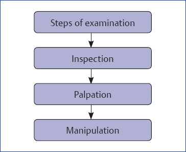
Flowchart 1.1 Steps of examination.
Inspection
Consider the following while inspecting foot and ankle:
♦ Alignment and position of foot with respect to ankle as well as alignment of ankle and foot with respect to leg, knee, and hip
♦ Shape and size of the foot
♦ Deformities, deviations, and prominences
♦ Skin
♦ Callosities and corns
♦ Varicosities
♦ Nails and hair
Palpation
Palpation is done by using the index finger, either self or that of the patient (Fig. 1.1), beginning sequentially from the medial aspect to the plantar, to the lateral, followed by the anterior, and finally ending at the posterior aspect.
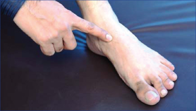
Fig. 1.1 Patient pointing at the painful area with his index finger.
The three important parts of palpation are mentioned in Box 1.5.
Box 1.5 Parts of palpation.
♦ Topographical palpation
♦ Neurological palpation
♦ Vascular palpation
For the right foot, start clockwise, and for the left foot start anticlockwise (Figs. 1.2 and 1.3).
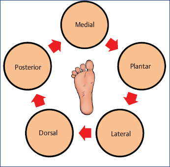

Fig. 1.3 Palpation of the left foot.
Topographical Palpation
Structures to be palpated in sequence are as follows:
♦ Skin
♦ Bone and joints
♦ Ligaments
♦ Muscles and tendons
♦ Arches
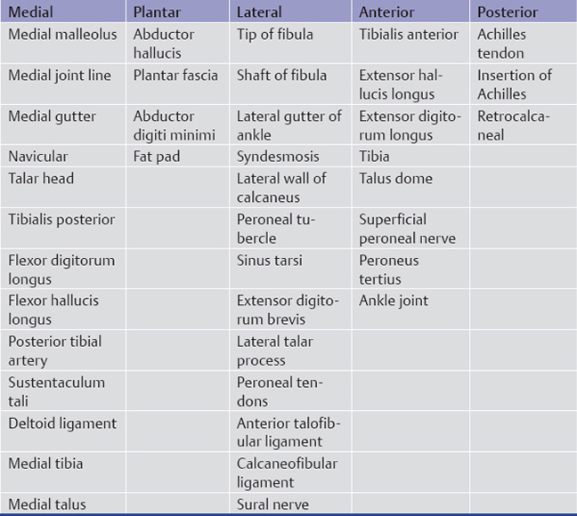
Topographical palpation areas of (1) ankle are depicted in Figs. 1.4 to 1.8 and are described in Table 1.1; (2) midfoot are depicted in Figs. 1.9 and 1.10 and are described in Table 1.2; (3) forefoot are depicted in Figs. 1.11 and 1.12 and are described in Table 1.3.
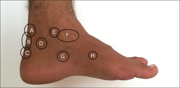
Fig. 1.4 Medial aspect of the foot and ankle. A, Achilles rupture/noninsertional tendonitis; B, insertional Achilles tendonitis; C, calcaneal apophysitis/pump bump; D, retrocalcaneal bursitis; E, tarsal tunnel syndrome/ tibial posterior tendon; F, medial/deltoid ankle sprain; G, entrapment of first branch of lateral plantar nerve; H, accessory navicular/master knot of Henry/medial plantar nerve entrapment.
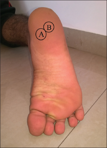
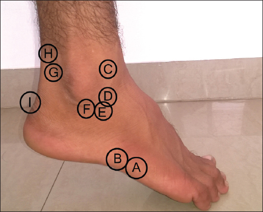
Fig. 1.6 Lateral aspect of the foot and ankle. A, Jones fracture; B, avulsion fracture of the fifth metatarsal; C, anterior ankle impingement; D, anterior talofibular ligament; E, sinus tarsi syndrome; F, calcaneofibular ligament; G, retrocalcaneal bursitis; H, Achilles tendonitis; I, calcaneal apophysitis/Sever’s disease/pump bump.
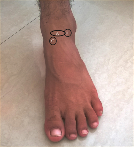
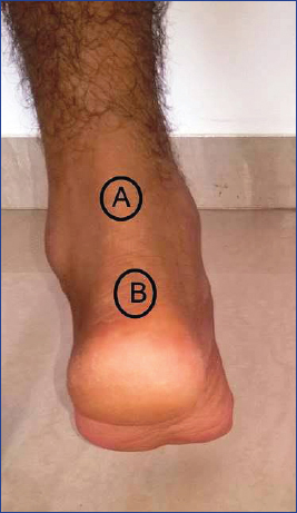
Fig. 1.8 Posterior aspect of the foot and ankle. A, tendo Achilles; B, insertion of tendo Achilles.
Related posts:


Stay updated, free articles. Join our Telegram channel

Full access? Get Clinical Tree


