Flexor Digitorum Longus Tendon Transfer
Annette D. Filiatrault
John A. Ruch
Steven A. Weiskopf
Complex digital deformities and metatarsophalangeal joint (MTPJ) instability encompass a wide range of pathology including crossover toe, severe hammer toe, subluxed or dislocated toe, metatarsalgia, capsulitis/synovitis, and plantar plate insufficiency or rupture. The surgical treatment of these problems requires a sequential process of correction, which may include proximal interphalangeal arthrodesis, soft tissue rebalancing, tendon transfers, plantar plate repair, or metatarsal osteotomy. The controversy that persists in the current day literature as to which procedures are the most appropriate for each deformity only highlights the fact that no one procedure has become the gold standard or fits every patient and deformity. The flexor digitorum longus (FDL) tendon transfer is a common and popular method of reestablishing mechanical stability to a deranged MTPJ with resultant digital deformity. It offers the advantage of changing the deforming force of the FDL to one of active stabilization and plantarflexion of the proximal phalanx at the MTPJ. It may also resist transverse plane forces by imparting both an active and passive stabilizing effect to the digit.
The transfer of the FDL for correction of digital deformities dates back to the early 1900s with Trethowan (1) referencing its use in 1925. Girdlestone used the procedure years before Taylor described it in 1951 as the transfer of the long and short flexor tendons to the extensor expansion, which became known as the Girdlestone-Taylor procedure (2,3). Parrish modified the procedure to include splitting the flexor tendon longitudinally and suturing the ends to themselves and to the dorsal extensor tendon expansion; it is this modification that most closely predates the surgical technique outlined in this chapter (4). Additional variations in the technique have been described by several authors and are discussed in a later subsection.
CLINICAL EXAM AND INDICATIONS
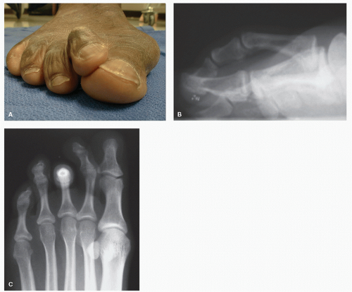
The clinical exam starts with a comprehensive biomechanical evaluation of the entire foot. Once the biomechanical pathology is identified, focus may shift to the digital and MTPJ level. Each joint should be evaluated for reducibility and plane of involvement (Fig. 16.1A). The Kelikian push-up test involves loading the plantar metatarsal head, which generates tension in the plantar fascia that pulls the toe plantarward if the mechanism is intact. The vertical stress test (or digital Lachman test) evaluates the MTPJ plantar plate integrity by placing a dorsally subluxating force of the proximal phalanx on the metatarsal head (5,6). Subluxation may be painful and greater than 2 mm of displacement, particularly compared to the opposite foot, and may indicate rupture of the plantar plate (Fig. 16.1B) (5,6). Standard radiographs taken weight-bearing in relaxed stance reveal the planes and severity of MTPJ derangement. The digital deviation angle (the angle created by the longitudinal axis of the proximal phalanx and the longitudinal axis of the metatarsal shaft) may assist in assessing the degree of toe subluxation (6). The radiographs are evaluated for subluxation or dislocation of the toe, metatarsal protrusion, planes of deformity, the amount of joint arthritis, and related deformities such as hallux valgus. Severe sagittal plane deformity can be seen radiographically as the “gun-barrel sign,” which is shown in Figure 16.1C (5). Magnetic resonance imaging, ultrasonography, or the injection of radiopaque dye (arthrography) for evaluation of the plantar plate and adjacent structures may be performed as adjuncts to radiographs as the clinician feels necessary.
As there is no gold standard for all digital deformities, it may be difficult to clearly define the absolute indications for the flexor tendon transfer. It may be utilized in a variety of situations and in combination with additional procedures. The procedure has been described as the final stage in the sequential repair of the complex hammer toe as well as an isolated procedure. In general, it is best considered as a useful adjunct after sequential hammer toe correction has failed to fully reduce the dorsal tendency of the proximal phalanx on the metatarsal head (5).
SURGICAL TECHNIQUE
The procedure (5) begins with a dorsolinear incision from the metatarsal neck across the MTPJ and ending at the middle phalanx, staying central over these structures to avoid laceration of the neurovascular bundles. The incision may be curved slightly over the MTPJ depending on the deformity. Anatomic dissection is performed at the subcutaneous level, coagulating any crossing tributaries as needed for hemostasis, until the deep fascia is exposed at the proximal interphalangeal joint (PIPJ) and extending this dissection over the long extensor tendon and capsule over the MTPJ. The PIPJ is disarticulated with a transverse tenotomy/capsulotomy at the joint level and is prepared for arthrodesis or arthroplasty. Next, a sequential release of the MTPJ is preformed when required. The extensor digitorum longus and brevis tendons are noted at the MTPJ level, and the extensor hood is released along the medial and lateral aspects of the combined extensor apparatus at that level. The extensor tendon complex should be left intact at the middle one-third area of the proximal phalanx. A Z-plasty lengthening of the extensor complex is performed by making a longitudinal incision between the digitorum longus and brevis tendons (Fig. 16.2A) and then transecting the long tendon distally at the
point where it joins the extensor brevis tendon and the brevis tendon proximal to the MTPJ (Fig. 16.2B). The tendon ends are retracted and an MTPJ release is performed sometimes including use of a McGlamry metatarsal elevator to release plantar adhesions.
point where it joins the extensor brevis tendon and the brevis tendon proximal to the MTPJ (Fig. 16.2B). The tendon ends are retracted and an MTPJ release is performed sometimes including use of a McGlamry metatarsal elevator to release plantar adhesions.
There are varied ways to harvest and transfer the FDL to the dorsal aspect of the proximal phalanx. In the author’s approach, dorsal transfer of the FDL at the level of the PIPJ, the FDL is isolated from the FDB, and the FDL is longitudinally split into two slips. The two slips are delivered dorsal around each side of the proximal phalangeal shaft and are secured superiorly under physiologic tension.
Prior to isolating the FDL tendon at the PIPJ, the proximal phalanx is prepared for transfer by making an incision through the deep fascial attachments along the inferior surface of the proximal phalanx. A freer elevator is passed along the inferior surface of the proximal phalanx, between the periosteum and flexor tendons, creating a plane for easy transfer (Fig. 16.2C). The inferior fibers of the extensor sling are incised along the elevator medially and laterally (Fig. 16.2D and E).
Stay updated, free articles. Join our Telegram channel

Full access? Get Clinical Tree


