CHAPTER 9 External Snapping Hip Syndrome
The snapping hip, or coxa saltans, is characterized by a snapping phenomenon that occurs around the hip in association with hip motion. It has been classified into three different types1:
HISTORY AND PHYSICAL EXAMINATION
In some cases, the snapping phenomenon may be visible under the skin and, in some other cases, it may be palpated over the area of the greater trochanter. In symptomatic patients, the snapping phenomenon is accompanied by pain in the area of the greater trochanter. The snapping phenomenon is always voluntary and the patient often volunteers to demonstrate it.1 Asymptomatic snapping (snapping without pain) must always be considered a normal occurrence.4 Clinical diagnosis is evident; the snapping will occur with flexion and extension of the hip. The snapping phenomenon is also described by some patients as the ability to “dislocate the hip.” This is often demonstrated by actively rotating the affected hip while tilting the pelvis in standing position. The voluntary dislocators are more frequently painless and should only be treated with stretching exercises of the iliotibial band. Symptomatic external snapping hip syndrome is always accompanied with pain and tenderness in the posterior greater trochanteric region. The pain is often secondary to greater trochanteric bursitis and may also be related to tendinosis of the trochanteric insertion of the gluteus medius. When Trendelenburg gait is also found, an associated abductor muscle tear must be suspected; this is an indication for surgical treatment.
DIAGNOSTIC IMAGING
Anteroposterior pelvis x-rays should always be done to identify bony abnormalities, calcifications, or other pathology. The snapping phenomenon can be documented with dynamic ultrasonography and associated pathology such as tendinitis, bursitis or muscle tears can be detected.5,6 Iliopsoas bursitis and abductor muscle tears can be diagnosed using magnetic resonance imaging (MRI).7,8
TREATMENT OPTIONS
Conservative Treatment
Most symptomatic cases improve with stretching physical therapy, nonsteroidal anti-inflammatory drugs (NSAIDs), and corticosteroid infiltration of the greater trochanter bursa.1 When conservative treatment fails, surgical release is indicated. Open iliotibial band release or lengthening has been the traditional surgical option to treat the external snapping hip syndrome.9–12 Recently, we have described a technique for endoscopic iliotibial band release for the treatment of external snapping hip syndrome13 that provides access to the peritrochanteric space.


Arthroscopic Technique
The patient is positioned laterally, similar to the setup for total hip replacement. Surgical drapes must allow for free range of motion of the lower extremity so that the snapping phenomenon is reproduced with flexion and extension of the hip. Snapping usually occurs with flexion of more than 90 degrees. Reproduction of the snapping phenomenon during surgery is important to evaluate when the release of the iliotibial band is complete (Figs. 9-1 and 9-2). No traction is necessary to access the peritrochanteric space, greater trochanteric bursa, and iliotibial band. When there is a combination of periarticular pathology and hip joint pathology, arthroscopic access to the hip joint is necessary. The patient is positioned for hip arthroscopy, preparing for traction to access the central compartment and dynamic positioning for the hip periphery. I favor the lateral position for hip arthroscopy. When arthroscopy of the hip joint is complete, the foot is taken out of the traction device, the perineal post is lowered, and the peritrochanteric space is accessed. This allows for flexion and extension for reproduction of the snapping phenomenon. A more recent positioning method for hip arthroscopy is the Spider device (Tenet Medical, Calgary, Canada) which allows for full range of motion without releasing the foot for the traction device (Figs. 9-3 and 9-4).
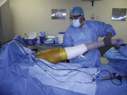
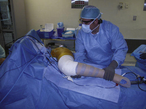
FIGURE 9-2 Hip flexion. It is necessary to flex the hip up to 90 degrees to reproduce the snapping phenomenon.
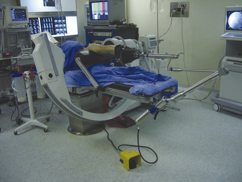
FIGURE 9-3 Patient positioned in lateral decubitus position for arthroscopy of the left hip. Note the horizontal perineal post and the traction device attached to the Spider positioner (Tenet Medical, Alberta, Canada). The image intensifier is horizontally positioned under the table to provide an anteroposterior view of the left hip.

Stay updated, free articles. Join our Telegram channel

Full access? Get Clinical Tree


