Fig. 1
Radiographs (anteroposterior, lateral, axial patella, and long leg view) of the left knee showed a primary cruciate-substituting guided-motion TKR with resurfaced patella. A varus mechanical leg axis was seen. In addition, a rotational malalignment of the tibial TKR component was suspected
Questions
1.
What is your differential diagnosis now?
2.
What are your next steps in diagnostics?
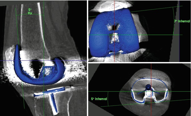
CT was performed to accurately determine the position of the TKR components. Seven-degree internal rotation of the femoral TKR component and 5° internal rotation of the tibial TKR component were found. The mechanical leg axis was in 1° varus (Fig. 2).


Fig. 2
Measurements of TKR component position using a customized and validated software. Five-degree flexion (left) of the femoral TKR component and 7° internal rotation of the femoral (right bottom) and 5° internal tibial rotation (right up)
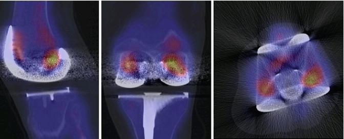
SPECT/CT showed no evidence of loosening or infection (Fig. 3). The mildly increased bone tracer uptake at the medial posterior femoral condyle is an incipient granuloma.


Fig. 3
99mTc-HDP-SPECT/CT images of the left knee showed no increased tracer uptake around the femoral and tibial TKR component indicating a well-fixed TKR and no infection. The mildly increased bone tracer uptake at the medial posterior femoral condyle is an incipient granuloma
Questions
1.
What is your diagnosis and proposed treatment?
2.
How would you address the femoral component malposition?
3.
How would you address the tibial component malposition?
In this case, an iliotibial band (ITB) traction syndrome was present after a cruciate-substituting guided-motion TKR. In addition, there was an internally rotated femoral and tibial TKR component.
A complete revision of the TKR as well as a release of the iliotibial band was discussed in detail with the patient. However, due to her advanced age and significant comorbidities, a primarily nonsurgical approach was chosen.
The patient underwent a series of local cortisone injections at Gerdy’s tubercle and the iliotibial band, which gave her a complete pain relief for 3 months, after which the injections need to be repeated.
Stay updated, free articles. Join our Telegram channel

Full access? Get Clinical Tree


