34
Delayed Treatment of Flexor Tendons: Staged Tendon Reconstruction
Lawrence H. Schneider
History and Clinical Presentation
An 18-year-old student lacerated both flexor tendons in zone 2 of his right dominant index finger on broken glass. His primary treatment was by direct repair of both flexor tendons 3 days postinjury, and he was started on a mobilization program in a hand therapy unit. Unfortunately, this did not result in functional pull-through of his flexor tendons. He then underwent flexor tenolysis in an attempt to salvage function, a procedure that was also not successful. He was first seen on my hand surgery service at 4 months postoperative complaining of restricted motion in the operated finger.
PEARLS
- The patient must demonstrate an ability to comply with a detailed rehabilitative program.
- Staged reconstructions are not a perfect technique, but they are the only way to restore active motion at the interphalangeal joints of the finger in these difficult cases of flexor system disruption.
- Never hesitate to build pulleys! Pulleys need to be strong. Although there are many techniques for pulley reconstruction, I prefer to wrap the tendon material around the proximal phalanx at A2 as was done in this case. At A4 I have most frequently used local tissue sutured over the implant, often with the addition of tendon material.
PITFALLS
- Complications are not infrequent.
- This is not a simple procedure for patient or surgeon.
- Severe flexion contractures especially after stage 1 are poor prognostic signs.
- Double digital nerve injury is a very poor preoperative sign. When present, it may be an indication for amputation or fusion (if the finger is in a nonfunctional position).
Physical Examination
The right hand showed no active flexor tendon function at either the flexor digitorum superficialis (FDS) or flexor digitorum profundus (FDP) of the right index finger (Fig. 34–1). Passive motion was almost full with only a mild flexion deformity at the proximal interphalangeal (PIP) joint. Sensory nerve function was intact. The skin was relatively pliable and soft considering that he had had an injury and two prior operations. The remainder of the hand was normal.
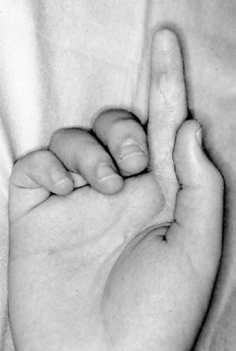
Figure 34–1. Right index finger held in extended position while attempted to make a full fist even after two operative procedures.
Diagnostic Studies
Except for routine radiographs in cases with associated bone injury or arthritis, there are no diagnostic studies needed. Although there is some interest in the use of ultrasound and magnetic resonance imaging (MRI) in the evaluation of the flexor tendon system, I have not used these studies for these cases.
Diagnosis
Flexor System Laceration of Right Index Finger, Status Postoperative Repair, and Tenolysis with Loss of Flexor Tendon Function
Patients with injuries of the flexor system in which there has been marked scarring, disruption of the supporting pulley mechanism, and joint contracture not responsive to therapy measures often become candidates for tendon reconstruction using a staged approach. These patients often have a history of failed prior surgery, with adhesions preventing tendon gliding after a direct repair or tendon rupture after a tenolysis attempt. At the first stage, the scarred tendon is excised and the implant, attached only at its distal end, is placed in the flexor system of the finger. The implant as devised by Hunter and Salisbury consists of a Dacron woven tape encased in silicone rubber. The tape gives body to the implant to enhance its passive gliding and further provides a better hold for the distal sutures at its attachment to the profundus stump. Joint release, pulley reconstruction, skin revision, and nerve repair are performed at this first stage, as needed. Postoperatively the patient is placed in a passive exercise program to regain joint motion while allowing a smooth synovial-like sheath to form in response to the implant. Then at the second stage, performed 3 months later, the implant is replaced by a tendon graft. After this second stage an intense therapy program is initiated.
Nonsurgical Management
Many of these patients present with joint contractures and may benefit from prereconstruction hand therapy to regain full passive motion in the finger preoperatively. This patient had almost full range of motion (ROM), so this therapy was not necessary here. There is no other nonsurgical management for this problem.
Surgical Management
The patient was an appropriate candidate for exploration, with proposed staged flexor tendon reconstruction. As is common in our service, these operations are started under local anesthesia to see whether the repair can be salvaged by flexor tenolysis, and if not, as in this case, then a staged tendon reconstruction is undertaken with implant placement at this first stage.
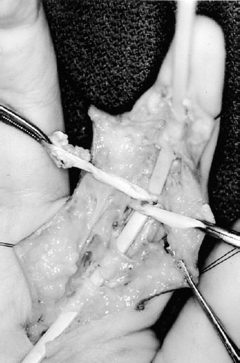
At exploration, it appeared that the repair had ruptured and the entire flexor system was involved in a heavy scar mass. Scarred tendon is sharply excised. Pulley destruction was noted at A2 and A3. Generally all remnants of the pulley system are saved, as even scar tissue can be used to support the implant. Even scarred pulleys are usable over the implant, but in this case A2 needed to be reconstructed. This was done by use of the technique where tendon material, which is generally available from the remnants of the discarded tendon, is wrapped two to three times around the proximal phalanx (Fig. 34–2). In reconstruction of A2, overlying the proximal phalanx, the tendon material is placed around the implant and deep (palmar) to the extensor mechanism. It is advantageous to carry out the pulley reconstruction over a temporary sizer tendon implant (Fig. 34–3). In this case the A1 pulley was preservable, and A4, although scarred, was salvageable. At completion of the pulley reconstruction, the sizer implant is removed and a new implant is broken out of its package, moistened in sterile solution, threaded into the flexor system, and attached distally to the stump of the profundus. In this passive program the proximal end of the implant is placed proximally in the distal forearm and not attached to any motor tendon but left free in the interval between the profundus and superficialis musculotendinous junctions in zone 5 (the distal forearm). At this first stage, scar excision and joint releases are performed as well as nerve repairs and skin revisions, as necessary. After the tourniquet is released and bleeding is controlled, wound closure is performed. The wound is dressed and the extremity is placed in a dorsal splint with the wrist in slight flexion, and the fingers in flexion at the MP joints and in extension for 1 week, at which time a passive mobilization program is begun.
The interval between the two stages is devoted to regaining passive mobility of the involved joints while the soft tissues are healing and a synovial-like sheath is formed in response to the implant. The goal is to grade the finger up to a point where it will accept a tendon graft at the second stage with a reasonable chance to succeed. Three months is sufficient to allow the sheath to form and the finger to heal completely.