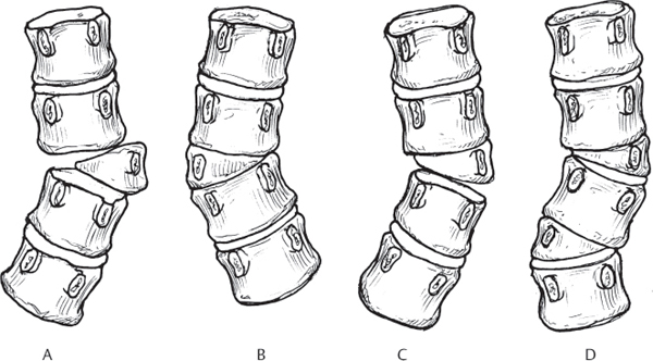
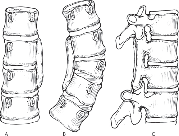
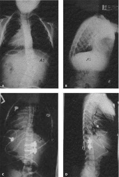
36 Congenital scoliosis is a spinal deformity caused by incompletely formed or incompletely segmented vertebrae and has as its essential characteristic asymmetric growth. Other associated abnormalities may also be present in patients with congenital scoliosis. Klippel-Feil syndrome is seen in ~ 25% of these patients, and some form of renal abnormality in 30%. Cardiac abnormalities can be found in ~ 12% of these patients and some type of dysraphism or other neuraxis abnormalities can be seen in ~ 15%. It is very important to identify these associated problems because they may have significant impact on the timing and/or the treatment of the spinal abnormality. Knowing the precise anomaly in the case of a congenital spine deformity can help in prognosticating the natural history of the condition. This may help in treatment planning or advising the patient and family with regard to the future and the need for treatment. Congenital deformities can be classified as type I: failure of formation (Fig. 36.1), type II: failures of segmentation (Fig. 36.2), or a combination of both. If healthy growth plates exist on a hemivertebra or between vertebrae connected by a uni-lateral bar, relentless progression of the deformity can be anticipated. However, the vertebral abnormality (and there can be multiple ones along the entire spinal column) may simply stunt the growth of the spine without causing major coronal or sagittal plane deformities. Therefore, stunting of trunk growth may occur because of the nature of the abnormality and have little or nothing to do with treatment. Fig. 36.1 Classification: type I congenital spine deformity, failure of formation. (A) Segmented, unincarcerated hemivertebra. (B) Nonsegmented hemivertebra. (C) Partially segmented hemivertebra. (D) Hemimetameric shift. Fig. 36.2 Classification: type II congenital spine deformity, failure of segmentation. (A) Uni-lateral bar with unsegmented adjacent vertebra. (B) Unilateral bar with contralateral segmented hemivertebra. (C) Posterior bar. The physical examination of a patient with a congenital spine deformity is important and revealing. Even infants with congenital scoliosis can manifest a severe deformity and secondary abnormalities, such as an elevated shoulder or rib hump. The exam may also demonstrate other signs of associated conditions, such as a short stiff neck, a hairy patch on the back, a heart murmur, or even an abdominal mass, which may reflect the presence of hydronephrosis. There also may be a significantly deformed chest and other musculoskeletal abnormalities. Appropriate spinal imaging techniques should be used to define the skeletal anomaly and also to rule out often-associated neuraxis abnormalities and abnormalities of other organ systems such as the kidneys or heart. Plain radiography in the upright position in the posteroanterior or anteroposterior (PA or AP) and lateral projections helps to define the nature of the deformity and the overall spinal balance and position of the trunk and is the standard image to be used for subsequent radiographic follow-up. Plain radiographs of the neck also can help rule out Klippel-Feil syndrome. Computed tomography (CT) with three-dimensional (3D) reconstruction has become the standard method of demonstrating the pathoanatomy and is extremely valuable in surgical planning. Magnetic resonance imaging (MRI) demonstrates the neuraxis, whose evaluation is absolutely essential in the presence of a recognizable neurologic deficit and/or prior to surgery as a preoperative evaluation. Ultrasound of the abdomen is important to delineate anomalies of the genitourinary tract, and additional workup of cardiac and renal function should be done prior to any surgical treatment. With regard to prognosis and natural history, active growth is the enemy of congenital scoliosis. Winter has noted that 25% of these deformities do not worsen, 50% worsen moderately, and 25% progress a severe amount. To determine which of these three categories a child falls into, a period of “controlled” observation should be instituted as long as there are no impending neurologic problems that may affect the child’s prognosis. Nonoperative measures are ineffective in the treatment of a deformity comprised of congenital vertebral abnormalities. Surgery for congenital scoliosis can be divided into one of two categories: growth-preserving procedures and trunk-shortening procedures. An example of a trunk-shortening procedure is posterior spinal fusion with or without instrumentation. It is important to understand that in a very skeletally immature patient, anterior fusion must generally accompany any posterior fusion to prevent the crankshaft phenomenon, which would tend to negate the improvement seen initially with posterior fusion. Hemivertebra excision is technically a trunk-shortening procedure, but shortening is minimal if only two vertebrae are fused together and good correction is achieved. Growth-preserving techniques include hemiepiphysiodesis/hemiarthrodesis (H/H), growing rods, and vertical expandable prosthetic titanium rib (VEPTR) as well as vertebral body stapling. H/H is used to treat congenital scoliosis secondary to a hemivertebra that does not have an opposite unsegmented bar. The goal of this procedure is to prevent curve progression and, as a secondary benefit, to gain slow spontaneous correction (though somewhat unpredictable), analogous to that seen with phy-seal stapling of a long bone. Generally only a few levels are involved, thereby limiting the amount of trunk shortening. However, in a situation appropriate for H/H, a hemivertebra excision would often be a better alternative that has the advantage of definitive and acute correction and elimination of the cause of the asymmetric growth (Fig. 36.3A–D). Surgery to preserve trunk height should not be performed if it cannot control a deformity that is severe and progressing. Even if a trunk-shortening procedure is chosen, it should not be delayed to the point that a complicated salvage operation is required because of the magnitude of the deformity. Vertebral stapling on the convex side of the curve is a new form of treatment. VEPTR is designed to correct chest deformity caused by fused ribs and to provide some corrective force to the congenital curve without fusing the spine. More experience with spinal correction will be needed to assess its effectiveness fully. However, it is clear that VEPTR can improve the space available for the lung and therefore lung function. Certain forms of treatment, such as H/H and vertebral stapling as well as VEPTR, need to be instituted early, before the deformity is “too big,” so that growth can help in the spontaneous correction of the deformity and excessive forces on the bone/implant interface can be avoided. The treatment for this heterogeneous group of spinal anomalies must be individualized for each patient. The outcome is largely dependent on the pattern of congenital deformity and the magnitude of the spinal curve; the nature and severity of associated anomalies of the neurologic, cardiac, and renal systems; and complications of treatment. Fig. 36.3 Radiographs of thoracolumbar spine with a hemivertebra causing a scoliosis, treated by hemivertebra resection. (A) Preoperative AP. (B) Preoperative lateral. (C) Postoperative AP. (D) Postoperative lateral of the same patient after hemivertebra resection, showing complete correction of the scoliosis and normalization of the sagittal alignment achieved with the fusion of only one motion segment.
Congenital Scoliosis
![]() Classification
Classification


![]() Workup
Workup
Physical Examination
Spinal Imaging
![]() Treatment
Treatment
![]() Outcome
Outcome

Stay updated, free articles. Join our Telegram channel

Full access? Get Clinical Tree


