Computer-Navigated Total Knee Arthroplasty

Chapter 116 Computer-Navigated Total Knee Arthroplasty


Computer-assisted surgery (CAS) has emerged as one of the most important technologies in orthopedic surgery, and many of the initial applications have focused on adult reconstructive surgery of the knee. The goals of this chapter are as follows: (1) provide a brief history of CAS of the knee and the evolution of basic concepts; (2) present the rationale for the use of CAS in knee surgery; (3) describe the hardware and software components of CAS systems; (4) illustrate the measured resection technique for total knee replacement surgery; and (5) present CAS applications in knee surgery that will be available in the near future.


History of Computer-Assisted Knee Surgery and Evolution of Basic Concepts


Although a large volume of important work that became the foundation for computer-assisted knee surgery was being carried out throughout the 20th century, the initial clinical applications for knee surgery began in the 1980s.109 In 1986, Kaiura, from the University of Washington presented a thesis on robotic-assisted total knee arthroplasty (TKA).39,62 This work led to the design of one of the first computer robotic assistive systems for TKA, described by Matsen and colleagues86 in 1993.137 In the early 1990s, Kienzle and associates65 and Stulberg134 also described a computer-assisted robotic total knee replacement system. The desired position of the femoral and tibial cutting blocks was determined on a three-dimensional model derived from a computed tomography (CT) scan obtained preoperatively. The robot was secured to the operating table and to the bones and then positioned a drill to make holes for the pins over which the femoral and tibial cutting blocks were placed. The surgeon then performed the cuts with a standard oscillating saw. The accuracy of block placement with this system was within 1 mm and 1 degree. This work also introduced a method for determining the center of the femoral head by means of a kinematic registration technique. This technique was subsequently incorporated into all current navigation systems. Dynamic reference frames that were tracked by a camera were placed on the femur and the hip was put through a range of motion. The center of the sphere described during the circumduction maneuver represented the center of the femoral head.


By the early 1990s, the principles on which current computer-assisted total knee replacement systems are based had been established and validated, and important steps had been taken to identify how critical anatomic landmarks for knee surgery could be accurately acquired. It was, however, apparent to those working on these projects at that time, especially orthopedic surgeons, that robotic-based computer-assisted systems were too cumbersome, complex, and potentially too unsafe to be of substantial use to an active knee replacement surgeon in the foreseeable future.


Surgical navigation systems, however, appeared then to offer an attractive alternative to a field such as knee surgery, which could benefit from the accuracy provided by computer-assisted techniques without having to deal with the drawbacks and complexity of robots. These systems allowed intraoperative tracking of the position of the surgical tools and the bones to which they were attached. The surgeon, not a robot, could control all phases of the procedure.


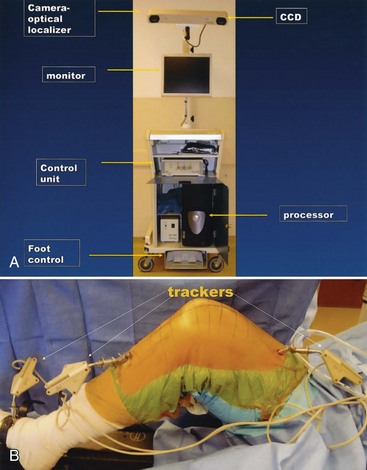
The rapid evolution of surgical navigation systems to support the performance of knee surgery was made possible by the availability, in the early 1990s, of optical and electromagnetic tracking systems (Fig. 116-1). Optical tracking systems have played a special role in the development of surgical navigation systems for knee surgery because of their accuracy and reliability. These tracking systems, also referred to as optical localizers, have charge-coupled devices (CCDs, or cameras) mounted on a rigid frame. These cameras measure the position and orientation of multiple tracking markers, also called trackers, or rigid bodies. Each tracker incorporates a set of light-emitting diodes (LEDs), or reflective spheres, mounted in precise relative positions. These trackers can be affixed to bones, tools, and implants. The optical tracker is therefore able to monitor the precise position of these objects at any point during the surgical procedure.


During the first half of the 1990s, a great deal of basic research was performed to develop surgical navigation systems using these optical tracking systems.109 The first clinical applications incorporating these efforts began in 1995 in the field of spine surgery. Although these applications were based on the acquisition of anatomic information by means of preoperative imaging techniques, they were the basis for the development in the late 1990s of the image-free systems currently most widely used in knee surgery.


Four types of surgical navigation models are used in computer-assisted orthopedic surgery: (1) preoperative image–based (e.g., CT scans)68,75; (2) intraoperative image–based (e.g., fluoroscopy)84,85; (3) image-free; and (4) individual templating.37,75,93,112,130 The anatomic information on which the surgical plan is made is acquired differently in each model. Although each model has its advantages and drawbacks, the image-free method for acquiring critical anatomic information has proved to be most amenable to the methods used currently to perform knee surgery. This method was first used clinically in 1993 to place grafts during anterior cruciate ligament (ACL) surgery.109 Image-free navigation was subsequently applied to TKA surgery by Leitner and colleagues.80 The first image-free computer-assisted TKA was performed in Grenoble, France, by Picard and associates in 1997.107 The system used became the first commercially available image-free navigation system for knee reconstructive surgery (the OrthoPilot).106 It identified critical anatomic landmarks using both kinematic (e.g., femoral circumduction, as described by Kienzle and coworkers in 198965) and surface registration techniques. Krackow and colleagues74 subsequently developed surgical navigation systems based on these concepts. There are now a large number of image-free navigation systems available for use with almost every total knee system.


Although the foundations for the CAS systems currently being used in knee surgery began to be developed a century ago, actual systems available for clinical use have been available for less than 10 years. The rapid emergence of these systems suggests that current models will undergo substantial evolution and modification in the next few years.


Rationale For Use


Successful surgical reconstruction of the knee requires proper patient selection, appropriate perioperative management, correct implant selection, and accurate surgical technique. The consequences of performing a knee reconstruction inaccurately have been well documented for TKA, unicondylar arthroplasty, ACL reconstruction, and high tibial osteotomy.*


Although mechanical instrumentation has significantly increased the accuracy and reliability with which knee reconstructions are performed, errors in implant and limb alignment continue to occur, even when the procedures are performed by experienced surgeons. Moreover, the accuracy with which these procedures are performed is dependent on the knowledge and experience of the surgeon and the frequency with which the surgeon performs the procedure. CAS techniques have been developed to address the inherent limitations of mechanical instrumentation. The goals of integrating CAS with knee reconstruction techniques are to increase the accuracy of these procedures and reduce the proportion of alignment outliers that occur when these procedures are performed.


A surgeon can only use computer-assisted navigation safely and effectively if he or she is familiar with and comfortable with the procedure’s goals and the surgical techniques necessary to achieve those goals. Numerous reports have confirmed that when surgeons experienced in the manual performance of the knee reconstruction procedure use CAS techniques, average implant and limb alignment is improved and the incidence of outliers is reduced.* Moreover, a recent study has indicated that when experienced surgeons and coworkers use techniques to perform knee reconstructive procedures, their ability to perform these procedures manually improves.145


Errors can occur at numerous points during the performance of a knee reconstruction. Placement of the cutting blocks or ligament alignment jigs may be inaccurate. Attachment of these tools to bone may produce an error in their placement. The actual performance of the cut or drilling of the hole may be inaccurate (e.g., the saw blade may deflect). The final insertion of the implant may be inaccurate. Mechanical instrumentation does not provide a method for checking the accuracy of each of these steps of a knee reconstructive procedure. Another goal of integrating CAS with knee reconstruction is to provide the surgeon with a means to measure the accuracy with which each step of the procedure is performed.


Knee reconstructive procedures attempt to align limbs and implants correctly. They also seek to restore appropriate kinematic relationships and ligamentous stability to the knee.


Mechanical instrumentation cannot measure the precision with which knee kinematics and ligament stability are restored. CAS techniques make it possible to determine the presurgical kinematic relationships and ligamentous stability of the knee and help guide the surgeon to restore desired kinematic relationships and ligamentous balance.


Finally, CAS provides a unique and unprecedented opportunity to train residents and orthopedic surgeons to perform knee reconstruction procedures accurately.95,140,148 CAS as a training tool has now been used more frequently. Applications have been developed and are now being used to allow surgeons to carry out self-assessment evaluation of their surgical skills for performing TKA and ACL surgery. Applications are also being developed to test the skills of surgeons and residents to learn various knee reconstruction procedures. Possibly, the most compelling rationale for applying CAS to knee reconstruction will prove to be the potential to revolutionize how surgeons develop and evaluate their surgical skills.


Hardware and Software Requirements


A detailed description of the hardware and software needed to perform computer-assisted reconstructive knee surgery is beyond the scope of this chapter. However, it is important that knee surgeons understand the basic components of a computer-assisted orthopedic system so that they can use the system correctly, safely, and efficiently and make intelligent choices regarding the appropriateness of various systems for their surgical needs.


Hardware


Hardware devices common to CAS systems are (1) imaging devices; (2) computers, peripherals, and interfaces to allow them to function in the operating room; and (3) localizers and trackers (see Fig. 116-1).


Localizers and Trackers


A CAS knee navigation system can be thought of as an aiming device that enables real-time visualization of surgical action with an image of the operated structures. For this navigation to occur, it is necessary that the position and orientation of an instrument be visualized with respect to the anatomic structures to which it is attached. Although this objective could be met by attaching tools to a rigid multilinked arm attached to a pedestal, such a device would be unsuitable for knee surgery, in which the limb must be freely moved. Therefore, contactless systems are used to communicate between the extremity and computer system. Information can be transmitted by infrared light, electromagnetic field, or ultrasound. Each method has its advantages and drawbacks. All these methods allow several objects (e.g., two bones) to be viewed simultaneously.


Optical Localization


Optical localization via infrared light is currently the most widely used method of communication between the operated extremity and computer. Two types of optical tracking are used, active and passive. Systems with active tracking use markers (also called trackers, or rigid bodies) with LEDs that send out light pulses to a camera (optical localizer). Three or (for redundancy) more of these LEDs are attached to screws or wires that are rigidly attached to the femur and tibia. The camera system to which the light is sent consists of two planar or three linear CCDs that are rigidly mounted onto a solid housing (Polaris, Northern Digital, Ontario, Canada, is a commonly used camera system). Passive systems use reflecting spheres placed on tracking markers that are attached to screws or pins rigidly implanted in the femur and tibia. Infrared flashes sent by LED arrays on the camera housing illuminate the spheres. The two planar or three linear CCDs observe the reflections and interpolate the spatial location of each light source. It is important for the surgeon and staff to realize that the arrays on the tracking markers, whether active or passive, are specific to each CAS system. One company’s trackers cannot be used on another company’s CAS system, even though the trackers may appear to be similar (see Fig. 116-1).


The advantages of optical localizing systems are that they are reliable, flexible, and highly accurate and have good operating room compatibility. A disadvantage of these systems is that it is necessary to provide free line of sight between the LED spheres and the CCD arrays on the camera (optical localizer). Active trackers may require cables to power and synchronize the LEDs. These cables may be cumbersome. Active trackers can be driven by batteries, which eliminates the need for cables, but these batteries require recharging, thus making their use for sequential procedures more difficult. Passive trackers do not require cords. However, automatic tool identification is more difficult for passive systems because all spheres in view reflect the light flashes equally. Unique identification of each tracker is possible with the sequentially pulsed LEDs of active trackers. The reflecting spheres must constantly be kept clean to obtain accurate signal transmission. Moreover, the spheres are disposable and therefore a source of additional expense for each procedure.


Software


The function of software in CAS systems is to integrate medical images and mathematic algorithms with surgical tools and surgical techniques. A relatively small number of software components underlie most CAS image-free systems. These components include registration, navigation, procedure guidance, and safety.


Image-free CAS knee systems use as their preoperative plan the concepts of limb and implant alignment that are currently used with manual instrumentation (e.g., restoration of the mechanical axis). To accomplish this, anatomic and kinematic information about a patient must be transmitted to the software on the computer and geometrically transformed by registration algorithms. Because bones are rigid and assumed to be unlikely to deform during the procedure, the algorithms used are termed rigid. These algorithms also require that the trackers attached to bones do not move during the procedure. Fiducial-based registration is a type of rigid registration. Therefore, the objects to which the LEDs are attached may be referred to as fiducials, trackers, or rigid bodies. Fiducial registration requires that at least three sets of markers be implanted into each bone or attached to each tool to determine the object’s position and orientation. Therefore, each tracker must have at least three LEDs or reflecting spheres. Some CAS knee systems currently use shaped-based registration as an alternative to fiducial-based registration. These systems measure the shape of the bone surface intraoperatively and match the acquired shape to a surface model created from medical images stored in the computer. The registration process for image-free knee navigation systems requires that information be acquired via kinematic techniques (e.g., circumducting the leg to determine the center of the femoral head) or surface registration techniques (e.g., touching bone landmarks with a probe).


Once the software takes anatomic and kinematic input from the extremity and geometrically transforms it, the surgeon is presented with a user interface that depicts the steps of the knee procedure in sequence. One of the most important objectives of software development in CAS knee applications is to depict procedure sequences that are familiar to surgeons and with which they have previously become comfortable using manual instrumentation.


Measured Gap Resection Technique


A surgeon can only use computer-assisted navigation safely and effectively if he or she is familiar with and comfortable with the procedure’s goals and surgical techniques necessary to achieve those goals. The goals of TKA surgery are to align the extremity and implant(s) accurately and to produce a stable, balanced knee joint. These goals can be achieved using one of two surgical strategies: (1) a gap balancing approach, in which equal collateral ligament tension in flexion and extension is sought prior to and as a guide to final bone cuts; or (2) a measured gap resection approach, in which bone landmarks are used to guide resections equal to the distal and posterior thicknesses of the femoral component. Collateral ligaments are then balanced with the trial implants in place. If done properly, the two techniques should produce identical results with regard to stability and alignment. A navigation system for assisting the performance of a total knee replacement should provide separate software programs to support each surgical approach. Surgeons should use the approach with which they are most familiar and comfortable.


Ligaments function properly with the desired isometry in the measured resection technique if the origin of the collateral ligaments is at or closely related to the axis of tibiofemoral flexion-extension. For circular condylar femoral geometry and dished tibial plateau geometry, the centers of femoral rotation coincide with the centers of the femoral surface geometry. It is essential to the optimum performance of the measured resection technique that the size of the femoral implant closely approximates the size of the femur. It is important to realize that the measured resection technique can be carried out by starting with a femoral or tibial resection. The femoral implant size and rotation can be determined using a posterior referencing or anterior referencing technique. Navigation software should allow the surgeon to choose among these options.


Preferred Approach


I prefer the navigated measured resection approach for the following reasons:


Although the measured resection technique can be performed accurately beginning on either the femur or tibia, I prefer to start the procedure on the femur for the following reasons:


Although the measured resection technique can be performed accurately using an anterior or posterior referencing approach to establish the size and rotation of the femur, I prefer the anterior referencing approach because the posterior condylar anatomy is highly variable and unpredictable and locating consistent posterior condylar points is not reliable. As a result, the posterior condylar line is not a reliable guide for establishing the rotation or size of the femoral component. Moreover, precise placement of the femoral implant on the anterior cortex is difficult with the posterior referencing technique. The accuracy of this technique is further compromised if less invasive approaches are used that may make access to the posterior condyles even more difficult. The anterior referencing approach allows the anterior cortex of the femur to be identified accurately and makes precise rotational alignment of the femoral component using the plane of patellar tracking possible. However, if the anterior referencing technique is used, it is critical that an accurate determination of femoral component size be made before positioning the alignment guide. Both navigation and manual tools should be used to make this determination.

Stay updated, free articles. Join our Telegram channel

Aug 26, 2016 | Posted by in ORTHOPEDIC | Comments Off on Computer-Navigated Total Knee Arthroplasty

Full access? Get Clinical Tree

Get Clinical Tree app for offline access